Review
. 2021 Apr 1;17(4):e1009394.
doi: 10.1371/journal.ppat.1009394. eCollection 2021 Apr.

Transport mechanisms at the malaria parasite-host cell interface

Affiliations
Review

Transport mechanisms at the malaria parasite-host cell interface

Josh R Beck et al. PLoS Pathog. .

Abstract

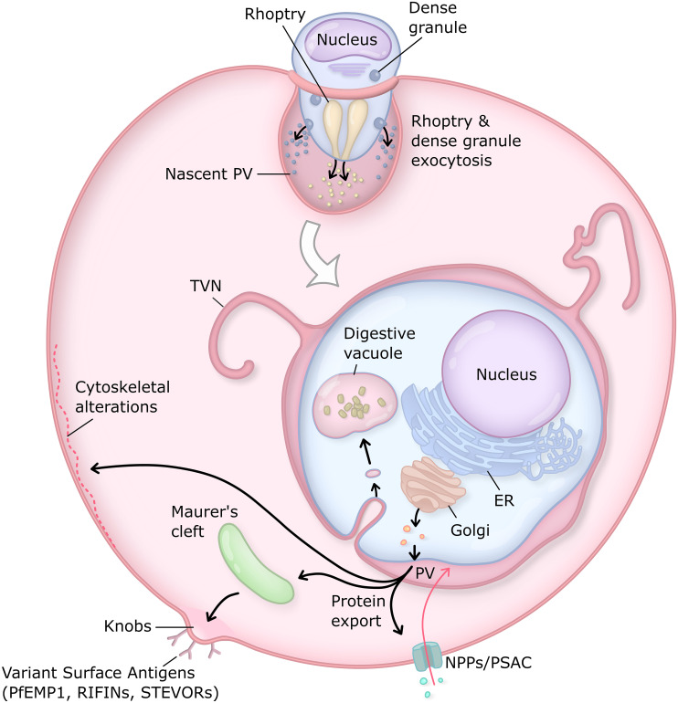
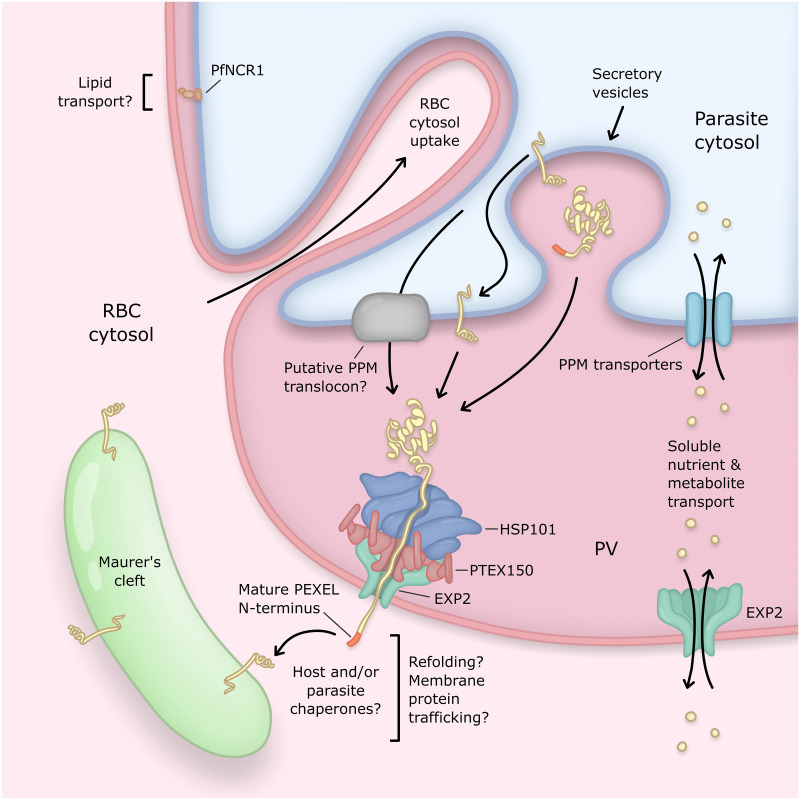
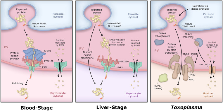
Obligate intracellular malaria parasites reside within a vacuolar compartment generated during invasion which is the principal interface between pathogen and host. To subvert their host cell and support their metabolism, these parasites coordinate a range of transport activities at this membrane interface that are critically important to parasite survival and virulence, including nutrient import, waste efflux, effector protein export, and uptake of host cell cytosol. Here, we review our current understanding of the transport mechanisms acting at the malaria parasite vacuole during the blood and liver-stages of development with a particular focus on recent advances in our understanding of effector protein translocation into the host cell by the Plasmodium Translocon of EXported proteins (PTEX) and small molecule transport by the PTEX membrane-spanning pore EXP2. Comparison to Toxoplasma gondii and other related apicomplexans is provided to highlight how similar and divergent mechanisms are employed to fulfill analogous transport activities.


Conflict of interest statement

The authors have declared that no competing interests exist.

Figures

Fig 1
Fig 1. The Plasmodium intraerythrocytic PV.
The PV is generated through invagination of the erythrocyte membrane during invasion. Discharge of the rhoptry and dense granule secretory organelles during and immediately after invasion rapidly establishes transport activities at the PVM. This is followed by a major wave of protein export during the early stages of intraerythrocytic development to remodel the host cell. During this time, the parasite exomembrane system is also generated, including TVN extensions of the PV thought to provide greater surface area for nutrient uptake and Maurer’s clefts, which serve as trafficking platforms for parasite exported effectors en route to the host membrane. Near the end of this period, a major increase in host membrane permeability is facilitated by activation of NPPs including PSAC. In addition to these changes in erythrocyte permeability, key host modifications include cytoskeletal rigidification and the formation of raised knobs on the erythrocyte surface for display of adhesins that mediate iRBC sequestration and immune evasion. In P. falciparum, the responsible variant surface antigens include PfEMP1, the RIFINs, and the STEVORs, each encoded by large, multigene families. Endocytic uptake of hemoglobin-rich host cytosol and degradation in the digestive vacuole liberates amino acids to support parasite metabolism and frees space for expansive growth within the erythrocyte. ER, endoplasmic reticulum; iRBC, infected red blood cell; NPPs, new permeability pathways; PfEMP1, P. falciparum erythrocyte membrane protein 1; PSAC, Plasmodial surface anion channel; PV, parasitophorous vacuole; PVM, PV membrane; RIFIN, repetitive interspersed family protein; STEVOR, sub-telomeric variable open reading frame protein; TVN, tubulovesicular network.
Fig 2
Fig 2. CryoEM structure of PTEX.
Top and side views of the HSP101 (a, d), PTEX150 (b, e), and EXP2 (c, f) cryoEM maps are shown. Protomers of each protein are colored as increasingly dark gradients of blue (HSP101), salmon (PTEX150), or aquamarine (EXP2). Also visible in the HSP101 portion of the map are molecules of ATPγS (magenta) bound to each NBD as well as density corresponding to the polypeptide backbone of cargo within the HSP101 channel (pink). The cargo is engaged by NBD2 pore loops colored light blue (protomers 1–3) and yellow (protomers 4–6). The high-resolution cryoEM map of the full PTEX complex is shown (g), with the same color scheme as in panels (a–f). The same cryoEM map at a lower threshold is shown overlaid with the high-resolution cryoEM map (h) to display additional lower-resolution features, including an additional density above HSP101 which most likely corresponds to the HSP101 NTDs, which are not resolved in the high-resolution structure. Also visible is the detergent belt, as well as long helical densities extending from the PTEX150 (668–823) region. These densities appear to connect to additional densities on top of the HSP101 M-domains. The cryoEM map at a lower threshold is shown alone (i) to allow better visualization of the additional densities. cryoEM, cryo-electron microscopy; EXP2, exported protein 2; HSP101, heat shock protein 101; M-domains, Middle domains; NBD, nucleotide-binding domain; NTD, N-terminal domain; PTEX, Plasmodium Translocon of EXported proteins.
Fig 3
Fig 3. Mechanism of effector protein export by PTEX.
Model for PVM translocation mechanism is shown based on the PTEX engaged and resetting states observed by cryoEM. Similar to other HSP100s, pore loops projecting from the HSP101 NBDs interact with cargo in the channel. Conformational changes in the HSP101 hexamer enable these loops to grasp successive portions of the cargo, pull it into the channel, and hold it in place to prevent backsliding. Repeated cycles mediate stepwise unfolding and threading of cargo through HSP101 and across the PVM via the PTEX150 flange-like adaptor and EXP2 membrane-spanning pore. cryoEM, cryo-electron microscopy; EXP2, exported protein 2; HSP101, heat shock protein 101; NBD, nucleotide-binding domain; PTEX, Plasmodium Translocon of EXported proteins; PVM, PV membrane; RBC, red blood cell.
Fig 4
Fig 4. Transport activities at the PV of the blood-stage malaria parasite.
The PV is the principal host–parasite interface and the site of several distinct transport activities mediating exchange of proteins and small molecules between the parasite and host compartments. The PV is compartmentalized into regions of close apposition between PVM–PPM (top left) and regions with greater luminal space (bottom right). Machinery involved in protein export and transport of solutes localizes to the latter regions. Parasite effector proteins destined for export into the host cell are delivered to the PV by secretory vesicles and then translocated across the PVM by the PTEX. Most exported proteins contain a PEXEL motif which is processed by the ER-localized aspartic protease Plasmepsin 5 to license export. Soluble exported proteins can be directly accessed by the PTEX AAA+ unfoldase HSP101 in the PV lumen, while exported integral membrane proteins are first incorporated into the PPM and then require a translocation step for PPM extraction that appears to involve unfolding power additional to HSP101. HSP101 is coupled to the membrane-spanning EXP2 pore via a flange-shaped adaptor known as PTEX150. Mechanisms for effector refolding, trafficking, and membrane insertion beyond the PVM are largely unknown and likely involve host and/or exported parasite chaperones. The EXP2 pore also serves a secondary role to transport small molecules across the PVM, likely independent of the PTEX complex. PfNCR1, a protein similar to the NPC1 protein involved in cholesterol egress from late endosomes, localizes to regions of close PVM–PPM apposition, suggesting that these may be sites of lipid transport. Parasites also endocytose large amounts of the erythrocyte cytosol through cytosomal invaginations of the PVM–PPM. ER, endoplasmic reticulum; EXP2, exported protein 2; HSP101, heat shock protein 101; NPC1, Niemann–Pick C1; PfNCR1, P. falciparum Niemann-Pick Type C1-related protein; PEXEL, Plasmodium EXport ELement; PPM, parasite plasma membrane; PTEX, Plasmodium Translocon of EXported proteins; PV, parasitophorous vacuole; PVM, PV membrane.
Fig 5
Fig 5. Comparison of PV transport in Plasmodium blood and liver-stages and Toxoplasma.
In contrast to the blood-stage, HSP101 has not been detected in the liver-stage PV, suggesting fundamental differences in the export mechanism within the hepatocyte. While EXP2 has recently been implicated in sporozoite invasion of hepatocytes, it is unknown whether EXP2 and/or PTEX150 contribute to protein export or small molecule transport during intrahepatic development. While EXP2 orthologs GRA17 and 23 are required for small molecule transport across the Toxoplasma PVM, they are not involved in the export of dense granule effector proteins from the PV. Instead, a distinct set of proteins are required for export of these effectors including the membrane proteins MYR1–4 which are believed to form a novel translocon (possible MYR translocon organization shown here is purely speculative for purposes of illustration). Export also requires the putative phosphatase GRA44 and the chaperone GRA45, which may insert the MYR proteins into the PVM. Export additionally depends on the activity of ROP17, a kinase injected from the rhoptries into the host cytosol during invasion. ROP17 acts at the PV surface but the phosphorylated target(s) is not known. TgPPMC3, an additional phosphatase localized to the PV and involved in the export of a subset of effectors, is not represented in the cartoon. Processing of a TEXEL motif by the aspartic protease ASP5 is important for export of some Toxoplasma dense granule effectors while others lack the motif and are not processed by ASP5, although their export still requires ASP5 for unclear reasons. In contrast to the Plasmodium blood-stage, most ASP5 substrates are non-exported PV resident proteins, including MYR1, GRA44, and GRA45. EXP2, exported protein 2; HSP101, heat shock protein 101; PTEX, Plasmodium Translocon of EXported proteins; PV, parasitophorous vacuole; PVM, PV membrane; TEXEL, Toxoplasma EXport ELement.

References

    1. Kwiatkowski DP. How malaria has affected the human genome and what human genetics can teach us about malaria. Am J Hum Genet. 2005;77(2):171–92. Epub 2005/07/06. 10.1086/432519 . - DOI - PMC - PubMed
    1. de Koning-Ward TF, Dixon MW, Tilley L, Gilson PR. Plasmodium species: master renovators of their host cells. Nat Rev Microbiol. 2016;14(8):494–507. 10.1038/nrmicro.2016.79 . - DOI - PubMed
    1. Lingelbach K, Joiner KA. The parasitophorous vacuole membrane surrounding Plasmodium and Toxoplasma: an unusual compartment in infected cells. J Cell Sci. 1998;111(Pt 11):1467–75. . - PubMed
    1. Suss-Toby E, Zimmerberg J, Ward GE. Toxoplasma invasion: the parasitophorous vacuole is formed from host cell plasma membrane and pinches off via a fission pore. Proc Natl Acad Sci U S A. 1996;93(16):8413–8. Epub 1996/08/06. 10.1073/pnas.93.16.8413 - DOI - PMC - PubMed
    1. Sherling ES, van Ooij C. Host cell remodeling by pathogens: the exomembrane system in Plasmodium-infected erythrocytes. FEMS Microbiol Rev. 2016;40(5):701–21. 10.1093/femsre/fuw016 - DOI - PMC - PubMed

Publication types

MeSH terms

LinkOut - more resources