Review
. 2021 Apr 2;185(3):765-780.
doi: 10.1093/plphys/kiaa061.

Shouting out loud: signaling modules in the regulation of stomatal development

Affiliations
Review

Shouting out loud: signaling modules in the regulation of stomatal development

Arvid Herrmann et al. Plant Physiol. .

Abstract

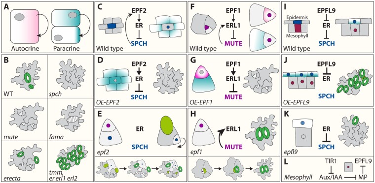
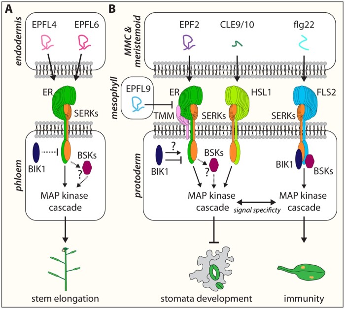
Stomata are small pores on the surface of land plants that facilitate gas exchange for photosynthesis while minimizing water loss. The function of stomata is pivotal for plant growth and survival. Intensive research on the model plant Arabidopsis (Arabidopsis thaliana) has discovered key peptide signaling pathways, transcription factors, and polarity components that together drive proper stomatal development and patterning. In this review, we focus on recent findings that have revealed co-option of peptide-receptor kinase signaling modules-utilized for diverse developmental processes and immune response. We further discuss an emerging connection between extrinsic signaling and intrinsic polarity modules. These findings have further enlightened our understanding of this fascinating developmental process.


Figures

Figure 1
Figure 1
Peptide–receptor signaling throughout stomata development. Overview of the different receptor peptide signaling modules and their main downstream components regulating cell-state transitional steps within the stomatal cell lineage. Members of the EPF family, EPF1, EPF2, and EPFL9/STOMAGEN, are secreted from stomatal precursors and underlying mesophyll cells into the apoplast and competitively bind to three cell surface LRR–RLKs, ERECTA (ER), ER-LIKE 1 (ERL1), and ERL2. Together with the LRR receptor protein, TMM and BRI1-ASSOCIATED RECEPTOR KINASE/SOMATIC EMBRYOGENESIS RECEPTOR KINASE (BAK/SERK) family RLKs, they form a ternary receptor complex. This complex activates the downstream MAPK cascade (arrow, P+) that is composed of YODA (YDA) MAPKKK, two redundant MAPKKs (MKK4 and MKK5) and two, redundant MAPKs (MPK3 and MPK6) possibly through two functional redundant members of the BSK family (question marks; cytoplasm). The activated MAPK cascade in return controls the consecutive activities of three bHLH TFs and their partner bHLH proteins SCREAM (SCRM) and SCRM2 (nucleus). While SPEECHLESS (SPCH) and MUTE are negatively regulated through TF inhibition by the MAPK cascade, FAMA is activated by the latter to inhibit further symmetric cell division and promote cell differentiation. Each TF pair is specifically expressed during the timely progression of stomata development (color indicated). In the model plant Arabidopsis, stomata development occurs by the initiation of an asymmetric division of protodermal cells called MMCs (blue). The ACD generates a small meristemoid (purple) and a large SLGC (light grey). The meristemoid can undergo one to three rounds of asymmetric divisions in an inward-spiral manner to produce additional amplicons surrounded by SLGCs. Stomata development ends when the late meristemoid differentiates into a GMC (light green), which then will further divide symmetrically to generate a pair of GCs (dark green).
Figure 2
Figure 2
Overview of stomata mutant phenotypes and signaling modes of action during stomata development. (A), Different modes of action during cell signaling. During autocrine signaling, a cell secretes a messenger (arrow), which leads to changes in the same cell after binding to cell surface receptors (light red). Paracrine signaling from a cell induces a cellular response in a nearby cell (arrow) in a non-cell autonomous manner (light blue). (B), Mutant phenotypes of key stomatal components. While loss of function mutations in the bHLH TFs SPCH and MUTE lead to phenotypes only consisting of epidermal cells in case of spch and arrested meristemoids in case of mute, piled up symmetric divisions in GMCs can be observed for fama mutants. This indicates that each individual TF is required for a specific cell differentiation step during stomata development ontogenesis. Although an increased number of ACDs in the er single mutant can be observed, only the erf triple mutant (er erl1 erl2) or loss of tmm exhibit severe stomatal clustering. Uncoupling the functional redundancy of the ERf members identified specific peptide–receptor pairs, which contribute to different steps during stomata development (C–H). SPCH-induced EPF2-dependent paracrine signaling by MMCs (light blue) is perceived by ER in neighboring protodermal cells to inhibit SPCH (dark blue) activity (C). Modulation in EPF2 activity can cause an epidermis devoid of stomata when overexpressed (D) or piled up asymmetric division in epf2 mutants (E) since SPCH activity is not downregulated by the EPF–ER signaling module. MUTE (purple) activity depends on both EPF1-dependent autocrine (light blue) and paracrine (light red) signaling. MUTE directly induces ERL1 expression, while at the same time ERL1 perceives EPF1 signal in SLGCs (paracrine) as well meristemoids (autocrine) to inhibit MUTE activity (F). This negative feedback loop ensures the right amount of MUTE activity provided by the EPF1–ERL1 signaling module for proper GC differentiation. While overexpression of EPF1 causes meristemoid arrest due to excessive activation of the ERL1-driven signaling pathway (G), loss of EPF1 results in stomata clustering due to excessive MUTE activity (H). I, EPFL9, also known as STOMAGEN, is expressed in the mesophyll (dark red) and positively regulates stomatal development through a competitive mode of action with EPF1 and EPF2 for receptor binding on ERf members. While overexpression or exogenous application of EPFL9/STOMAGEN induces paracrine signaling, leading to numerous clustered stomata (J), epfl9/stomagen confers reduced stomatal density (K). Intriguingly, the molecular control of EPFL9/STOMAGEN expression within the mesophyll is tied to auxin signaling. Repression of EPFL9/STOMAGEN is ensured by ARF5/MONOPTEROS (MP), which in return is under the control of specific AUX/IAA BODENLOS (L).
Figure 3
Figure 3
Intrinsic polarity cues contribute to stomatal development. The plant-specific polarity protein BASL (green) exhibits a dual localization pattern. While BASL accumulation can be detected in the nucleus during stomatal lineage progression, it additionally forms a polarized crescent in MMCs and meristemoids in the region distal to the future division plane and away from the migrated nucleus. This crescent localization is dependent on both MPK3/6 (A), as well as on members of the plant-specific BREVIS RADIX (BRX) (light red) family (B). Right before MMC asymmetric division (C), BASL and POLAR (orange) recruit the BIN2 (light blue) to the cellular cortex. This BASL-POLAR-BIN2 polarity module enables the inhibition of the YDA MAPK cascade via direct phosphorylation of YDA. This leads to an elevated accumulation of SPCH, hence promoting stomatal asymmetric division. To prevent further ACD in SLGCs, SPCH must be down regulated. To do so, BIN2 disassociates from POLAR after phosphorylating the latter and re-localizes into the nucleus to inhibit SPCH activity while POLAR is degraded (D). Furthermore, a positive feedback loop between BASL and the YODA MAPK cascade restricts further stomatal ACD in the SLGC through direct phosphorylation of SPCH. The specific localization pattern of the BASL-POLAR-BIN2 polarity module further fine tunes stomatal development (E–G).
Figure 4
Figure 4
Signal specificity is maintained between different LRR–RLKs with shared intermediate components. ER mediates different developmental processes upon different peptide-ER-co-receptor formation. (A) During stem elongation EPFL4 and EPFL6 are secreted from endodermis cells into the apoplast to be perceived by ER in phloem cells in a noncell autonomous manner. Upon EPFL4/6-ER-SERKs formation, with the additional possibility of a higher order complex with BSKs (question mark) as well the RLCK BOTRYTIS-INDUCED KINASE1 (BIK1), a MAP kinase cascade is activated to transduce the extracellular information into the cell to further promote downstream effectors for stem elongation. Although stomata development depends also on the formation of an EPF/EPFL–ERf–SERKs complex, possibly with BSKs, to activate a MAPK cascade to overall inhibit stomata development (B), this peptide–receptor-co-receptor complex additionally requires the co-receptor TMM. Whether BIK1 also plays a role during stomata development is still unknown (question mark). Stomata development can be further fine-tuned through CLE9/10 peptides, which are perceived by the LRR receptor kinase HAESA-LIKE 1 (HSL1) and its coreceptor BAK1, enabling the activation of the same components of the MAPK cascade. It is unclear how the MAPK cascade is able to cope with the signal output of two different peptide–receptor signaling complexes for the same developmental process as well as create signal specificity between shared pathways, such as the flg22–FLS1–SERKs signaling module for plant innate immunity.

Similar articles

Cited by

References

    1. Aan den Toorn M, Albrecht C, de Vries S (2015) On the origin of SERKs: bioinformatics analysis of the somatic embryogenesis receptor kinases. Mol Plant 8:762–782 - PubMed
    1. Abrash E, Anleu Gil MX, Matos JL, Bergmann DC (2018) Conservation and divergence of YODA MAPKKK function in regulation of grass epidermal patterning. Development 145:dev165860. - PMC - PubMed
    1. Abrash EB, Bergmann DC (2010) Regional specification of stomatal production by the putative ligand CHALLAH. Development 137:447–455 - PubMed
    1. Abrash EB, Davies KA, Bergmann DC (2011) Generation of signaling specificity in Arabidopsis by spatially restricted buffering of ligand-receptor interactions. Plant Cell 23:2864–2879 - PMC - PubMed
    1. Albert I, Hua C, Nürnberger T, Pruitt RN, Zhang L (2020) Surface Sensor Systems in Plant Immunity. Plant Physiol 182:1582–1596 - PMC - PubMed

Publication types

MeSH terms

Substances