Review
. 2021 Apr 2;185(3):663-681.
doi: 10.1093/plphys/kiaa082.

Function of membrane domains in rho-of-plant signaling

Affiliations
Review

Function of membrane domains in rho-of-plant signaling

Marija Smokvarska et al. Plant Physiol. .

Abstract

In a crowded environment, establishing interactions between different molecular partners can take a long time. Biological membranes have solved this issue, as they simultaneously are fluid and possess compartmentalized domains. This nanoscale organization of the membrane is often based on weak, local, and multivalent interactions between lipids and proteins. However, from local interactions at the nanoscale, different functional properties emerge at the higher scale, and these are critical to regulate and integrate cellular signaling. Rho of Plant (ROP) proteins are small guanosine triphosphate hydrolase enzymes (GTPases) involved in hormonal, biotic, and abiotic signaling, as well as fundamental cell biological properties such as polarity, vesicular trafficking, and cytoskeleton dynamics. Association with the membrane is essential for ROP function, as well as their precise targeting within micrometer-sized polar domains (i.e. microdomains) and nanometer-sized clusters (i.e. nanodomains). Here, we review our current knowledge about the formation and the maintenance of the ROP domains in membranes. Furthermore, we propose a model for ROP membrane targeting and discuss how the nanoscale organization of ROPs in membranes could determine signaling parameters like signal specificity, amplification, and integration.


Figures

Figure 1
Figure 1
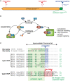
ROP structure and GTPase cycle. A, Schematic representation of the linear architecture of type-I ROPs. The residue number (in superscript) is given for ROP6 as an example. B, Schematic representation of the ROP GTPase cycle. C, Alignment of the C-terminal hypervariable region of the 11 Arabidopsis ROPs, showing the differences between type-I and type-II ROPs. Cationic residues are highlighted in green, acidic residues are highlighted in pink, the CaaX prenylation motif is in blue (CaaL: geranylgeranylation, CaaM: farnesylation) and the GC–CG S-acylation motif in red. For comparison purposes, the sequence of the C-terminal tail of human Cdc42 and K-Ras4B is included, as the archetypal representative of the Rho and Ras family, respectively. GEF, GTPase Exchange Factor; Pi, inorganic phosphate.
Figure 2
Figure 2
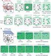
ROP microdomains in different cell types. A, In a trichoblast cell, ROP2/4/6 accumulate in the root hair initiation domain (RHID). During hair elongation, ROP2 (red) is present at the root hair tip whereas ROP10 (blue) accumulates at the shank. B, In the pollen tube tip, ROP1 is present in a microdomain. Its accumulation fluctuates over time during pollen tube growth. C, During xylem differentiation, ROP11 is present in microdomains that lead to the formation of cell wall pits.
Figure 3
Figure 3
ROP self-organization through a reaction–diffusion system. A, Two chemical species (here ROP-GDP and ROP-GTP bound) are under a reaction–diffusion process when long-range inhibition by a higher diffusion of the inhibitor and local activation by a slower diffusion of the activator exist. B, Changing various parameters in the reaction–diffusion equation leads to an array of two dimension patterns. C, In the case of cell wall pit formation, ROP11 is globally inhibited by the cytoplasmic GAP3. It gets activated by GEF4, which decreases its diffusion. As GEF4 makes dimers, it induces a local recruitment of new ROP11 molecules.
Figure 4
Figure 4
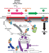
Summary of ROP6 and ROP6 mutant localization depending on the background and activation status. Schematic representation of xFP-ROP6 (where x can be different fluorescent protein) localization as seen by A) confocal microscopy (median view, red), and B) TIRF microscopy (surface view, blue). The ROP6 mutant versions or the background/treatment are indicated on top. In each condition, the localization is indicated in green, with a dark shade of green indicating strong accumulation, while a lighter green indicates weaker accumulation. In B, “Resting” indicates the ROP6 localization in the absence of treatment, while “stimulated” indicates ROP6 localization following auxin treatment or osmotic stress. PM, plasma membrane; ND, nanodomain; inh PI4P, inhibition of PI4P synthesis using a pan-PI4Kinase inhibitor; pss1, phosphatidylserine synthase1; PS, phosphatidylserine; G domain, GTPase domain; n, nucleus. Note that “lipid binding” refers to interaction with anionic lipids via the ROP6 polybasic C-terminal tail.
Figure 5
Figure 5
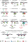
A multistep model for the role of lipid modifications and lipid interactions in the regulation of ROP6 localization and function. Note that this is a hypothetical model with some steps that have not been formally demonstrated (e.g. step 3). The exact order in which the events at the plasma membrane unfold is also currently unknown and this scenario represents one possibility among others. It is, for example, possible that nanoclustering and stabilization are concomitant or that interaction with phosphatidylserine is required upstream of S-acylation. PS, phosphatidylserine; ND, nanodomain; red +, positive charge present in ROP polybasic region; blue zig zag line, prenyl chain; magenta zig zag line, acyl chain; GEF, GTPase exchange factor; GDI, guaniside nucleotide dissociation inhibitor.
Figure 6
Figure 6
Possible scenarios for the function of nanoclustering in ROP signaling. Note that most of these scenarios have not been formally demonstrated and they remain to be explored. In addition, these scenarios are not mutually exclusive and not exhaustive. A, Nanodomains appear to spatially segregate active (GDP-loaded) and inactive (GTP-loaded) ROPs. However, only a portion (∼30% to 40%) of activated ROPs are localized in nanodomains and this model does not explain by itself why nanodomains are required for downstream signaling. B, Nanodomains could be a favorable membrane environment for ROPs to meet with their effectors, explaining the requirement for nanodomains in signaling. C, ROPs may also recruit their effectors specifically in nanodomains to induce signaling. D, Nanodomains may act as signaling platforms to trigger specific output, as seen in response to auxin treatments or osmotic stress (Platre, 2019; Smokvarska, 2020). E, It is possible that ROP nanodomain formation allows the clustering of downstream effectors, which could induce low energy interactions between them. Such multivalent interaction therefore explains the requirement for nanoclustering in signaling. F, It is possible that multiple ROPs cluster within the same nanodomains, thereby scaffolding several ROP effectors required for signaling. G, It is also possible that multiple ROPs localize to different nanodomains, which could ensure the propagation of specific cytosolic outputs. H, The stabilization of ROPs in nanodomains could increase their dwell time at the plasma membrane, which may be required to stabilize their effector at the cell surface and/or allow them sufficient time to catalyze their reaction. I, The clustering of ROPs in nanodomains may induce the formation of phase-separated cytosolic condensates below the plasma membrane, which could be required for effector function. J, The clustering of ROPs in nanodomains may quantitatively control the strength of the output signal even when the input signal is constant. According to this model, variation in membrane lipids could act like a rheostat that tunes the signaling capacity of cells. Such a model has been proposed for the membrane lipid phosphatidylserine during the ROP6 response to auxin (Platre et al., 2019). PM, plasma membrane, ND, nanodomain. Effectors are represented as a U-shaped protein.

References

    1. Barbosa ICR, Rojas-Murcia N, Geldner N (2019) The Casparian strip—one ring to bring cell biology to lignification? Curr Opin Biotechnol 56:121–129 - PubMed
    1. Batistič O (2012) Genomics and localization of the Arabidopsis DHHC-cysteine-rich domain S-acyltransferase protein family. Plant Physiol 160:1597–1612 - PMC - PubMed
    1. Berken A, Wittinghofer A (2008) Structure and function of Rho-type molecular switches in plants. Plant Physiol Biochem PPB 46:380–393 - PubMed
    1. Bonello TT, Perez-Vale KZ, Sumigray KD, Peifer M (2018) Rap1 acts via multiple mechanisms to position Canoe and adherens junctions and mediate apical–basal polarity establishment. Development 145:dev157941. - PMC - PubMed
    1. Bourne HR, Sanders DA, McCormick F (1991) The GTPase superfamily: conserved structure and molecular mechanism. Nature 349:117–127 - PubMed

Publication types

MeSH terms

Substances

LinkOut - more resources