Comparative Study
. 2021 Apr 23;185(4):1574-1594.
doi: 10.1093/plphys/kiab010.

The role of GDP-l-galactose phosphorylase in the control of ascorbate biosynthesis

Affiliations
Comparative Study

The role of GDP-l-galactose phosphorylase in the control of ascorbate biosynthesis

Mario Fenech et al. Plant Physiol. .

Abstract

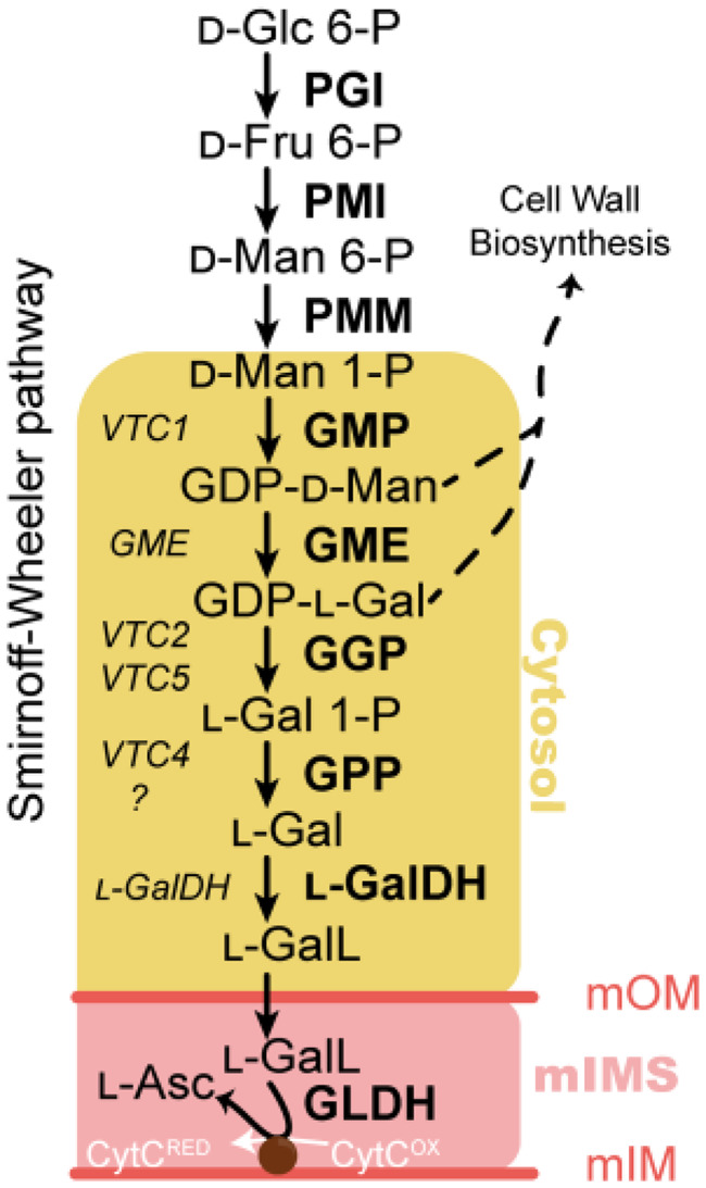
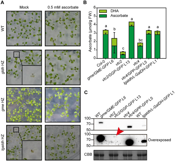
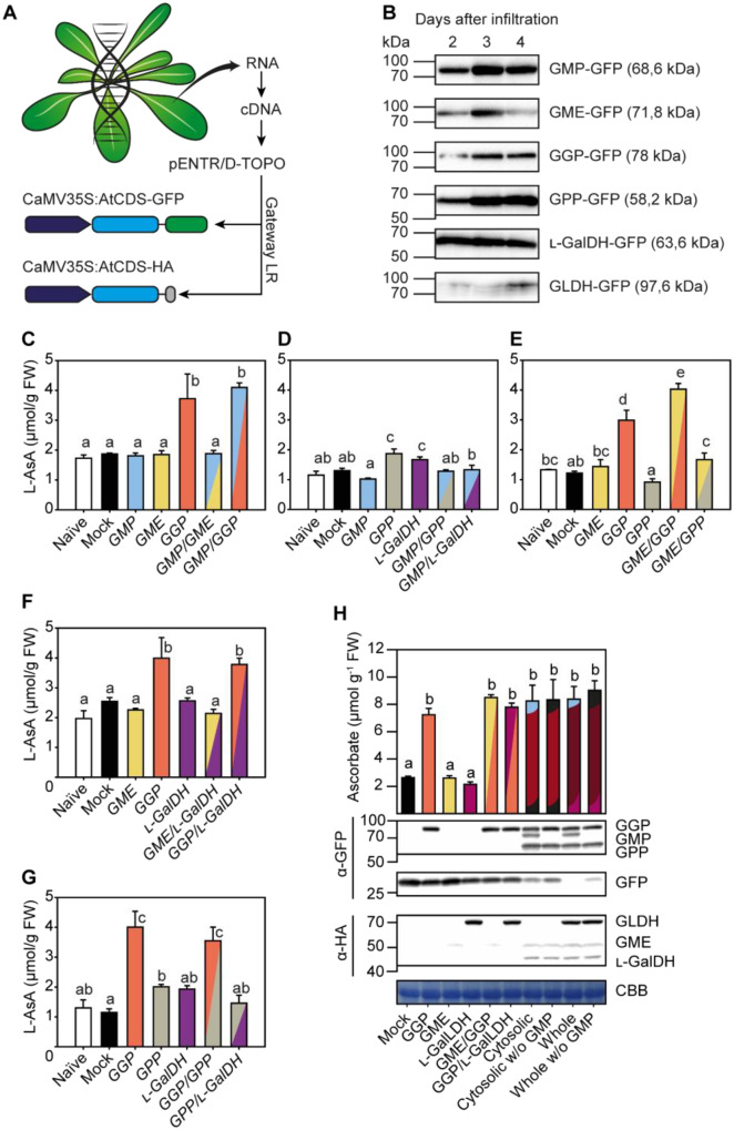
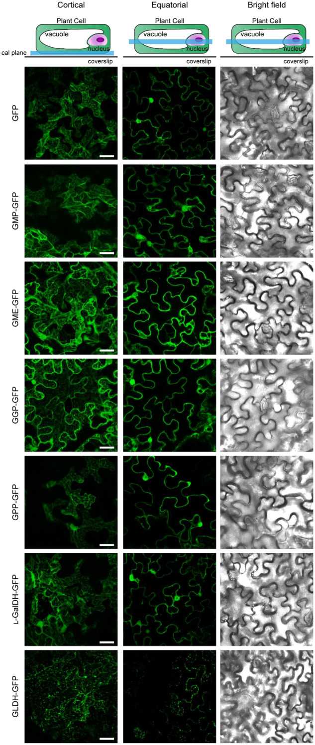
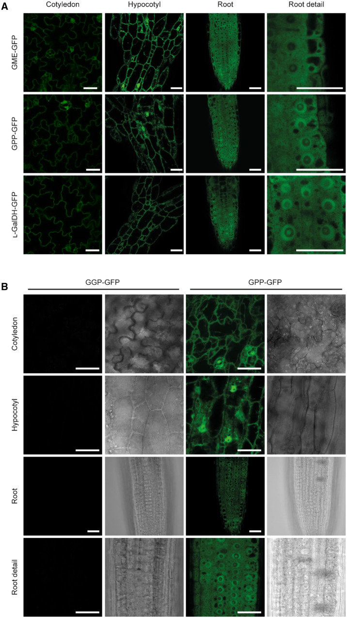
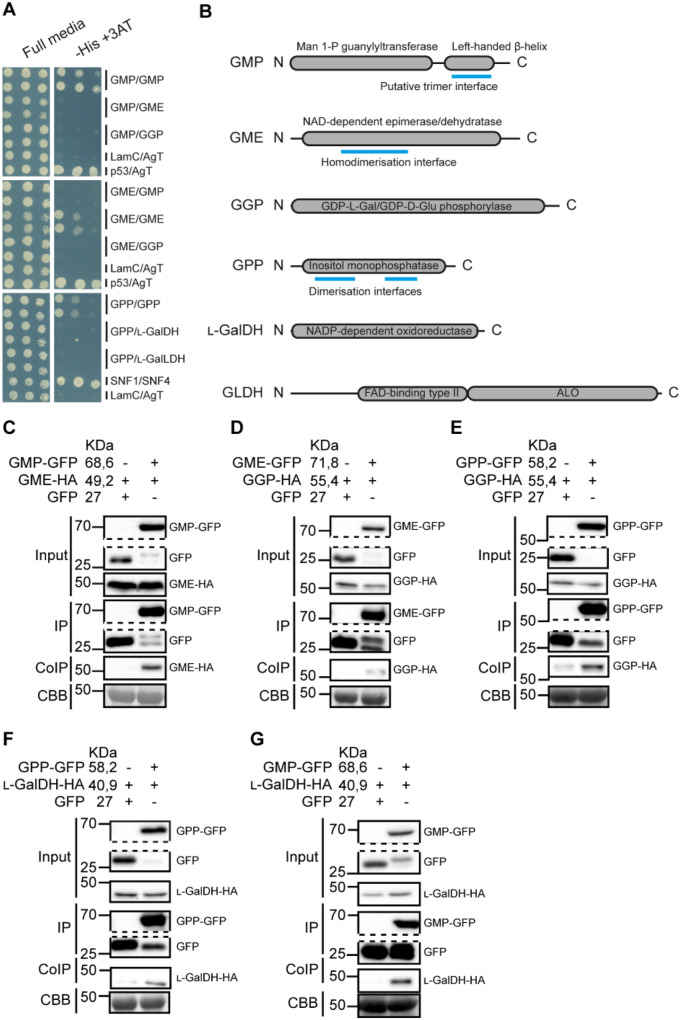
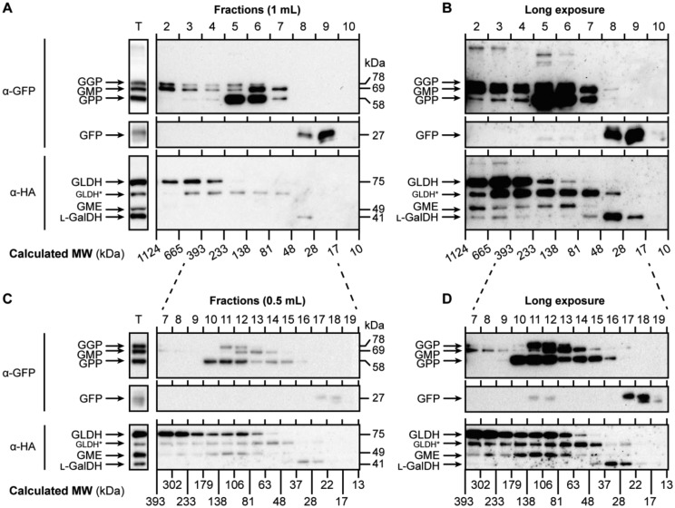
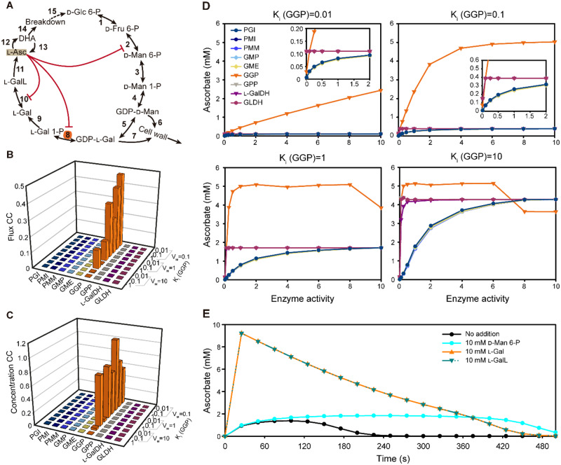
The enzymes involved in l-ascorbate biosynthesis in photosynthetic organisms (the Smirnoff-Wheeler [SW] pathway) are well established. Here, we analyzed their subcellular localizations and potential physical interactions and assessed their role in the control of ascorbate synthesis. Transient expression of C terminal-tagged fusions of SW genes in Nicotiana benthamiana and Arabidopsis thaliana mutants complemented with genomic constructs showed that while GDP-d-mannose epimerase is cytosolic, all the enzymes from GDP-d-mannose pyrophosphorylase (GMP) to l-galactose dehydrogenase (l-GalDH) show a dual cytosolic/nuclear localization. All transgenic lines expressing functional SW protein green fluorescent protein fusions driven by their endogenous promoters showed a high accumulation of the fusion proteins, with the exception of those lines expressing GDP-l-galactose phosphorylase (GGP) protein, which had very low abundance. Transient expression of individual or combinations of SW pathway enzymes in N. benthamiana only increased ascorbate concentration if GGP was included. Although we did not detect direct interaction between the different enzymes of the pathway using yeast-two hybrid analysis, consecutive SW enzymes, as well as the first and last enzymes (GMP and l-GalDH) associated in coimmunoprecipitation studies. This association was supported by gel filtration chromatography, showing the presence of SW proteins in high-molecular weight fractions. Finally, metabolic control analysis incorporating known kinetic characteristics showed that previously reported feedback repression at the GGP step, combined with its relatively low abundance, confers a high-flux control coefficient and rationalizes why manipulation of other enzymes has little effect on ascorbate concentration.


Figures

Figure 1
Figure 1
The ascorbate biosynthesis via GDP-D-mannose and L-galactose (the SW pathway) in A. thaliana. Yellow area refers to the cytosolic enzymes used in this work. mOM, mitochondrion outer membrane; mIMS, mitochondrion intermembrane space; mIM, mitochondrion inner membrane. In the left-hand side of the arrows, the genes encoding each enzyme are displayed, while in the right-hand side the encoded proteins/enzymatic activities are shown. Glc=glucose; Fru=fructose; Man=mannose; Gal=galactose; GalL=galactono-1,4-lactone; Asc=ascorbic acid; PGI=phosphoglucose isomerase; PMI=phosphomannose isomerase; PMM=phosphomannomutase; GMP=GDP-D-mannose pyrophosphorylase; GME=GDP-D-mannose 3′,5′ epimerase; GGP=GDP-L-galactose phosphorylase; GPP=L-galactose 1-phosphate phosphatase; L-GalDH=L-galactose dehydrogenase; L-GalLDH=L-galactono-1,4-lactone dehydrogenase; CytRED =cytochrome c reduced; CytOX =cytochrome c oxidized.
Figure 2
Figure 2
Characterization of C-terminal GFP fusions of ascorbate biosynthesis enzymes. A, The ascorbate complementation assay rescues growth arrest from gldh and lgaldh mutants but not gme lethality. Insets show homozygous mutants with arrested growth in media lacking ascorbate, but not in media supplemented with ascorbate. HZ, heterozygous. B, Complementation of ascorbate concentration by expression of GFP fusion proteins in ascorbate deficient Arabidopsis mutants. Data displayed correspond to mean±SD from three independent samples composed of three 6-week-old fully expanded rosettes grown in a short-day regime at 150-µmol photons m−2 s−1, one and a half hours after lights turned on. Different letters denote statistically significant differences for ascorbate concentration (one-way ANOVA, α = 0.05, post-hoc Tukey test). C, Immunoblot (α-GFP) of complemented lines shows that a small amount of GGP compared with other components restores ascorbate content. Red arrowhead indicates GGP-GFP. CBB, Coomassie Brilliant Blue.
Figure 3
Figure 3
The effect of transient expression of C-terminal GFP and HA fusions of ascorbate biosynthesis enzymes on ascorbate concentration in N. benthamiana leaves. A, Strategy followed to clone and to overexpress ascorbate biosynthesis genes from Arabidopsis translationally fused to GFP and HA at the protein C-terminus in N. benthamiana leaves. B, Immunoblots (α-GFP) of fusion protein accumulation 2, 3, and 4 d after agroinfiltration. Expression was driven by the 35S promoter. C–H, Leaf ascorbate concentration 3 d after agroinfiltration with different combinations of C-terminal GFP ad HA fusions of ascorbate biosynthesis enzymes. In H, “Cytosolic” refers to the coexpression of GMP, GME, GGP, GPP, and L-GalDH genes; “Whole” refers to the coexpression of those mentioned cytosolic genes plus mitochondrial L-GalLDH. For further details, constructs used in each infiltration are shown in Supplementary Table S1. Data displayed correspond to mean±SD from two leaves of at least three N. benthamiana plants infiltrated, collected at 3 d after infiltration. Different letters denote statistically significant differences for ascorbate concentration (one-way ANOVA, α = 0.05, post hoc Tukey test).
Figure 4
Figure 4
Subcellular localization of free GFP and C-terminal GFP fusions of ascorbate biosynthesis enzymes transiently expressed in N. benthamiana leaves under the control of the 35S promoter 3 d after agroinfiltration. GFP was visualized by laser scanning confocal microscopy. Scale bar = 30 µm.
Figure 5
Figure 5
Subcellular localization of ascorbate biosynthesis enzymes fused to GFP at the C-terminus and expressed under the control of their native promoters in Arabidopsis. A, The expression of GFP-tagged enzymes is ubiquitous in 4-d-old seedlings and their subcellular localization is compatible with the cytosol and nucleus except for GME, which only locates in cytosol. B, GFP signal of a functional GGP-GFP (line 13) protein was not detectable. GFP was visualized by laser scanning confocal microscopy. Scale bar = 30 µm.
Figure 6
Figure 6
Interaction of ascorbate biosynthesis enzymes assessed by Y2H analysis and Co-IP. A, The Y2HY2H assay detected no direct interaction between the enzymes, but GMP, GME, and GPP, which contain predicted dimerization/trimerization domains (B), form dimers/trimers. B, Protein scheme for each of the enzymes assayed in the Y2H assay showing length (in amino acids), annotated domains and predicted dimer/trimerization interfaces. C–G, The Co-IP assay reported association in vivo in N. benthamiana between consecutive steps as well as between the first (GMP) and the last (L-GalDH) steps occurring in the cytosol. GFP-tagged enzyme from N. benthamiana crude protein extracts containing two overexpressed consecutive enzymes, GFP and HA-tagged, was pulled-down using α-GFP agarose beads and coIP protein was detected using α-HA antibody. CBB: Coomassie Brilliant Blue.
Figure 7
Figure 7
The enzymes of the Smirnoff-Wheeler pathway elute together in high-molecular weight fractions of N. benthamiana protein extract. A, All the proteins transiently expressed were separated in 1 mL fractions and mainly accumulated between 393 and 48 kDa. B, Longer exposure of the blot presented in (A) shows that all the proteins eluted in high-molecular weight fractions. C, Fractions collected every 0.5 mL more accurately determined that the accumulation of proteins mainly occurs between 81 and 138 kDa but a longer exposure (D) enabled the detection of proteins in higher-molecular weight fractions than that. Proteins from N. benthamiana leaves were extracted as described in “Co-IP assay” section and separated as described in “Gel filtration chromatography” section. T=Total protein extract; MW=molecular weight. Calculated MWs were estimated using Dextran blue (2,000 kDa), Alcohol dehydrogenase (150 kDa), Albumin (66 kDa), and Carbonic Anhydrase (29 kDa); GLDH* points to a degradation product of GLDH (see Supplemental Figure S9).
Figure 8
Figure 8
A kinetic model of ascorbate biosynthesis and turnover. A, Scheme of the pathway modeled in COPASI showing intermediates catalytic steps (arrows) and feedback (red lines). The initial model parameters are shown in Supplementary Table S3. B, Metabolic control analysis at steady state was used to calculate flux control coefficients for each enzyme with various strengths of noncompetitive inhibition exerted by ascorbate (indicated by Ki values) on GGP (Step 8). C, Metabolic control analysis at steady state was used to calculate concentration control coefficients for ascorbate with various strengths of noncompetitive inhibition exerted by ascorbate (indicated by Ki values) on GGP (Step 8). D, The effect of varying enzyme activity on ascorbate concentration with various strengths of noncompetitive inhibition exerted by ascorbate (indicated by Ki values) on GGP (Step 8). E, Time course of the change in ascorbate concentration in response to the addition of D-mannose 6-phosphate (D-Man 6-P), L-galactose (L-Gal), and L-galactono-1,4-lactone (L-GalL). 1: PGI, 2: PMI, 3: PMM, 4: GMP, 5: GME, 6:GDP-D-mannose to cell wall polymers, 7: GDP-L-galactose to cell wall polymers, 8: GGP, 9: GPP, 10: L-GalDH, 11: GLDH, 12: Oxidation, 13: Reduction, 14: Turnover, and 15: Recycle.

References

    1. Ali B, Pantha S, Acharya R, Ueda Y, Wu LB, Ashrafuzzaman M, Ishizaki T, Wissuwa M, Bulley S, Frei M (2019) Enhanced ascorbate level improves multi-stress tolerance in a widely grown indica rice variety without compromising its agronomic characteristics. J Plant Physiol 240: 1–9 - PubMed
    1. Altschul SF, Madden TL, Schäffer AA, Zhang J, Zhang Z, Miller W, Lipman DJ (1997) Gapped BLAST and PSI-BLAST: a new generation of protein database search programs. Nucleic Acids Res 25: 3389–3402 - PMC - PubMed
    1. Amorim-Silva V, García-Moreno Á, Castillo AG, Lakhssassi N, Del Valle AE, Pérez-Sancho J, Li Y, Posé D, Pérez-Rodriguez J, Lin J, et al. (2019) TTL proteins scaffold brassinosteroid signaling components at the plasma membrane to optimize signal transduction in arabidopsis. Plant Cell 31: 1807–1828 - PMC - PubMed
    1. Asada K (1999) The water-water cycle in chloroplasts: scavenging of active oxygens and dissipation of excess photons. Annu Rev Plant Physiol Plant Mol Biol 50: 601–639 - PubMed
    1. Badejo AA, Jeong ST, Goto-Yamamoto N, Esaka M (2007) Cloning and expression of GDP-D-mannose pyrophosphorylase gene and ascorbic acid content of acerola (Malpighia glabra L.) fruit at ripening stages. Plant Physiol Biochem 45:665. - PubMed

Publication types