. 2021 Apr 1;22(1):18.
doi: 10.1186/s10194-021-01226-6.

Advantages of imaging photoplethysmography for migraine modeling: new optical markers of trigemino-vascular activation in rats

Affiliations

Advantages of imaging photoplethysmography for migraine modeling: new optical markers of trigemino-vascular activation in rats

Alexey Y Sokolov et al. J Headache Pain. .

Abstract

Background: Existent animal models of migraine are not without drawbacks and limitations. The aim of our study was to evaluate imaging photoplethysmography (PPG) as a method of assessing intracranial blood flow in rats and its changes in response to electrical stimulation of dural trigeminal afferents.

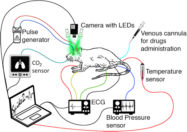
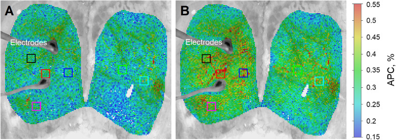
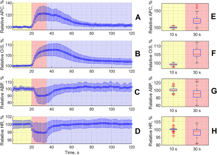
Methods: Experiments were carried out with 32 anesthetized adult male Wistar rats. Trigeminovascular system (TVS) was activated by means of electrical stimulation of dural afferents through a closed cranial window (CCW). Parameters of meningeal blood flow were monitored using a PPG imaging system under green illumination with synchronous recording of an electrocardiogram (ECG) and systemic arterial blood pressure (ABP). Two indicators related to blood-flow parameters were assessed: intrinsic optical signals (OIS) and the amplitude of pulsatile component (APC) of the PPG waveform. Moreover, we carried out pharmacological validation of these indicators by determining their sensitivity to anti-migraine drugs: valproic acid and sumatriptan. For statistical analysis the non-parametric tests with post-hoc Bonferroni correction was used.

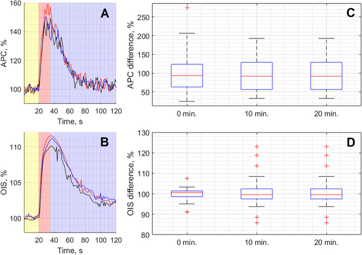
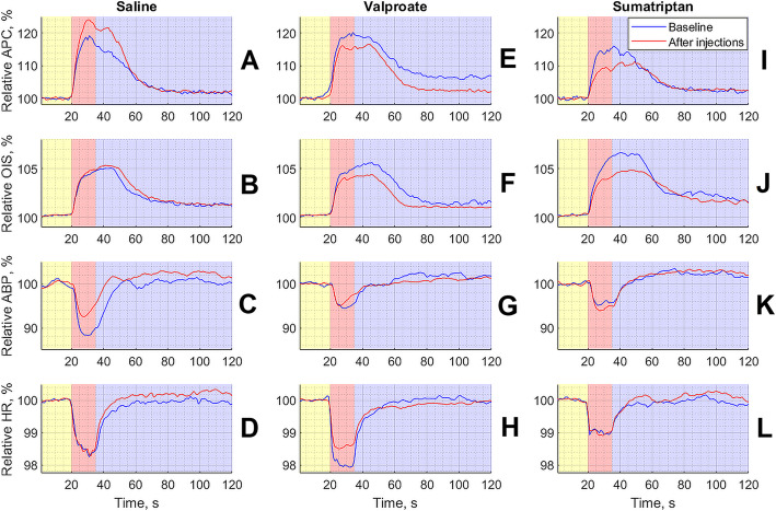
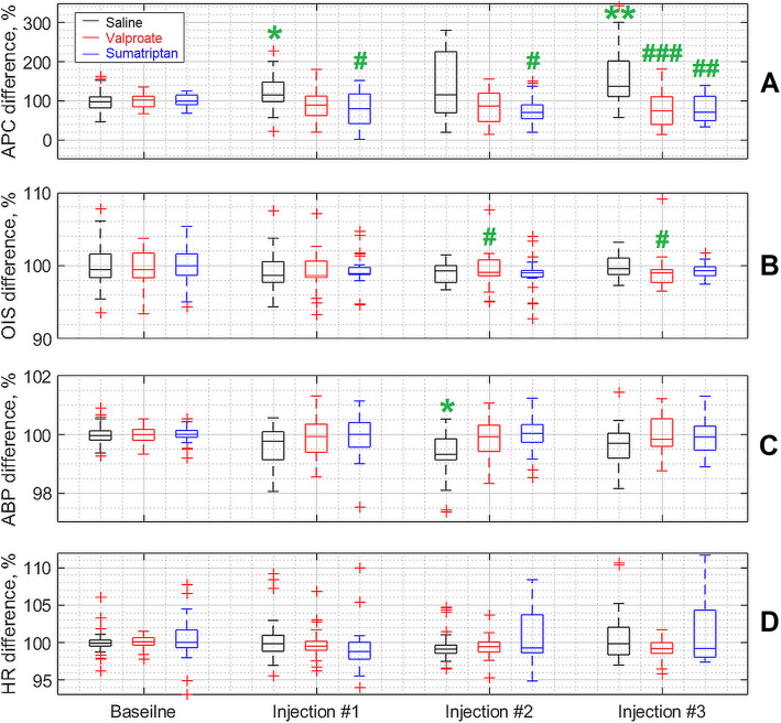
Results: Significant increase of both APC and OIS was observed due to CCW electrical stimulation. Compared to saline (n = 11), intravenous administration of both the sumatriptan (n = 11) and valproate (n = 10) by using a cumulative infusion regimen (three steps performed 30 min apart) lead to significant inhibitory effect on the APC response to the stimulation. In contrast, intravenous infusion of any substance or saline did not affect the OIS response to the stimulation. It was found that infusion of either sumatriptan or valproate did not affect the response of ABP or heart rate to the stimulation.

Conclusions: Imaging PPG can be used in an animal migraine model as a method for contactless assessment of intracranial blood flow. We have identified two new markers of TVS activation, one of which (APC) was pharmacologically confirmed to be associated with migraine. Monitoring of changes in APC caused by CCW electrical stimulation (controlling efficiency of stimulation by OIS) can be considered as a new way to assess the peripheral mechanism of action of anti-migraine interventions.

Keywords: Animal model; Electrical stimulation; Imaging photoplethysmography; Intracranial blood flow; Migraine; Sumatriptan; Trigemino‐vascular system; Valproic acid.


Conflict of interest statement

The authors declare that they have no financial and non-financial competing interest.

Figures

Fig. 1
Fig. 1
Schematic diagram of the experimental setup for monitoring the meningeal-blood-flow response and key physiological parameters to CCW electrical stimulation
Fig. 2
Fig. 2
Pseudo-colored maps of the electrical stimulation-evoked changes in APC over the cortical surface at 530 nm. a Spatial distribution of APC overlaid with the initial cortex image as estimated at the beginning of the pre-stimulation phase (10th s of the trial). b APC mapping during stimulation (30th s of the trial). The color bar on the right shows APC as a percentage, which is the same for both panels (a) and (b). Colored squares show the position of selected regions (big regions of interest) in which changes in mean APC were assessed to monitor the parameter’s dynamics
Fig. 3
Fig. 3
Relative change of the parameters measured in a contactless way by the digital camera (APC, graph a, and OIS, graph b) and invasively (ABP, graph c, and HR, graph d) in response to the CCW electrical stimulation. Here we show the data averaged over 96 baseline measurements carried out in three subsequent trials with three groups of animals before pharmacological interventions. Curves in the graphs show evolution of the mean value while the whiskers are the standard deviations. Yellow color indicates the pre-stimulation phase, the red one marks a 15-seconds period of the electrical stimulation, and the violet is the relaxation phase. Right-side panels (e, f, g and h) illustrate the statistical significance of the respective hemodynamic parameter assessed at the 10th s of the trial (pre-stimulation phase) and at the 30th s of the trial (stimulation phase)
Fig. 4
Fig. 4
Representative example of the responses of optical parameters APC (panel a) and OIS (panel b) to three subsequent CCW electrical stimulations repeated every 10 min for one of the rats during the baseline stage. Each curve in panels a and b shows dynamics of APC and OIS parameters averaged for the selected two most representative ROIs. The red curve was obtained in the first trial (0 min), the blue in the second trial (10 min), and the black in the third trial (20 min). The box plots in the panels c and d show cumulative changes in the responses to the stimulation of APC and OIS, respectively. In the panels c and d, the response was estimated as an integral of the respective curve shown in graph a or b (see details in the text). The data were obtained every 10 min for all rats (n = 32) in the baseline stage (before pharmacological interventions)
Fig. 5
Fig. 5
Summarized responses of both PPG and hemodynamic parameters to CCW electrical stimulation averaged over studied animals separately in each of three groups. Panels ad show responses for the group that received infusion of saline, panels eh for the group of valproate, and panels il for the group of sumatriptan. In all panels, yellow, red, and violet highlighting indicates pre-, during-, and post-stimulation phase, respectively. The time scale in seconds is shown in X-axis of each graphs, whereas the normalized parameters in a percentage are shown in Y-axes. The blue curve in each graph is the response of the measured parameter during the baseline stage, and the red curve is the response after an infusion of a respective substance. All curves were calculated as mean over three steps of the substance infusions
Fig. 6
Fig. 6
Dynamics of changes in parameters of systemic hemodynamics and PPG due to CCW electrical stimulation. Each box plot is a composite of indicators of a specific parameter for a 30-minute interval before (baseline stage) and after each of three infusion steps of the testing substance. The black, red and blue boxes correspond to the saline, valproate and sumatriptan infusion group, respectively. Green stars indicate statistical differences in relation to the baseline stage, whereas green grids represent differences between testing substance and saline. One symbol corresponds to p < 0.05, two of them correspond to p < 0.01, and three of them correspond to p < 0.001. All statistical significance is indicated with Bonferroni corrections for multiplicity

References

    1. Headache Classification Committee of the International Headache Society (2018) The International Classification of Headache Disorders, 3rd edition. Cephalalgia 38:1–211. doi: 10.1177/0333102417738202 - PubMed
    1. Stovner LJ, Nichols E, Steiner TJ, et al. Global, regional, and national burden of migraine and tension-type headache, 1990–2016: a systematic analysis for the Global Burden of Disease Study 2016. Lancet Neurol. 2018;17:954–976. doi: 10.1016/S1474-4422(18)30322-3. - DOI - PMC - PubMed
    1. Burch RC, Buse DC, Lipton RB. Migraine: Epidemiology, Burden, and Comorbidity. Neurol Clin. 2019;37:631–649. doi: 10.1016/j.ncl.2019.06.001. - DOI - PubMed
    1. Øie LR, Kurth T, Gulati S, Dodick DM. Migraine and risk of stroke. J Neurol Neurosurg Psychiatry. 2020;91:593–604. doi: 10.1136/jnnp-2018-318254. - DOI - PMC - PubMed
    1. Bonafede M, Sapra S, Shah N, et al. Direct and Indirect Healthcare Resource Utilization and Costs Among Migraine Patients in the United States. Headache. 2018;58:700–714. doi: 10.1111/head.13275. - DOI - PubMed