. 2021 Apr 1;22(1):97.
doi: 10.1186/s12931-021-01677-0.

CREB-dependent LPA-induced signaling initiates a pro-fibrotic feedback loop between small airway basal cells and fibroblasts

Affiliations

CREB-dependent LPA-induced signaling initiates a pro-fibrotic feedback loop between small airway basal cells and fibroblasts

Shyam Nathan et al. Respir Res. .

Abstract

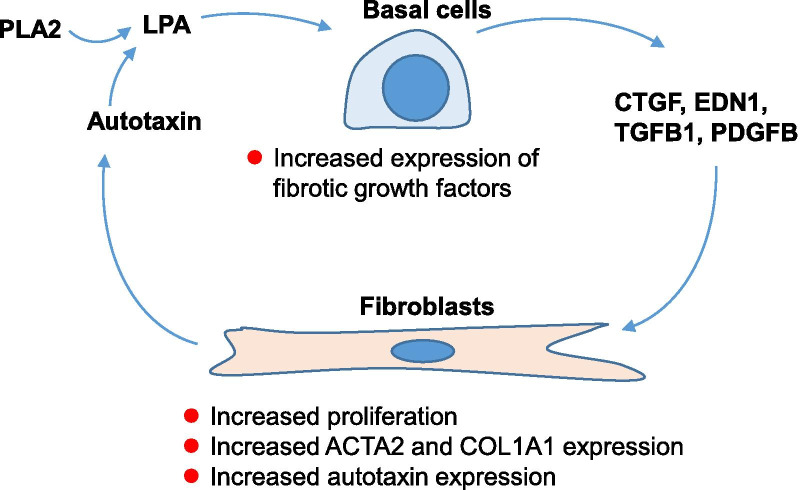
Background: Lysophosphatidic acid (LPA), generated extracellularly by the action of autotaxin and phospholipase A2, functions through LPA receptors (LPARs) or sphingosine-1-phosphate receptors (S1PRs) to induce pro-fibrotic signaling in the lower respiratory tract of patients with idiopathic pulmonary fibrosis (IPF). We hypothesized that LPA induces changes in small airway epithelial (SAE) basal cells (BC) that create cross-talk between the BC and normal human lung fibroblasts (NHLF), enhancing myofibroblast formation.

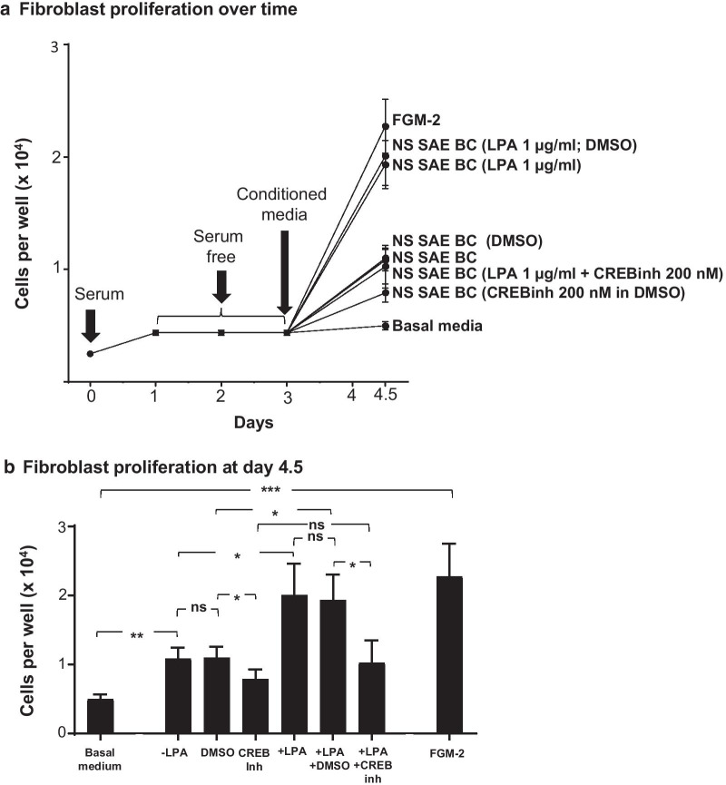
Methods: To assess LPA-induced signaling, BC were treated with LPA for 2.5 min and cell lysates were analyzed by phosphokinase array and Western blot. To assess transcriptional changes, BC were treated with LPA for 3 h and harvested for collection and analysis of RNA by quantitative polymerase chain reaction (qPCR). To assess signaling protein production and function, BC were washed thoroughly after LPA treatment and incubated for 24 h before collection for protein analysis by ELISA or functional analysis by transfer of conditioned medium to NHLF cultures. Transcription, protein production, and proliferation of NHLF were assessed.

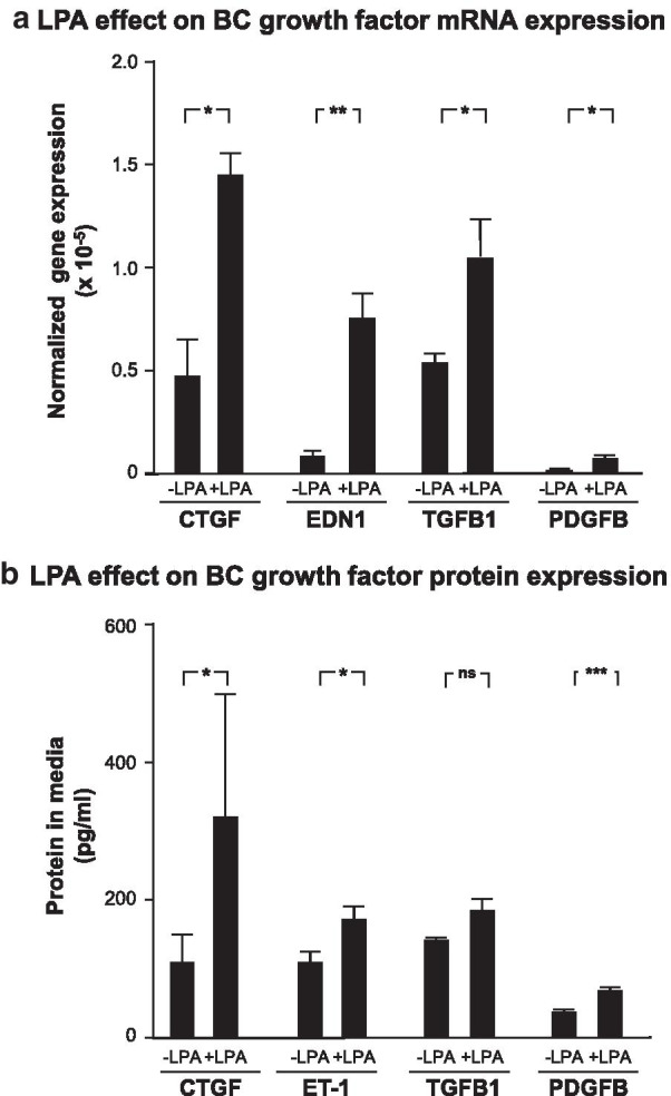
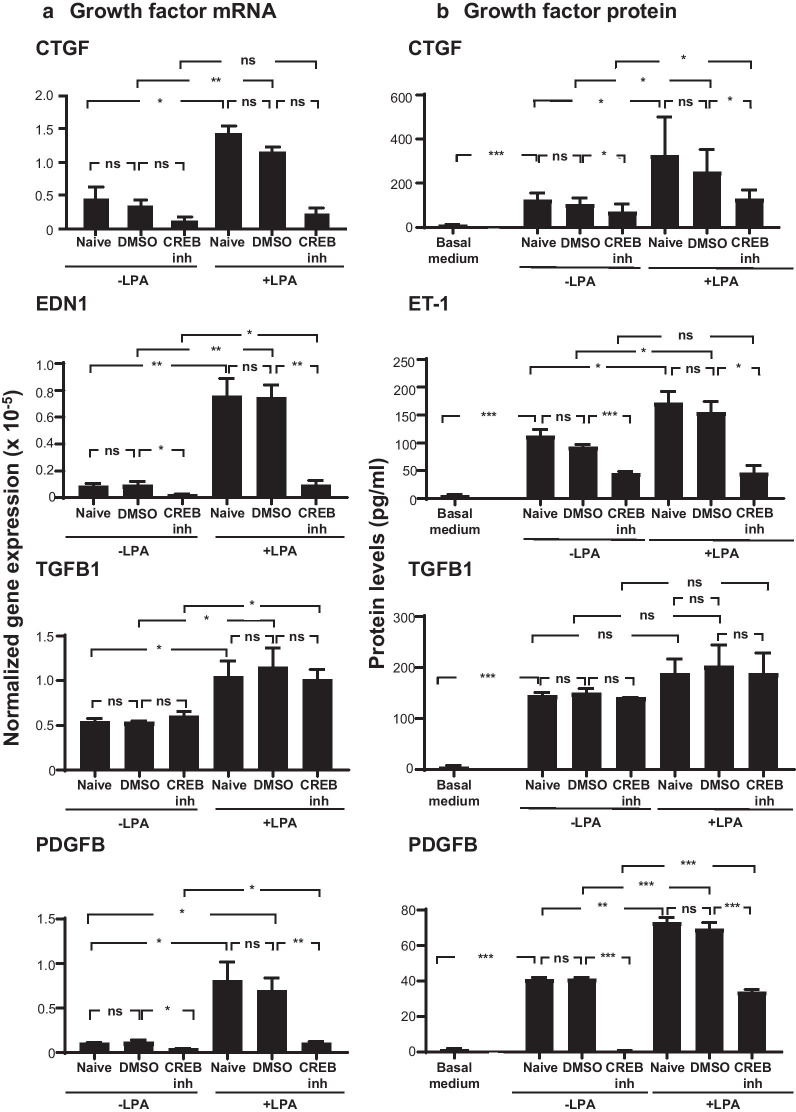
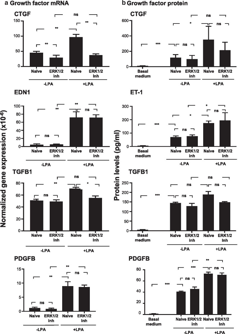
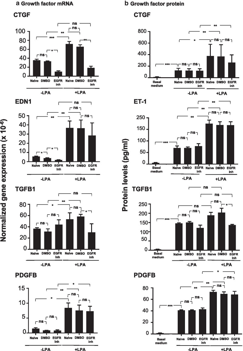
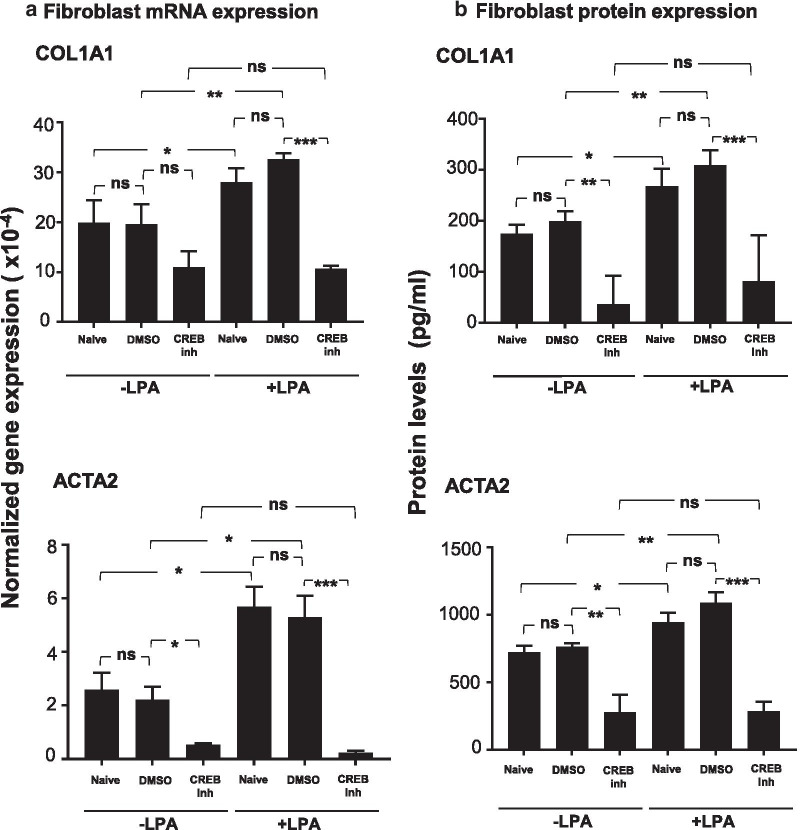
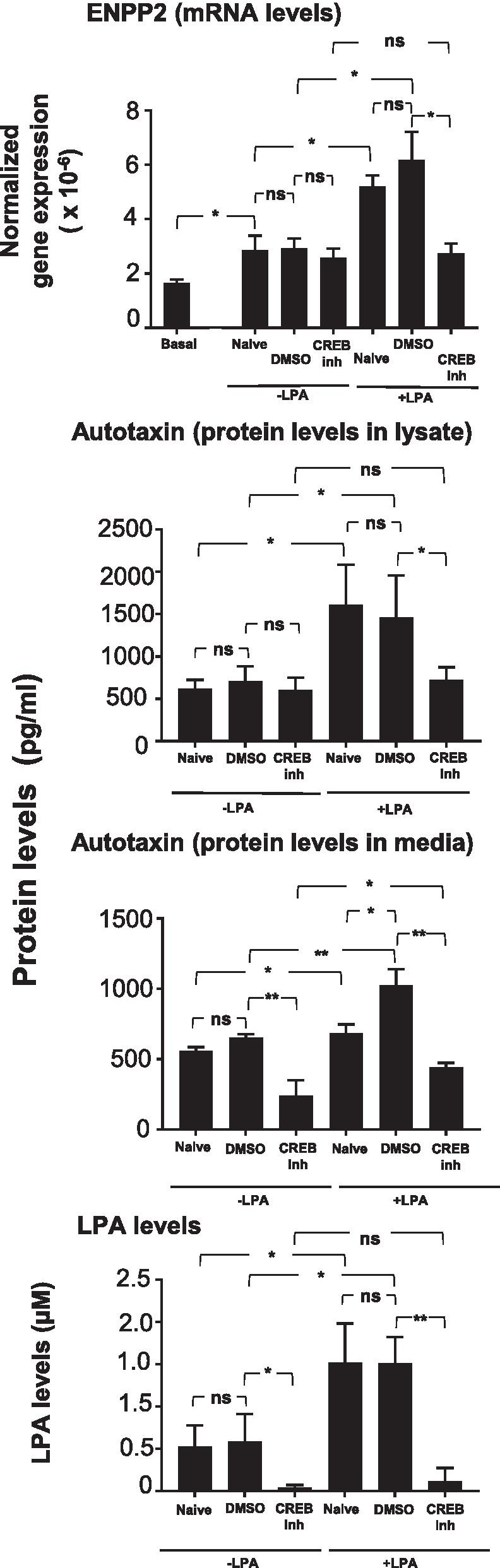
Results: LPA treatment induced signaling by cAMP response element-binding protein (CREB), extracellular signal-related kinases 1 and 2 (Erk1/2), and epithelial growth factor receptor (EGFR) resulting in elevated expression of connective tissue growth factor (CTGF), endothelin-1 (EDN1/ET-1 protein), and platelet derived growth factor B (PDGFB) at the mRNA and protein levels. The conditioned medium from LPA-treated BC induced NHLF proliferation and increased NHLF expression of collagen I (COL1A1), smooth muscle actin (ACTA2), and autotaxin (ENPP2) at the mRNA and protein levels. Increased autotaxin secretion from NHLF correlated with increased LPA in the NHLF culture medium. Inhibition of CREB signaling blocked LPA-induced changes in BC transcription and translation as well as the pro-fibrotic effects of the conditioned medium on NHLF.

Conclusion: Inhibition of CREB signaling may represent a novel target for alleviating the LPA-induced pro-fibrotic feedback loop between SAE BC and NHLF.

Keywords: ACTA2; Airway basal cell; Autotaxin; COL1A1; CREB; Fibroblast; Idiopathic pulmonary fibrosis; Intracellular signaling; Lung; Lysophosphatidic acid.


Conflict of interest statement

The authors declare that they have no competing interests.

The authors have declared that no conflict of interest exists.

Figures

Fig. 1
Fig. 1
Effect of lysophosphatidic acid (LPA) on fibrotic growth factor expression in the small airway epithelial basal cells (SAE BC). Primary SAE BC from each of 3 non-smoking individuals were plated in triplicate in the presence or absence of 1.0 μg/ml LPA and evaluated for expression of connective tissue growth factor (CTGF), endothelin-1 (EDN1/ET-1), transforming growth factor beta (TGFB1), and platelet derived growth factor family member B (PDGFB). a mRNA. After a 3 h LPA exposure, RNA was harvested and evaluated by qRT-PCR normalized to 18S RNA. b Protein. After a 3 h LPA exposure, LPA was removed and cells were incubated with basal media without growth factor supplements for 24 h. Conditioned media was harvested and evaluated for growth factor content by ELISA. Each sample was assessed in triplicate; data are expressed as the mean value of the 3 donors ± SE. *p < 0.05, **p < 0.01, ***p < 0.001
Fig. 2
Fig. 2
Signaling pathways activated by LPA in SAE BC. Primary basal cells were plated in triplicate in the absence or presence of 1.0 μg/ml LPA for 2.5 min and evaluated for expression of 45 protein kinases. a Human phosphokinase array revealing changes in kinase phosphorylation levels following LPA treatment. 1-Erk1/2, 2-EGFR (Y1086), 3-CREB and 4-p70 S6 kinase (T389). b Quantification of protein kinase array by Image J software showing top 4 protein kinases with enhanced phosphorylation following LPA treatment. The main pathways stimulated by LPA included Erk1/2, CREB, p70 S6 kinase, and EGFR. Data are expressed as the ratio of the mean signal intensity in the presence of LPA to the mean signal intensity in the absence of LPA of the 3 donors ± SEM. Significance of each set of intensities was determined using a Chi square test. *p < 0.05, **p < 0.01, ***p < 0.001. c Western analysis of CREB, ERK1/2, P70 S6 kinase, and EGFR kinase phosphorylation in the absence or presence of LPA confirming phosphorylation of CREB, ERK1/2, and EGFR. Shown is data of BC ± LPA for 3 subjects
Fig. 3
Fig. 3
Effect of CREB inhibitor (CREB inh) on growth factor mRNA and protein levels. Primary basal cells from each of three non-smoking donors were plated in triplicate in the presence or absence of 1 μg/ml LPA, 200 nM CREB inhibitor 666-15 and a combination of LPA and CREB inhibitor 666-15. a mRNA expression levels. Shown are connective tissue growth factor (CTGF), endothelin-1 (EDN1), transforming growth factor beta (TGFB1) and platelet derived growth factor B (PDGFB) mRNA levels determined after 3 h of LPA exposure with and without CREB inhibitor (666-15) using qRT-PCR using 18S RNA to normalize the samples. b Protein levels. Shown are protein levels in conditioned media of LPA with and without CREB inhibitor 666-15, LPA with or without CREB inhibitor. After 3 h, the stimuli/inhibitor were removed and cells were incubated with non-supplemented basal media for 24 h. Conditioned media was harvested and evaluated for growth factor levels by ELISA. Data are expressed as the mean value of the 3 donors ± SEM. ND not detected. *p < 0.05, **p < 0.01, ***p < 0.001
Fig. 4
Fig. 4
Effect of ERK1/2 inhibitor (ERK1/2 inh) on growth factor expression. Primary basal cells from each of 3 non-smoking donors were plated in triplicate in the presence or absence of 1 μg/ml LPA, 5 µM ERK1/2 inhibitor (LY32149966) and a combination of LPA and ERK1/2 inhibitor (LY32149966). a mRNA. Shown are expression levels of connective tissue growth factor (CTGF), endothelin-1 (EDN1), transforming growth factor beta (TGFB1) and platelet derived growth factor B (PDGFB) determined after 3 h of LPA exposure with and without ERK1/2 inhibitor (LY32149966) using qRT-PCR using 18S RNA to normalize the samples. b Protein. Shown are growth factor levels in the conditioned media. After 3 h of LPA with and without CREB inhibitor (666-15) exposure, LPA with or without ERK1/2 inhibitor was removed and cells were incubated with un-supplemented basal media for 24 h. Conditioned media was harvested and evaluated for growth factor content by ELISA. Data are expressed as the mean value of the 3 donors ± SEM. ND—not detected. *p < 0.05, **p < 0.01, ***p < 0.001
Fig. 5
Fig. 5
Effect of EGFR inhibitor (EGFR inh) on growth factor mRNA levels. Primary basal cells (passage 3) from each of three non-smoking donors were plated in triplicate in the presence or absence of 1 μg/ml LPA, 10 µM EGFR inhibitor (AG1478), and a combination of LPA and EGFR inhibitor. a mRNA expression levels shown are mRNA levels of connective tissue growth factor (CTGF), endothelin-1(EDN1), transforming growth factor beta (TGFB1), and platelet derived growth factor B were determined after 3 h of LPA exposure with and without the inhibitor using qRT-PCR using 18S RNA to normalize the samples. b Protein levels in the conditioned media. After 3 h of LPA with and without EGFR inhibitor (AG1478) exposure, LPA with or without inhibitor was removed and cells were incubated with non-supplemented basal media for 24 h. Conditioned media was harvested and evaluated for growth factor content by ELISA. Data are expressed as the mean value of the 3 donors ± SEM. ND—not detected. *p < 0.05, **p < 0.01, ***p < 0.001
Fig. 6
Fig. 6
Effect of CREB inhibitor (CREB inh) on fibroblast proliferative response to basal cell conditioned medium. Primary normal human lung fibroblast (NHLF) were plated in triplicate wells and treated with conditioned media from each of 3 non-smoking donors obtained in the presence or absence of 1 μg/ml LPA. For each condition, conditioned medium collected from cultures that were naive or treated with vehicle control (DMSO) or 200 nM CREB inhibitor (666-15) were collected and transferred to NHLF that had been serum starved for 48 h. a Fibroblast growth. NHLF were counted after 24 h. Shown is data from BC of a single individual. b Summary data showing mean fibroblast cell proliferation exposed to BC conditioned media from 3 donors. Data are expressed as the mean value of the 3 donors ± SEM. *p < 0.05, **p < 0.01, ***p < 0.001
Fig. 7
Fig. 7
Effect of CREB inhibitor (CREB inh) on fibroblast expression in response to basal cell conditioned medium. Primary normal human lung fibroblasts (NHLF) were treated with conditioned media from each of 3 non-smoking donors obtained in the presence or absence of 1 μg/ml LPA. For each condition, conditioned medium collected from cultures that were naive or treated with vehicle control (DMSO) or 200 nM CREB inhibitor (666-15) were collected and transferred to NHLF. Fibroblast expression of collagen type 1 (COL1A1) and smooth muscle actin (ACTA2) was assessed after 24 h in conditioned medium. a mRNA levels. RNA was harvested and evaluated by qRT-PCR using 18S RNA to normalize the samples. b Protein levels. At the same time point, conditioned media was collected and assessed for COL1A1 secretion. Cells were solubilized with a RIPA buffer and evaluated for alpha smooth muscle myosin by ELISA. Data are expressed as the mean value of the 3 donors ± SEM. *p < 0.05, **p < 0.01, ***p < 0.001
Fig. 8
Fig. 8
Effect of CREB inhibitor (CREB inh) on autotaxin (ENPP2) expression, secretion, and activity in fibroblasts in response to medium conditioned by LPA-treated basal cells. Primary NHLF were treated with conditioned media from each of 3 non-smoking donors obtained in the presence or absence of 1.0 μg/ml LPA. For each condition, conditioned medium collected from cultures that were naive or treated with vehicle control (DMSO) or 200 nM CREB inhibitor (666-15) were collected and transferred to NHLF. Autotaxin, an enzyme responsible for the extracellular production of LPA, in encoded by ENPP2. Expression of the ENPP2 gene was assessed in fibroblasts 24 h after exposure to conditioned medium. Autotaxin protein levels and LPA levels were assessed in cell culture medium 24 h after exposure to conditioned medium. a ENPP2 mRNA levels. mRNA was measured by qRT-PCR using 18S RNA to normalize the samples. b Autotaxin protein levels. Protein levels were measured in cell culture medium by ELISA. c LPA levels. LPA was measured in cell culture medium by ELISA. Data are expressed as the mean value of the 3 donors ± SEM. *p < 0.05, **p < 0.01, ***p < 0.001
Fig. 9
Fig. 9
Model for basal cell-fibroblast interaction in IPF. There is a positive feedback relationship in which elevated levels of LPA induce the production of pro-fibrotic factors in basal cells leading to fibrotic changes in fibroblasts including elevated production and secretion of autotaxin, the enzyme that generates LPA. Interruption of the basal cell-fibroblast LPA feedback loop, potentially via inhibition of autotaxin inhibitors, LPAR receptors, CREB-dependent intracellular signaling or any other targets in the LPA signaling pathway, may slow lung fibrosis

Similar articles

Cited by

References

    1. Fulmer JD, Roberts WC, von Gal ER, Crystal RG. Small airways in idiopathic pulmonary fibrosis. Comparison of morphologic and physiologic observations. J Clin Invest. 1977;60:595–610. doi: 10.1172/JCI108811. - DOI - PMC - PubMed
    1. Crystal RG, Bitterman PB, Mossman B, Schwarz MI, Sheppard D, Almasy L, Chapman HA, Friedman SL, King TE, Jr, Leinwand LA, et al. Future research directions in idiopathic pulmonary fibrosis: summary of a National Heart, Lung, and Blood Institute working group. Am J Respir Crit Care Med. 2002;166:236–246. doi: 10.1164/rccm.2201069. - DOI - PubMed
    1. Raghu G, Collard HR, Egan JJ, Martinez FJ, Behr J, Brown KK, Colby TV, Cordier JF, Flaherty KR, Lasky JA, et al. An official ATS/ERS/JRS/ALAT statement: idiopathic pulmonary fibrosis: evidence-based guidelines for diagnosis and management. Am J Respir Crit Care Med. 2011;183:788–824. doi: 10.1164/rccm.2009-040GL. - DOI - PMC - PubMed
    1. Kim HJ, Perlman D, Tomic R. Natural history of idiopathic pulmonary fibrosis. Respir Med. 2015;109:661–670. doi: 10.1016/j.rmed.2015.02.002. - DOI - PubMed
    1. Martinez FJ, Collard HR, Pardo A, Raghu G, Richeldi L, Selman M, Swigris JJ, Taniguchi H, Wells AU. Idiopathic pulmonary fibrosis. Nat Rev Dis Primers. 2017;3:17074. doi: 10.1038/nrdp.2017.74. - DOI - PubMed

MeSH terms