Review
. 2021 Apr 1;23(1):44.
doi: 10.1186/s12968-020-00671-7.

Society for Cardiovascular Magnetic Resonance 2019 Case of the Week series

Affiliations
Review

Society for Cardiovascular Magnetic Resonance 2019 Case of the Week series

Arun Dahiya et al. J Cardiovasc Magn Reson. .

Abstract

The Society for Cardiovascular Magnetic Resonance (SCMR) is an international society focused on the research, education, and clinical application of cardiovascular magnetic resonance (CMR). The SCMR web site ( https://www.scmr.org ) hosts a case series designed to present case reports demonstrating the unique attributes of CMR in the diagnosis or management of cardiovascular disease. Each clinical presentation is followed by a brief discussion of the disease and unique role of CMR in disease diagnosis or management guidance. By nature, some of these are somewhat esoteric, but all are instructive. In this publication, we provide a digital archive of the 2019 Case of the Week series as a means of further enhancing the education of those interested in CMR and as a means of more readily identifying these cases using a PubMed or similar search engine.

Keywords: Cardiac tumor; Cardiomyopathy; Eosinophilic granulomatosis; MRI.


Conflict of interest statement

There are no competing interests.

Figures

Fig. 1.
Fig. 1.
Case 1. 12 lead electrocardiogram (ECG). Sinus rhythm with T-wave inversion in most precordial leads (V2−V6) and some inferior limb leads (III and aVF)
Fig. 2
Fig. 2
Case 1. T2-weighted short tau inversion recovery (STIR) imaging of the left ventricular (LV) short axis from base to apex. Diffuse increase in subendocardial signal intensity suggestive of subendocardial edema
Fig. 3
Fig. 3
Case 1. Short axis stack T1 weighted (high resolution-magnitude) late gadolinium enhancement (LGE) imaging. Subendocardial enhancement in the mid to distal LV cavity
Fig. 4
Fig. 4
Case 1. Phase sensitive inversion recovery (PSIR). a Three chamber view with subtle subendocardial LGE of the mid and apical anteroseptal and inferolateral wall segments. b Four chamber view with subtle subendocardial LGE of the mid and apical inferoseptal and anterolateral wall segments
Fig. 5
Fig. 5
Case 1. Native T1, post-contrast T1, and extracellular volume (ECV) map in 4-chamber view. Native T1 (a) and ECV map (b) with abnormal measured native T1 and ECV in region of interest (apical septum) compared to remote region (basal septum). Pre-contrast T1 (c), post-contrast T1 (d), and ECV (e) map in 4-chamber view
Fig. 6
Fig. 6
Case 1. Chest computed tomography (CT) coronal view. Nonspecific sub pleural pulmonary infiltrates in the left upper lobe (yellow arrow). CT guided needle biopsy of this lesion revealed eosinophilic infiltrates consistent with acute eosinophilic pneumonia
Fig. 7
Fig. 7
Case 1. Side by side comparison of pre- and post-treatment imaging. a 12 lead ECG. b T2-weighted STIR imaging LV short axis. c LGE short axis images. d GE 4 chamber image. e LGE 3 chamber image
Fig. 8
Fig. 8
Case 2. Twelve lead ECG. Sinus rhythm with left bundle branch block
Fig. 9
Fig. 9
Case 2. Systolic displacement vectors in a the patient's and b a healthy subject’s mid-ventricle
Fig. 10
Fig. 10
Case 2. Torsion in the LV and a mid-ventricular slice. a Healthy subject. b Abnormal torsion in the patient
Fig. 11
Fig. 11
Case 3. Transthoracic echocardiography (TTE) apical LV views with and without ultrasound enhancing agent
Fig. 12
Fig. 12
Case 3. Transesophageal echocardiographic (TEE) mid-esophageal 2 chamber and transgastric projections
Fig. 13
Fig. 13
Case 3. Diffusion weighted brain MRI showing multifocal infarctions consistent with embolism
Fig. 14
Fig. 14
Case 4. Cine balanced steady state free precession (bSSFP) image shows the right pulmonary veins (yellow circle) and left pulmonary veins (yellow arrow) infiltrated by the mediastinal mass
Fig. 15
Fig. 15
Case 4. Axial T2 fat suppression image. Pericardial infiltration over the atria, yet, no effusion and no atrial extension. The inferior vena cava (IVC) entrance to the right atrium is spared
Fig. 16
Fig. 16
Case 4. Axial T2 image. Large mediastinal metastatic mass (yellow arrows) infiltrating the left atrium (LA) and encasing the main pulmonary artery (MPA). The mass appears separable from the ascending aorta (AO). The right lower pulmonary vein (PV) appears infiltrated by mass (red arrow)
Fig. 17
Fig. 17
Case 4. Axial T1 image before and after contrast. Increased signal intensity in the post-contrast study relative to the pre-contrast study
Fig. 18
Fig. 18
Case 4. LGE with myocardial nulling and long inversion time. In comparison to the long TI image, there is still enhancement in the mass with no jet-black signal seen in the long TI image
Fig. 19
Fig. 19
Case 4. Large liver mass indicated by yellow arrows
Fig. 20
Fig. 20
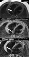
Case 5. T1 weighted with and without fat saturation and T2 weighted with fat saturation axial images. The atrial mass (white arrow) was isointense on T1w sequences (a, b) and hyperintense on T2w sequences (c)
Fig. 21
Fig. 21
Case 5. Axial first pass perfusion, early gadolinium enhancement, and LGE images. The atrial mass (white arrow) has contrast uptake on first pass perfusion (a), not well seen on early enhancement (b), and hyperintense on LGE (c)
Fig. 22
Fig. 22
Case 6. Twelve lead ECG. a Ventricular tachycardia on presentation. b Sinus tachycardia with T wave inversion of lateral leads
Fig. 23
Fig. 23
Case 6. Coronary CT angiogram (CTA). a Axial CTA with anomalous right coronary artery (blue arrow) with left coronary cusp origin just anterior to the origin of the left main coronary artery. b Volume rendered CTA with anomalous right coronary artery (blue arrow)
Fig. 24
Fig. 24
Case 6. Cine bSSFP 2 chamber end-diastole. Mid LV thickening with apical aneurysm
Fig. 25
Fig. 25
Case 6. LGE. 4 chamber view (a), 3 chamber view (b), and 2 chamber view (c) with dense scar of the mid to apical LV
Fig. 26
Fig. 26
Case 7. Twelve lead ECG. LA enlargement present
Fig. 27
Fig. 27
Case 7. Variable CMR techniques to evaluate the cardiac mass. a Axial T1 double inversion recovery with fat saturation. A large hyperintense mass in LA (white arrow). b Axial T2 double inversion recovery image. Hyperintense large LA mass (white arrow). c Coronal first pass angiogram. No mass enhancement present (white arrow). d Myocardial delayed enhancement four chamber view. Variable intensity of the mass, which could be an India ink artifact (white arrow)
Fig. 28
Fig. 28
Case 7. Operating room picture. Removal of myxoma by the LA stalk
Fig. 29
Fig. 29
Case 7. Resected tumor gross specimen. a Myxoma on a surgical towel. b Myxoma stalk easily seen. The entire myxoma measures approximately 6 cm × 4 cm
Fig. 30
Fig. 30
Case 8. Cine SSFP 4 chamber. Initial CMR with moderate pericardial effusion
Fig. 31
Fig. 31
Case 8. Short axis T2 double inversion recovery sequences. Myocardial enhancement suggesting edema (white arrows)
Fig. 32
Fig. 32
Case 8. LGE images. ac Four chamber stack with endocardial enhancement throughout the LV myocardium, particularly the lateral LV (white arrows). d Short axis mid image with myocardial LGE of the inferolateral wall (white arrows)
Fig. 33
Fig. 33
Case 8. Four chamber bSSFP cine image. Small pericardial effusion without significant pericardial thickening or pericardial enhancement. There was no evidence of ventricular interdependence. Interval resolution of the bilateral small pleural effusions had occurred

References

    1. Masi AT, Hunder GG, Lie JT, Michel BA, Bloch DA, Arend WP, Calabrese LH, Edworthy SM, Fauci AS, Leavitt RY, Lightfoot RW, McShane DJ, Mills JA, Stevens MB, Wallace SL, Zvaifler NJ. The American College of Rheumatology 1990 criteria for the classification of Churg-Strauss syndrome (allergic granulomatosis and angiitis) Arthritis Rheum. 1990;33:1094–1100. doi: 10.1002/art.1780330806. - DOI - PubMed
    1. Watts RA, Lane S, Scott DG. What is known about the epidemiology of the vasculitides? Best Pract Res Clin Rheumatol. 2005;19:191–207. doi: 10.1016/j.berh.2004.11.006. - DOI - PubMed
    1. Neumann T, Manger B, Schmid M, Kroegel C, Hansch A, Keiser WA, Reinhardt D, Wolf G, Hein G, Mall G, Schett G, Zwerina J. Cardiac involvement in Churg-Strauss syndrome: impact of endomyocarditis. Medicine (Baltimore) 2009;88(4):236. doi: 10.1097/MD.0b013e3181af35a5. - DOI - PubMed
    1. Dennert RM, van Paassen P, Schalla S, Kuznetsova R, Alzand BS, Staessen JA, Velthuis S, Crijns HJ, Tervaert JW, Heymans S. Cardiac involvement in Churg-Strauss syndrome. Arthritis Rheum. 2010;62:627–634. - PubMed
    1. Salemi VMC, Rochitte CE, Shiozaki AA, Joalbo MA, Parga JR, de Avila LF, Benvenuti LA, Cestari IN, Picard MH, Kim RJ, Mady C. Late gadolinium enhancement magnetic resonance imaging in the diagnosis and prognosis of endomyocardial fibrosis patients. Circ Cardiovasc Imaging. 2011;4:304–311. doi: 10.1161/CIRCIMAGING.110.950675. - DOI - PubMed

MeSH terms

Substances