. 2021 Apr 1;9(1):58.
doi: 10.1186/s40478-021-01162-1.

Oral administration of repurposed drug targeting Cyp46A1 increases survival times of prion infected mice

Affiliations

Oral administration of repurposed drug targeting Cyp46A1 increases survival times of prion infected mice

Tahir Ali et al. Acta Neuropathol Commun. .

Abstract

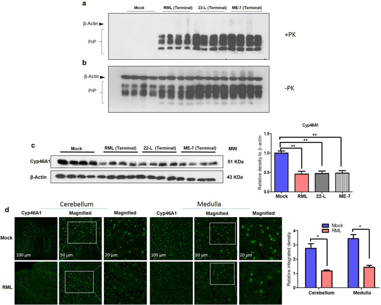
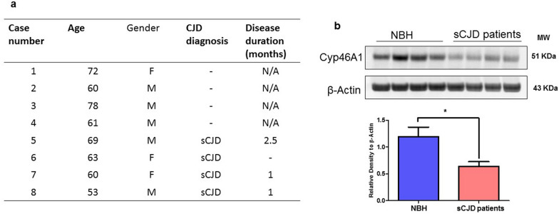
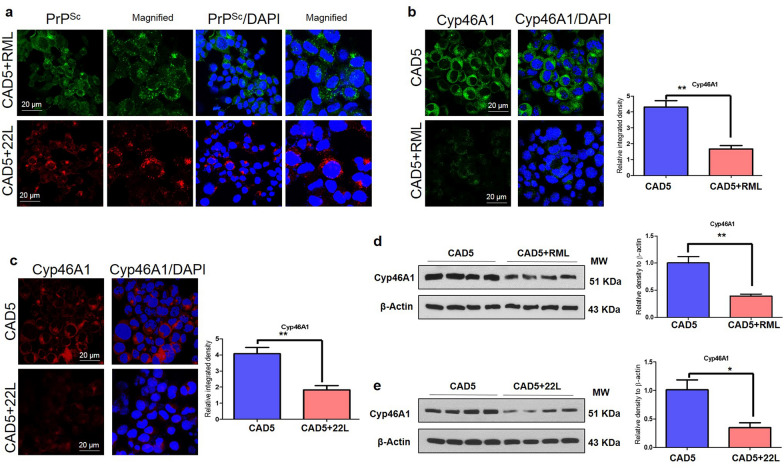
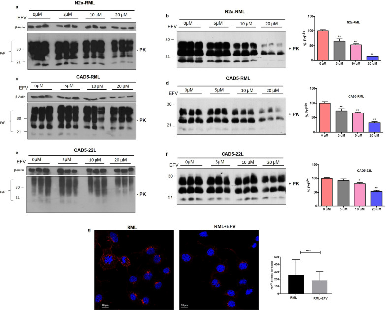
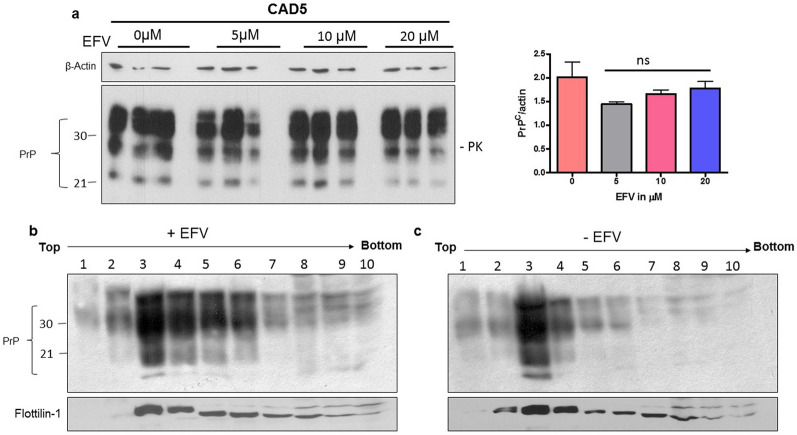
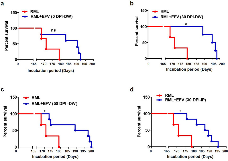
Prion diseases are fatal, infectious, and incurable neurodegenerative disorders caused by misfolding of the cellular prion protein (PrPC) into the infectious isoform (PrPSc). In humans, there are sporadic, genetic and infectious etiologies, with sporadic Creutzfeldt-Jakob disease (sCJD) being the most common form. Currently, no treatment is available for prion diseases. Cellular cholesterol is known to impact prion conversion, which in turn results in an accumulation of cholesterol in prion-infected neurons. The major elimination of brain cholesterol is achieved by the brain specific enzyme, cholesterol 24-hydroxylase (CYP46A1). Cyp46A1 converts cholesterol into 24(S)-hydroxycholesterol, a membrane-permeable molecule that exits the brain. We have demonstrated for the first time that Cyp46A1 levels are reduced in the brains of prion-infected mice at advanced disease stage, in prion-infected neuronal cells and in post-mortem brains of sCJD patients. We have employed the Cyp46A1 activator efavirenz (EFV) for treatment of prion-infected neuronal cells and mice. EFV is an FDA approved anti-HIV medication effectively crossing the blood brain barrier and has been used for decades to chronically treat HIV patients. EFV significantly mitigated PrPSc propagation in prion-infected cells while preserving physiological PrPC and lipid raft integrity. Notably, oral administration of EFV treatment chronically at very low dosage starting weeks to months after intracerebral prion inoculation of mice significantly prolonged the lifespan of animals. In summary, our results suggest that Cyp46A1 as a novel therapeutic target and that its activation through repurposing the anti-retroviral medication EFV might be valuable treatment approach for prion diseases.


Conflict of interest statement

The authors declare no competing financial interest.

Figures

Fig. 1
Fig. 1
Reduced Cyp46A1 protein levels at terminal stages of prion disease in mice. a, b PK-digested (PK +) and undigested (PK-) brain homogenates of mock and prion-infected mice (RML, 22-L and ME-7) were used for immunoblotting and PrP detection using mAb 4H11. No PK-resistant PrPSc (PrPres) was detected in + PK samples in the mock group, while PrPSc was abundantly detected in all prion-infected brain homogenates. c The same brain homogenates of mock and prion-infected mice (RML, 22-L and ME-7) at terminal stage (> 150 days post infection (DPI) were analyzed by immunoblot for Cyp46A1 levels. β-Actin was used as a loading control and was obtained after stripping of the same membrane. The histograms are represented as the means ± SEM (n = 4 mice/group) of three independent experiments. Significance = **p < 0.01; ANOVA followed by Turkey’s post hoc test. d Immunofluorescence images of Cyp46A1 (green) in the cerebellum and medulla regions of mouse brains. Fluorescence intensity was quantified; the histograms represent the means ± SEM of n = 3 mice per group, obtained from 3 independent experiments. Magnification: 10X. Scale bar = 100 μm, 50 μm, 20 μm. Significance = *p < 0.05; Student’s unpaired t test
Fig. 2
Fig. 2
Reduced Cyp46A1 levels in human sCJD brains. a Demographic details of the post-mortem brain of four different healthy individuals (NBH: normal brain homogenates) and four different sporadic CJD patients (sCJD). b Cyp46A1 levels in the brain homogenates of healthy individuals (NBH) and sCJD patients. β-Actin was used as a loading control and was obtained after stripping of the same membrane. The histogram represents the means ± SEM (n = 4 humans/group) of three independent experiments. Significance = *p < 0.05; Student’s unpaired t test
Fig. 3
Fig. 3
Cyp46A1 levels in prion-infected neuronal cells. a Immunofluorescence images of PrPSc (Green: Alexa Fluor 488; Red: Alexa Fluor™ 555; Blue: DAPI) for CAD5-RML and CAD5-22L. Magnification: 63X. Scale bar = 20 µm. b, c Immunofluorescence analysis of Cyp46A1 (Green: Alexa Fluor 488; Blue: DAPI) for CAD5 and CAD5-RML (Red: Alexa Fluor™ 555; Blue: DAPI) and for CAD5 and CAD5-22L cells. Fluorescence intensity was quantified; data are indicated as the mean ± SEM for n = 5 images per group from 3 independent experiments. Magnification: 63X. Scale bar = 20 µm. Significance = *p < 0.05; Student’s unpaired t test. d, e Immunoblot results of Cyp46A1 in CAD5, CAD5-RML and CAD5-22L cells. β-Actin was used as a loading control and was obtained after stripping the same membrane. The histograms depict the means ± SEM of three independent experiments. Significance = **p < 0.01; Student’s unpaired t test
Fig. 4
Fig. 4
EFV treatment reduced PrPSc in prion infected neuronal cells. Immunoblot images of PrP signals in cell lysates of RML-infected N2a cells (a, b), RML-infected CAD5 cells (c, d) and 22L-infected CAD5 cells (e, f) treated with EFV (5 µM, 10 µM, 20 µM) or vehicle (0 µM) for three days. For PrPSc detection, samples were digested with PK (+ PK). PrP was detected using mAb 4H11. β-actin served as a loading control. The densitometric analysis of PrPSc signals (+ PK) is shown as a percentage of the signals in the untreated control cells. The data are indicated as the mean ± SEM for n = 3 per group, and the number of independent experiments = 3, each performed in triplicate. Significanc e = **p < 0.001; One-way ANOVA with Dunnett’s multiple comparison test. g Immunofluorescence images of PrPSc (Red: TRITC; Blue: DAPI) in RML-N2a cells treated with or without EFV (20 µM). The histogram represents the means ± SEM of the intensity of PrPSc staining/nuclei obtained from 3 independent experiments. Magnification: 63X. Scale bar = 20 µm. Significance = ****p < 0.0001; Student’s unpaired t test
Fig. 5
Fig. 5
The effect of EFV treatment on PrPC level and lipid raft association in neuronal cells. a Non-infected CAD5 cell were treated with EFV (5 µM, 10 µM, 20 µM) or vehicle and PrPC in cell lysates was detected by immunoblot using mAb 4H11. β-Actin was used as a loading control. The data are indicated as the mean ± SEM for n = 3 per group, and the number of independent experiments = 3, each performed in triplicate. Significance = no significant (ns) differences b, c N2a-WT cells treated with EFV (+ EFV) or not (-EFV) were lysed in cold Triton-X 100 and subjected to flotation density gradients. Fractions 1–10 were collected from the top to the bottom of the gradient and analyzed by immunoblot using anti-PrP antibody 4H11 and anti-flotillin-1 as a lipid raft marker. b Representative immunoblot result of PrP in the fractions of cells treated with EFV. c Representative immunoblot result of PrP in the fractions of cells without EFV treatment
Fig. 6
Fig. 6
Oral EFV treatment delays prion disease and extends survival of prion-infected animals. a The representative Kaplan–Meier plot indicates the percent survival of RML-infected mice in the untreated group (n = 9 mice) and EFV-treated group RML + EFV (0 DPI-DW) (n = 7 mice), using log rank test for statistical analysis. b The representative Kaplan–Meier plot indicates the percent survival of RML-infected mice in the untreated group (n = 9 mice) and EFV-treated group RML + EFV (30 DPI-DW) (n = 9 mice), using log rank test for statistical analysis. c The representative Kaplan–Meier plot indicates the percent survival of RML-infected mice in the untreated group (n = 9 mice) and EFV-treated group RML + EFV (50 DPI-DW) (n = 8 mice), using log rank test for statistical analysis. d The representative Kaplan–Meier plot indicates the percent survival of RML-infected mice in the untreated group (n = 9 mice) and EFV-treated group RML + EFV (30 DPI-IP) (n = 7 mice), using log rank test for statistical analysis. NS p > 0.05, *p < 0.05. The x-axes depict the days post i.c. infection with RML prions

Similar articles

Cited by

References

    1. Prusiner SB. Novel proteinaceous infectious particles cause scrapie. Science. 1982;216:136–144. doi: 10.1126/science.6801762. - DOI - PubMed
    1. Aguzzi A, Heikenwalder M, Miele G. Progress and problems in the biology, diagnostics, and therapeutics of prion diseases. J Clin Investig. 2004;114:153–160. doi: 10.1172/JCI22438. - DOI - PMC - PubMed
    1. Watts JC, Balachandran A, Westaway D. The expanding universe of prion diseases. PLoS Pathog. 2006;2:e26. doi: 10.1371/journal.ppat.0020026. - DOI - PMC - PubMed
    1. Chen CC, Wang YH. Estimation of the exposure of the UK population to the bovine spongiform encephalopathy agent through dietary intake during the period 1980 to 1996. PLoS ONE. 2014;9:e94020. doi: 10.1371/journal.pone.0094020. - DOI - PMC - PubMed
    1. Hannaoui S, Arifin MI, Chang SC, et al. Cellulose ether treatment in vivo generates chronic wasting disease prions with reduced protease resistance and delayed disease progression. J Neurochem. 2020;152:727–740. doi: 10.1111/jnc.14877. - DOI - PMC - PubMed

Publication types

MeSH terms

Supplementary concepts

Grants and funding