. 2021 Jun;99(6):412-425.
doi: 10.1124/molpharm.120.000191. Epub 2021 Apr 1.

Inhibition of Human Uracil DNA Glycosylase Sensitizes a Large Fraction of Colorectal Cancer Cells to 5-Fluorodeoxyuridine and Raltitrexed but Not Fluorouracil

Affiliations

Inhibition of Human Uracil DNA Glycosylase Sensitizes a Large Fraction of Colorectal Cancer Cells to 5-Fluorodeoxyuridine and Raltitrexed but Not Fluorouracil

Eric S Christenson et al. Mol Pharmacol. 2021 Jun.

Abstract

Previous short-hairpin RNA knockdown studies have established that depletion of human uracil DNA glycosylase (hUNG) sensitizes some cell lines to 5-fluorodeoxyuridine (FdU). Here, we selectively inhibit the catalytic activity of hUNG by lentiviral transduction of uracil DNA glycosylase inhibitor protein into a large panel of cancer cell lines under control of a doxycycline-inducible promoter. This induced inhibition strategy better assesses the therapeutic potential of small-molecule targeting of hUNG. In total, 6 of 11 colorectal lines showed 6- to 70-fold increases in FdU potency upon hUNG inhibition ("responsive"). This hUNG-dependent response was not observed with fluorouracil (FU), indicating that FU does not operate through the same DNA repair mechanism as FdU in vitro. Potency of the thymidylate synthase inhibitor raltitrexed (RTX), which elevates deoxyuridine triphosphate levels, was only incrementally enhanced upon hUNG inhibition (<40%), suggesting that responsiveness is associated with incorporation and persistence of FdU in DNA rather than deoxyuridine. The importance of FU/A and FU/G lesions in the toxicity of FdU is supported by the observation that dT supplementation completely rescued the toxic effects of U/A lesions resulting from RTX, but dT only increased the IC50 for FdU, which forms both FU/A and FU/G mismatches. Contrary to previous reports, cellular responsiveness to hUNG inhibition did not correlate with p53 status or thymine DNA glycosylase expression. A model is suggested in which the persistence of FU/A and FU/G base pairs in the absence of hUNG activity elicits an apoptotic DNA damage response in both responsive and nonresponsive colorectal lines. SIGNIFICANCE STATEMENT: The pyrimidine base 5-fluorouracil is a mainstay chemotherapeutic for treatment of advanced colorectal cancer. Here, this study shows that its deoxynucleoside form, 5-fluorodeoxyuridine (FdU), operates by a distinct DNA incorporation mechanism that is strongly potentiated by inhibition of the DNA repair enzyme human uracil DNA glycosylase. The hUNG-dependent mechanism was present in over 50% of colorectal cell lines tested, suggesting that a significant fraction of human cancers may be sensitized to FdU in the presence of a small-molecule hUNG inhibitor.


Conflict of interest statement

The authors report no conflicts of interest.

Figures

None
Graphical abstract
Fig. 1.
Fig. 1.
Metabolism of FU and FdU and inhibitors of hUNG. (A) Metabolic reactions of FU and FdU leading to the RNA and DNA pathways for cellular toxicity. (B) hUNG can be inhibited in human cells by expression of the small PBS2 bacteriophage protein UGI (blue), which targets the active site of hUNG [Protein Data Bank (PDB) 1UGH]. (C) Small-molecule inhibitors of hUNG have been characterized that also target the same site as UGI (PDB 3FCI). dUDP, deoxyuridine diphosphate; dR-1P, DNA-dependent RNA polymerase; FdUDP, 5-fluorodeoxyuridine diphosphate; FUDP, 5-fluorouridine diphosphate; FUMP, 5-fluorouridine monophosphate; FUTP, 5-fluorouridine triphosphate; NDPK, nucleoside diphosphate kinase; NMPK, nucleoside monophosphate kinase; OPRT, orotate phosphoribosyltransferase; polε, DNA polymerase ε; polδ, polymerase δ; RNAP III, RNA polymerase III; RNR, ribonucleotide reductase; TK, thymidine kinase.
Fig. 2.
Fig. 2.
Inhibition of hUNG activity by UGI inducible expression increases potency of FdU but not FU for a subset of colorectal cancer cell lines. (A) hUNG activity assay performed on protein lysates from parental (WT) colon cancer cell lines or the constructs containing doxycycline-inducible UGI. This assay uses a 5′-FAM–labeled 19-mer ssDNA substrate containing a central uracil base. When hUNG activity is present, a 5′-FAM–labeled 9-mer product is produced after sample processing, which is resolved from substrate by denaturing polyacrylamide gel electrophoresis. (B) Dose-response curves for responsive colon cancer cell lines exposed to FdU under hUNG active (black) and inhibited (red) conditions. (C) Dose-response curves for nonresponsive colon cancer cell lines exposed to FdU under hUNG active (black) and inhibited (red) conditions. (D) Dose-response curves for FdU responsive colon cancer cell lines exposed to FU under hUNG active (black) and inhibited (red) conditions. Each dose-response curve was repeated at least three times beginning from a fresh culture of each cell line. The error bars on each data point are the S.D. of three technical replicates from the same biologic replicate. Overall, three biologic replicates were performed (Supplemental Table 2). WT, parental cells; I, doxycycline induced; NI, not induced.
Fig. 3.
Fig. 3.
LC/MS measurements of U and FU bases in DNA of R and NR lines in the presence or absence of hUNG inhibition or FdU treatment. (A) Genomic U content of responsive DLD1 and HT29 lines and nonresponsive LoVo and SW620 lines in the presence and absence of FdU treatment and hUNG inhibition as indicated. (B) Genomic FU content of responsive DLD1 and HT29 lines and nonresponsive LoVo and SW620 lines in the presence and absence of FdU treatment and hUNG inhibition as indicated. In these experiments, cells were synchronized at the G1/S border by serum starvation for 48 hours, followed by return to normal media for 2 hours and supplementation with 5 μM FdU for 24 hours before harvesting genomic DNA. The error bars on each measurement represent the S.D. from two biologic replicates from two distinct experiments started from a fresh culture of each cell line/condition.
Fig. 4.
Fig. 4.
Cell killing by RTX for a subset of FdU responsive colorectal cancer cell lines in the presence and absence of hUNG activity. (A) Dose-response curves for RTX for the responsive colon cancer lines that demonstrated increased sensitivity to FdU upon hUNG inhibition. (B) Dose-response curves for the nonresponsive colon cancer cell lines that demonstrated no change in sensitivity to FdU upon hUNG inhibition. Each dose-response curve was repeated at least twice and was begun from a fresh culture of each cell line. The error bars on each measurement represent the S.D. from three technical replicates of each cell line/condition.
Fig. 5.
Fig. 5.
Comparison of FdU, FU, or RTX responses of hUNG R and NR colon cancer lines. (A) FdU IC50 values for R and NR lines in absence (black) and presence (red) of hUNG inhibition. (B) FU IC50 values for R and NR lines in the absence and presence of hUNG inhibition. (C) RTX AUC values for R and NR in the absence and presence of hUNG inhibition. Error bars are the S.D. of three (FdU) and two (FU, RTX) biologic replicates from distinct experiments started from a fresh cell culture. A Student’s t test was performed to compare the difference between the FdU IC50 in the six responsive and six nonresponsive lines when hUNG was active (P value = 0.002). The change in raltitrexed AUC with hUNG inhibition between the FdU responsive and nonresponsive cell lines was compared using a Student’s t test (P value = 0.03).
Fig. 6.
Fig. 6.
Differential effects of dT and dU supplementation on toxicities of FdU, FU, and RTX in the hUNG responsive DLD1 and HT29 cell lines. (A) LC/MS measurements of U and FU bases in DNA in DLD1 and HT29 cells treated with FdU in the absence and presence of hUNG activity. These measurements were performed with and without supplementation with 100 μM dU or dT as indicated in the legend. Two biologic replicate measurements were made. (B) Supplementation with 100 μM dT increases the IC50 of FdU and removes the response to hUNG inhibition (red curves). In contrast, dT supplementation completely rescues the toxicity of RTX while having no impact on FU toxicity. Dose-response curves for these cell lines under hUNG active and inhibited conditions are shown by the dashed lines for comparison. (C) Supplementation with 100 μM dU increases the IC50 of FdU, but the response to hUNG inhibition (red curves) is retained. In contrast, dU supplementation with FU does not alter toxicity in the DLD1 cell line but slightly enhances toxicity in the HT29 line. Supplementation with 100 μM dU during treatment with RTX increases the IC50 by 100-fold but retains the increased AUC under conditions of hUNG inhibition (red curves) observed with RTX alone (see text). The supplementation experiments with FdU were performed in biologic triplicates (three technical replicates for each), and supplementation experiments with FU and RTX were performed with biologic duplicates (three technical replicates for each).
Fig. 7.
Fig. 7.
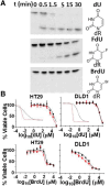
hUNG activity on U, FU, and BrU DNA bases and cell toxicities of dU and BrdU. (A) The 19-mer substrates containing U, FU, or BrU were reacted with 50 pM of hUNG catalytic domain for the indicated times, and the reaction products were resolved by gel electrophoresis after processing. The excision of the base results in a 5′-FAM–labeled 9-mer product. (B) HT29UGI and DLD1UGI cells were dosed with dU and BrdU in the presence (black) and absence (red) of hUNG activity. After 72 hours of nucleoside exposure, cell viability was measured using the MTS assay. Each concentration-response curve was repeated at least twice, beginning from a fresh culture of each cell line. The error bars on each measurement represent the S.D. from three technical replicates of each cell line/condition.
Fig. 8.
Fig. 8.
Sensitivity of cells to FdU upon loss of TDG. (A) Western blot demonstrating loss of TDG protein expression with two independent clones generated with unique CRISPR guides targeting the TDG coding sequence (DLD1UGI cell line). A negative control guide sequence is also shown. (B) Western blot establishing reduced TDG expression after introduction of two unique TDG shRNA coding sequences into HT29UGI and DLD1UGI cells. Expression in the control guide shRNA and the parental cell lines (wt) are also shown. (C) DLDUGI cells containing two unique CRISPR TDG knockouts were exposed to a spectrum of FdU concentrations in a hUNG active (black data) and inhibited (red data) background. Cells were exposed to drug for 72 hours before determination of viability using the MTS assay, and shRNA negative controls are also shown. (D) HT29UGI and DLD1UGI cells expressing shRNA targeting TDG were exposed to a spectrum of FdU concentrations in a hUNG active (black data) and inhibited (red data) background. Cells were exposed to drug for 72 hours before determination of viability using the MTS assay and shRNA negative controls are also shown. Each dose-response curve was repeated at least twice, beginning from a fresh culture of each cell line. The error bars on each measurement represent the S.D. from three technical replicates of each cell line/condition. WT, parental cells; Ctrl, control cells containing noncoding shRNA construct; sumo-TDG, sumoylated TDG.

References

    1. Barretina J, Caponigro G, Stransky N, Venkatesan K, Margolin AA, Kim S, Wilson CJ, Lehár J, Kryukov GV, Sonkin D, et al. (2012) The Cancer Cell Line Encyclopedia enables predictive modelling of anticancer drug sensitivity. Nature 483:603–607. - PMC - PubMed
    1. Benson AB III, Venook AP, Cederquist L, Chan E, Chen Y-J, Cooper HS, Deming D, Engstrom PF, Enzinger PC, Fichera A, et al. (2017) Colon cancer, version 1.2017, NCCN clinical practice guidelines in oncology. J Natl Compr Canc Netw 15:370–398. - PubMed
    1. Bulgar AD, Weeks LD, Miao Y, Yang S, Xu Y, Guo C, Markowitz S, Oleinick N, Gerson SL, Liu L (2012) Removal of uracil by uracil DNA glycosylase limits pemetrexed cytotoxicity: overriding the limit with methoxyamine to inhibit base excision repair. Cell Death Dis 3:e252. - PMC - PubMed
    1. Chen WY, Wang DH, Yen RC, Luo J, Gu W, Baylin SB (2005) Tumor suppressor HIC1 directly regulates SIRT1 to modulate p53-dependent DNA-damage responses. Cell 123:437–448. - PubMed
    1. Cole AR, Ofer S, Ryzhenkova K, Baltulionis G, Hornyak P, Savva R (2013) Architecturally diverse proteins converge on an analogous mechanism to inactivate Uracil-DNA glycosylase. Nucleic Acids Res 41:8760–8775. - PMC - PubMed

Publication types

MeSH terms