Review
. 2021 Apr 1;134(7):jcs254292.
doi: 10.1242/jcs.254292.

Data science in cell imaging

Affiliations
Review

Data science in cell imaging

Meghan K Driscoll et al. J Cell Sci. .

Abstract

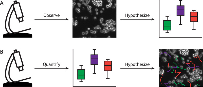
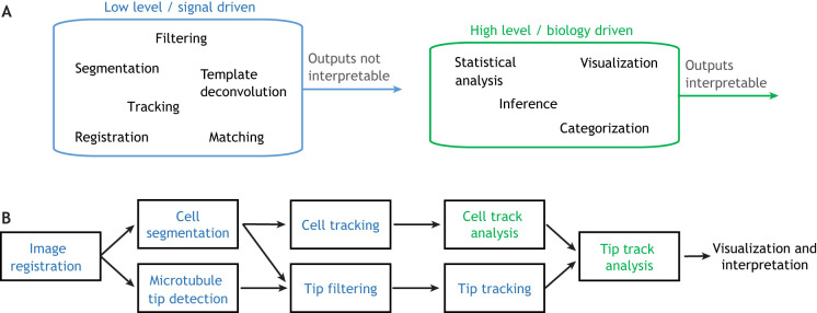
Cell imaging has entered the 'Big Data' era. New technologies in light microscopy and molecular biology have led to an explosion in high-content, dynamic and multidimensional imaging data. Similar to the 'omics' fields two decades ago, our current ability to process, visualize, integrate and mine this new generation of cell imaging data is becoming a critical bottleneck in advancing cell biology. Computation, traditionally used to quantitatively test specific hypotheses, must now also enable iterative hypothesis generation and testing by deciphering hidden biologically meaningful patterns in complex, dynamic or high-dimensional cell image data. Data science is uniquely positioned to aid in this process. In this Perspective, we survey the rapidly expanding new field of data science in cell imaging. Specifically, we highlight how data science tools are used within current image analysis pipelines, propose a computation-first approach to derive new hypotheses from cell image data, identify challenges and describe the next frontiers where we believe data science will make an impact. We also outline steps to ensure broad access to these powerful tools - democratizing infrastructure availability, developing sensitive, robust and usable tools, and promoting interdisciplinary training to both familiarize biologists with data science and expose data scientists to cell imaging.

Keywords: Data science; Deep learning; Imaging; Machine learning; Microscopy.


Conflict of interest statement

Competing interestsThe authors declare no competing or financial interests.

Figures

Fig. 1.
Fig. 1.
Image analysis workflows. (A) In a typical microscopy-heavy research project, scientists acquire and observe images, and then form and quantitatively test hypotheses based on their observations. (B) We propose that in the future it will be necessary to flip this procedure; first acquiring and quantifying images and only then interacting with the data to further form and test hypotheses.
Fig. 2.
Fig. 2.
Hierarchy of image analysis tasks. (A) Image analysis pipelines can be decomposed into low-level (blue) and high-level (green) tasks, with low-level tasks generally preceding high-level tasks. (B) An example image-analysis pipeline for microtubule tracking with low-level tasks shown blue and high-level tasks shown green. Here, images are first registered, or aligned across frames, to account for microscope movement. Next, the cell is segmented, or distinguished from the background, and the microtubule tips are detected. The cell segmentation is used to filter tips by location, removing spurious detections outside the cell, and the cell segmentation and tip detections are separately tracked across frames. Finally, using information derived from a cell-tracking analysis, the tip tracks are analyzed to generate biological insight. In this example, only the track analyses are high-level tasks, since they are the only tasks whose outputs can be directly interpreted to gain biological insight.

References

    1. Aguet, F., Antonescu, C. N., Mettlen, M., Schmid, S. L. and Danuser, G. (2013). Advances in analysis of low signal-to-noise images link dynamin and AP2 to the functions of an endocytic checkpoint. Dev. Cell 26, 279-291. 10.1016/j.devcel.2013.06.019 - DOI - PMC - PubMed
    1. Amat, F., Lemon, W., Mossing, D. P., McDole, K., Wan, Y., Branson, K., Myers, E. W. and Keller, P. J. (2014). Fast, accurate reconstruction of cell lineages from large-scale fluorescence microscopy data. Nat. Methods 11, 951-958. 10.1038/nmeth.3036 - DOI - PubMed
    1. Attwood, T. K., Blackford, S., Brazas, M. D., Davies, A. and Schneider, M. V. (2019). A global perspective on evolving bioinformatics and data science training needs. Brief. Bioinform. 20, 398-404. 10.1093/bib/bbx100 - DOI - PMC - PubMed
    1. Bagonis, M. M., Fusco, L., Pertz, O. and Danuser, G. (2019). Automated profiling of growth cone heterogeneity defines relations between morphology and motility. J. Cell Biol. 218, 350-379. 10.1083/jcb.201711023 - DOI - PMC - PubMed
    1. Bannon, D., Moen, E., Schwartz, M., Borba, E., Kudo, T., Greenwald, N., Vijayakumar, V., Chang, B., Pao, E., Osterman, E.et al. (2021). DeepCell Kiosk: scaling deep learning-enabled cellular image analysis with Kubernetes. Nat. Methods 18, 43-45. 10.1038/s41592-020-01023-0 - DOI - PMC - PubMed

Publication types

MeSH terms

LinkOut - more resources