. 2021 Apr 2;372(6537):91-94.
doi: 10.1126/science.abb9032.

Incorporation of a nucleoside analog maps genome repair sites in postmitotic human neurons

Affiliations

Incorporation of a nucleoside analog maps genome repair sites in postmitotic human neurons

Dylan A Reid et al. Science. .

Abstract

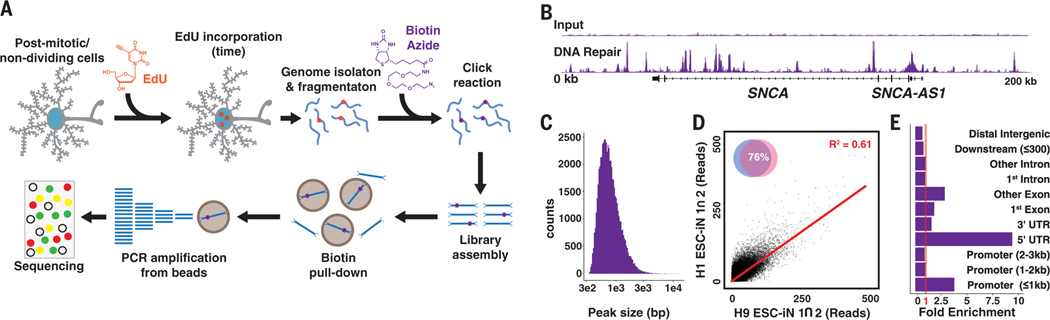
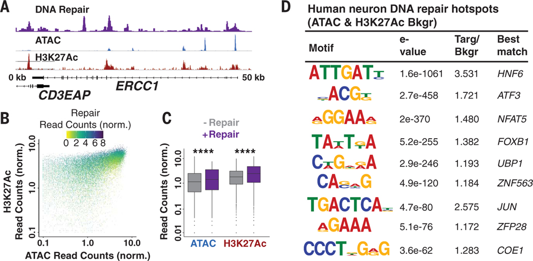
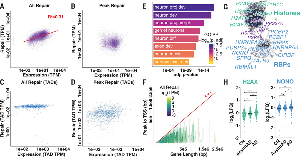
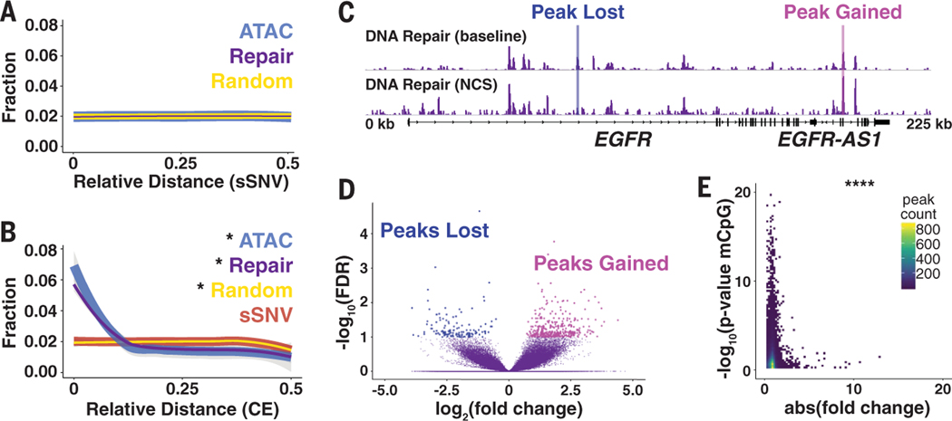
Neurons are the longest-lived cells in our bodies and lack DNA replication, which makes them reliant on a limited repertoire of DNA repair mechanisms to maintain genome fidelity. These repair mechanisms decline with age, but we have limited knowledge of how genome instability emerges and what strategies neurons and other long-lived cells may have evolved to protect their genomes over the human life span. A targeted sequencing approach in human embryonic stem cell-induced neurons shows that, in neurons, DNA repair is enriched at well-defined hotspots that protect essential genes. These hotspots are enriched with histone H2A isoforms and RNA binding proteins and are associated with evolutionarily conserved elements of the human genome. These findings provide a basis for understanding genome integrity as it relates to aging and disease in the nervous system.


Figures

Fig. 1.
Fig. 1.. EdU incorporated into the genomes of postmitotic neurons by DNA repair can be mapped by next-generation sequencing.
(A) Repair-seq workflow: Neurons are cultured with EdU for 24 hours, the genomes are isolated and fragmented with sonication, a click reaction adds biotin to the EdU, and biotin-DNA fragments are enriched on streptavidin beads and subsequently amplified (12). PCR, polymerase chain reaction. (B) DNA repair peaks from the SNCA locus in EdU-fed neurons compared with input genomes sequenced to the same depth show a site with substantial enrichment. (C) Histogram of DNA repair hotspot peak widths. bp, base pairs. (D) Comparison of intersecting peaks in two H1 and two H9 ESC-iN samples. (E) Fold enrichment of DRHs over predicted genome distribution. UTR, untranslated region.
Fig. 2.
Fig. 2.. Chromatin accessibility controls the placement of repair hotspots.
(A) Repair-seq, ATAC-seq, and H3K27Ac ChIP-seq data at the ERCC1 locus demonstrate overlap between DNA repair, chromatin accessibility, and histone acetylation. (B) Scatter plot of Repair-seq–normalized read counts compared to ATAC- and H3K27Ac-normalized read counts. (C) Box plots of ATAC and H3K27Ac peaks with and without DNA repair. The horizontal black lines represent the medians, whereas the whiskers are displayed at the largest and smallest values no more than 1.5 times the interquartile range from quartiles 3 and 1, respectively. ****P < 2.2 × 10⁻¹⁶ by Kruskal-Wallis test. (D) DNA sequence motifs identified de novo and predicted as enriched in DRHs relative to randomized sequence. Targ/Bkgr, target/background.
Fig. 3.
Fig. 3.. Transcriptional output correlates with total DNA repair in genes but not repair hotspots.
(A and B) All DNA repair–associated reads in genes (A) and repair peak–associated reads in genes (B) compared with RNA-associated reads from total RNA-seq. TPM, transcripts per kilobase million. (C and D) All DNA repair–associated reads in genes (C) and repair peak–associated reads in genes (D) compared with RNA-associated reads from total RNA-seq in length-normalized TADs. (E) Select biological process (BP) GO terms for genes containing DRHs. Terms are neuron projection development, neuron development, neuron projection morphogenesis, genesis of neurons, neuron differentiation, axon development, neurogenesis, and nervous system development. (F) Line plot of transcription start sites (TSSs) to DRHs in each gene compared with total gene length (colored by total DNA repair level). (G) String network representation of peptides enriched for histones (green), RNA binding proteins (RBPs; blue), and some chaperones and ubiquitin (purple). (H) LFQ proteomics data for H2AX and NONO abundance in cognitively normal (CN), asymptomatic Alzheimer’s disease (AsymtAD), and Alzheimer’s disease (AD). Horizontal black lines represent mean log2(LFQ). ns, not significant; *P < 0.5; **P < 0.01; and ****P < 0.0001 by analysis of variance (ANOVA) with Tukey’s multiple comparison test.
Fig. 4.
Fig. 4.. Repair hotspots protect evolutionarily constrained regions of the human genome from epigenetic drift.
(A) Relative distance measurement from postmortem human neuron sSVNs to nearest DRH or randomly generated peaks. (B) Relative distance measurement from GERP CEs to nearest sSNV, DRH, ATAC, or random peaks. (C) Representative browser view of DRHs at baseline and 24 hours after 10 min of NCS treatment demonstrates that peaks are lost and gained. (D) Volcano plot for NCS differential peaks using a false discovery rate (FDR) of <0.1 from four samples. (E) Heat map of the DRH stability (absolute fold change after NCS treatment) compared with epigenetic clock mCpG sites from sorted human neurons. *P < 0.01 by Jaccard distance test;****P < 8.52 × 10⁻⁵ by hypergeometric test.

Comment in

References

    1. Mattson MP, Magnus T, Nat. Rev. Neurosci 7, 278–294 (2006). - PMC - PubMed
    1. Jackson SP, Bartek J, Nature 461, 1071–1078 (2009). - PMC - PubMed
    1. Chow HM, Herrup K, Nat. Rev. Neurosci 16, 672–684 (2015). - PubMed
    1. McKinnon PJ, Nat. Neurosci 16, 1523–1529 (2013). - PMC - PubMed
    1. Nouspikel T, Hanawalt PC, Mol. Cell. Biol 20, 1562–1570 (2000). - PMC - PubMed

Publication types