Review
. 2021 Apr 1;12(4):339.
doi: 10.1038/s41419-021-03614-x.

Regulated cell death pathways in doxorubicin-induced cardiotoxicity

Affiliations
Review

Regulated cell death pathways in doxorubicin-induced cardiotoxicity

Effimia Christidi et al. Cell Death Dis. .

Abstract

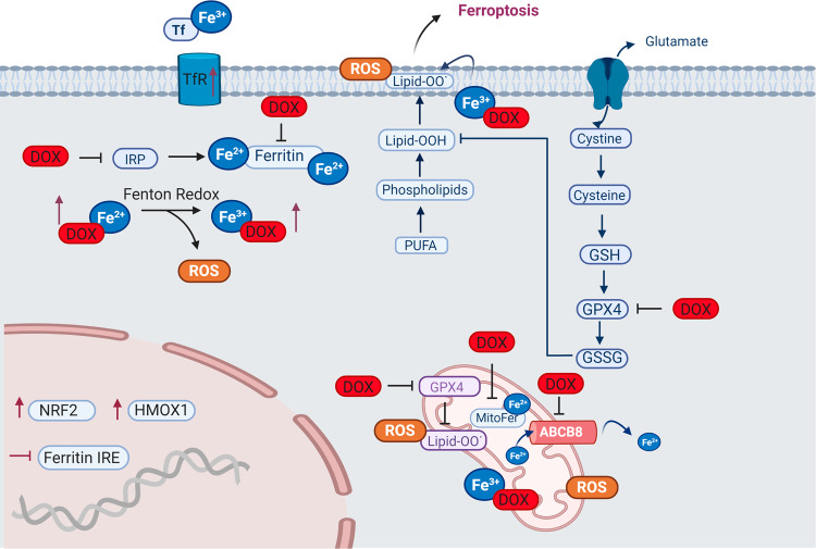
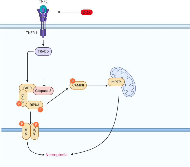
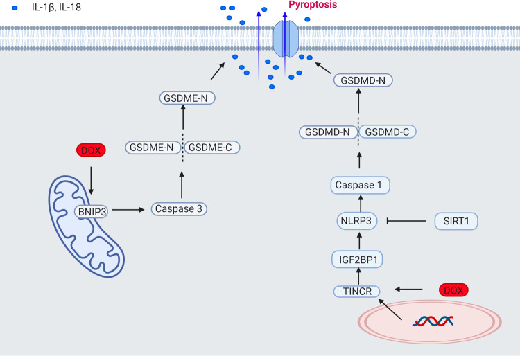
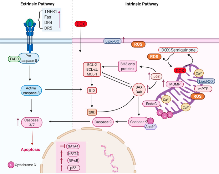
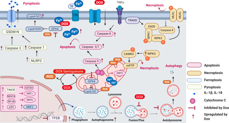
Doxorubicin is a chemotherapeutic drug used for the treatment of various malignancies; however, patients can experience cardiotoxic effects and this has limited the use of this potent drug. The mechanisms by which doxorubicin kills cardiomyocytes has been elusive and despite extensive research the exact mechanisms remain unknown. This review focuses on recent advances in our understanding of doxorubicin induced regulated cardiomyocyte death pathways including autophagy, ferroptosis, necroptosis, pyroptosis and apoptosis. Understanding the mechanisms by which doxorubicin leads to cardiomyocyte death may help identify novel therapeutic agents and lead to more targeted approaches to cardiotoxicity testing.


Conflict of interest statement

The authors declare no competing interests.

Figures

Fig. 1
Fig. 1. Role of doxorubicin in autophagy-related cardiomyocyte death.
Schematic representation of autophagy during doxorubicin treatment. Doxorubicin disrupts autophagy by: inducing initiation through AMPK activation and/or mTOR inhibition, the formation of the phagophore, upregulation of Atg proteins, and by blocking lysosomal proteolysis resulting in accumulation of autophagosomes and autolysosomes and reactive oxygen species. DOX doxorubicin, AMPK 5′ AMP-activated protein kinase, mTOR mammalian target of rapamycin, TFEB transcription factor EB, Atg autophagy-related protein, LC3II microtubule-associated protein 1A/1B-light chain 3, ROS reactive oxygen species.
Fig. 2
Fig. 2. Doxorubicin-induced ferroptosis.
Schematic representation of doxorubicin-induced ferroptosis pathway. Doxorubicin treatment results in iron overload through upregulation of TfR and inactivation of ferritin. Free iron complexes with doxorubicin and through the Fenton reaction create reactive oxygen species (ROS). Doxorubicin induces lipid peroxidation by inhibiting cytosolic and mitochondrial GPX4 resulting in ferroptosis. In the mitochondria, doxorubicin causes iron overload by blocking MitoFer and ABCB8. In the nucleus, activation of NRF2 results in upregulation of HMOX1 leading to heme degradation and resulting in excess free iron and ferroptosis. Tf transferrin, TfR transferrin receptor, IRP iron response regulatory protein, IRE Iron response element, NRF2 nuclear factor erythroid 2-related factor, HMOX1 heme oxygenase 1, PUFA polyunsaturated fatty acids, Lipid-OO lipid peroxides, GSH reduced glutathione, GPX4 glutathione peroxidase, GSSG glutathione disulfide, H2O2 oxygen peroxide, MitoFer mitochondria ferritin. ABCB8 ATP-binding cassette sub-family B member 8, ROS reactive oxygen species.
Fig. 3
Fig. 3. Doxorubicin-induced necroptosis.
Schematic representation of the doxorubicin-induced necroptosis pathway. Doxorubicin causes upregulation of TNFα, activating TRADD and FADD, and upon caspase 8 inhibition and activation of RIPK1, RIPK3, and MLKL induces cell death via necroptosis. Doxorubicin can also activate necroptosis via the RIPK1 independent pathway, where RIPK3 activates CAMKII and mitochondrial permeability transition pore (mPTP) resulting in membrane potential and integrity loss. TNFα tumor necrosis factor-alpha, TRADD tumor necrosis factor receptor type 1 associated death domain protein, FADD Fas-associated protein with death domain, RIPK receptor-interacting serine/threonine-protein kinase, CAMKII calcium/calmodulin-dependent protein kinase II association domain, MLKL mixed lineage kinase domain-like, mPTP mitochondrial permeability transition pore.
Fig. 4
Fig. 4. Doxorubicin-induced pyroptosis.
Schematic representation of doxorubicin-induced pyroptosis in the heart. Doxorubicin induces pyroptosis via the upregulation of TINCR, which recruits IGF2BP and increases the expression of NLRP3 leading to activation of caspase-1, the cleavage of GMDSD-N and the release of IL-1β, IL-18. Pyroptosis is also induced via BNIP3 activation in the mitochondria, which activates caspase 3 and causes GSDME-dependent pyroptosis. Sirtuin 1 activation inhibits NLRP3 and protects cardiomyocytes from doxorubicin-induced pyroptosis. BNIP3 BCL2 interacting protein 3, GSDMD gasdermin D, GSDME gasdermin E, TINCR terminal differentiation-induced NcRNA, IGF2BP1 insulin-like growth factor 2 mRNA-binding protein 1, NLRP3 NOD−, LRR−, and pyrin domain-containing protein 3, IL interleukin.
Fig. 5
Fig. 5. Doxorubicin-induced apoptosis.
Schematic representation of doxorubicin-induced apoptosis in the heart. Doxorubicin-induced upregulation of p53, Bax/Bak, and downregulation of GATA4 and Bcl-XL activating caspases 9, 3, and 7 resulting in apoptotic death. Mitochondrial calcium overload and activation of mitochondrial permeability transition pore (mPTP) lead to mitochondrial membrane potential loss, mitochondrial swelling, and outer membrane rupture allowing the release of endonuclease G (EndoG), cytochrome c and activation of caspase 9. Doxorubicin induces the extrinsic apoptotic pathway via the upregulation of death receptors and the activation of NFAT and NF-κΒ. DR: death receptor, TNFR1 tumor necrosis factor receptor 1, FADD Fas-associated protein with death domain, DR death receptor, lipid-OO lipid peroxides, EndoG endonuclease G, Ca2+ calcium, ROS reactive oxygen species, mPTP mitochondrial permeability transition pore, MOMP mitochondria outer membrane permeability, DOX doxorubicin, Bax Bcl-2-associated X protein, Apf-1 apoptosis protease factor-1, BID BH3 interacting-domain death, NF-κΒ nuclear factor-κB, NFAT4 nuclear factor of activated T-cells, GATA4 GATA-binding protein 4.
Fig. 6
Fig. 6. Role of Sirtuins in doxorubicin-induced cardiotoxicity.
Doxorubicin downregulates SIRT1, causing increased oxidative damage, loss of mitochondria integrity, AMPK, and NLRP activation resulting in increased apoptosis, autophagy, and pyroptosis. Berberine, FGF21, and resveratrol protect from DIC via SIRT1 activation. Doxorubicin downregulates SIRT2 via miRNA-140-5p, leading to reduced SIRT2 and NRF2 expression. SIRT2 has been implicated with iron homeostasis while NRF2 activation is involved in doxorubicin-induced ferroptosis. Doxorubicin-induced downregulation of SIRT3 causes increased oxidative damage and loss of mitochondria integrity leading to increased apoptosis. Berberine and honokiol protect from DIC via SIRT3 upregulation. Doxorubicin treatment reduces the expression of SIRT6 leading to repression of GATA4 and increased apoptosis. SIRT Sirtuin, DOX doxorubicin, FGF21 fibroblast growth factor 21, Nrf2 nuclear factor erythroid 2-related factor 2, GATA4 GATA-binding protein 4.
Fig. 7
Fig. 7. Doxorubicin-induced regulated cell death pathways.
Summary of regulated cell death pathways triggered by doxorubicin in the heart. Doxorubicin triggers ROS production by inducing initiation of autophagy and by blocking lysosomal proteolysis resulting in the accumulation of autophagosomes and autolysosomes. Doxorubicin undergoes both redox cycling forming dox-semiquinone moieties and Fenton reaction creating oxidative species. Excess iron and lipid peroxidation due to doxorubicin treatment results in ferroptosis. Doxorubicin activates NLRP inducing the release of Il-1β and Il-18 resulting in death due to pyroptosis. RIPK1 and RIPK3 activation due to doxorubicin treatment leads to phosphorylation of MLKL and necroptosis. Doxorubicin causes necroptosis through RIPK1 independent pathway by activating RIPK3 and CAMKII leading to mPTP and membrane potential loss. ROS trigger p53 activation and GATA4 downregulation stimulating the intrinsic apoptotic pathway. Doxorubicin treatment upregulates death receptors and together with the activation of NFAT and NF-κΒ the extrinsic apoptotic pathway is triggered. Pink color capsules represent apoptosis, brown color capsules represent necroptosis, blue color capsules represent ferroptosis and white capsules represent pyroptosis. DOX doxorubicin, ROS reactive oxygen species, Tf transferrin, TfR transferrin receptor, GPX4 glutathione peroxidase 4, mPTP mitochondria permeability transition pore, Lipid-OO lipid peroxides, NLRP3 NOD−, LRR−, and pyrin domain-containing protein 3, TINCR terminal differentiation-induced ncRNA, GSDM-N gasdermin, TNFα tumor necrosis factor-alpha, TRADD tumor necrosis factor receptor type 1 associated death domain protein, FADD Fas-associated protein with death domain, RIPK receptor-interacting serine/threonine-protein kinase, CAMKII calcium/calmodulin-dependent protein kinase II, NRF2 nuclear factor erythroid 2-related factor, TFEB Transcription factor EB, NF-κΒ nuclear factor-κB, NFAT4 nuclear factor of activated T-cells, ROS reactive oxygen species, SIRT sirtuin, GATA4 GATA-binding protein 4.

References

    1. Miller KD, et al. Cancer treatment and survivorship statistics, 2019. CA Cancer J. Clin. 2019;69:363–385. doi: 10.3322/caac.21565. - DOI - PubMed
    1. Global Burden of Disease Cancer Collaboration GB of DC. Fitzmaurice C, et al. The global burden of cancer 2013. JAMA Oncol. 2015;1:505–527. doi: 10.1001/jamaoncol.2015.0735. - DOI - PMC - PubMed
    1. Volkova M, Russell R. Anthracycline cardiotoxicity: prevalence, pathogenesis and treatment. Curr. Cardiol. Rev. 2012;7:214–220. doi: 10.2174/157340311799960645. - DOI - PMC - PubMed
    1. Swain SM, Whaley FS, Ewer MS. Congestive heart failure in patients treated with doxorubicin. Cancer. 2003;97:2869–2879. doi: 10.1002/cncr.11407. - DOI - PubMed
    1. Lefrak EA, Piťha J, Rosenheim S, Gottlieb JA. A clinicopathologic analysis of adriamycin cardiotoxicity. Cancer. 1973;32:302–314. doi: 10.1002/1097-0142(197308)32:2<302::AID-CNCR2820320205>3.0.CO;2-2. - DOI - PubMed

Publication types

Grants and funding