. 2021 Aug;34(8):1456-1467.
doi: 10.1038/s41379-021-00793-y. Epub 2021 Apr 1.

Pathophysiology of SARS-CoV-2: the Mount Sinai COVID-19 autopsy experience

Affiliations

Pathophysiology of SARS-CoV-2: the Mount Sinai COVID-19 autopsy experience

Clare Bryce et al. Mod Pathol. 2021 Aug.

Abstract

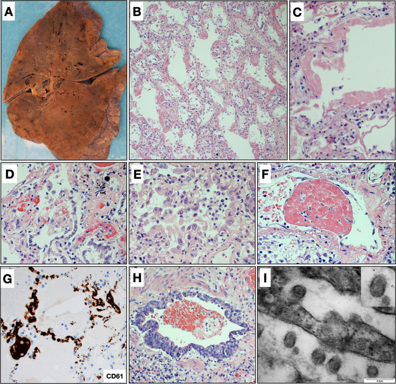
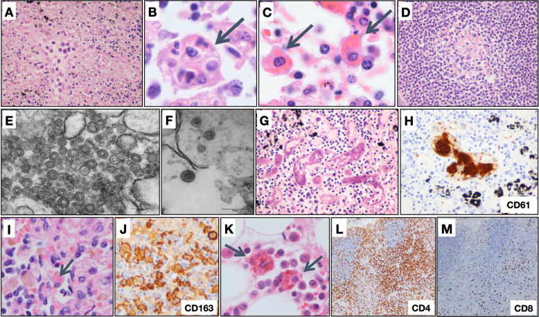
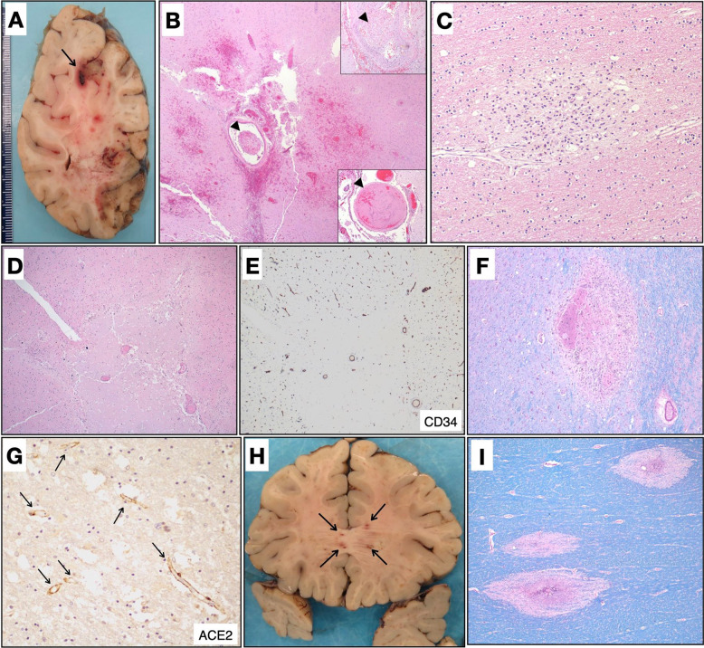
Severe acute respiratory syndrome coronavirus 2 (SARS-CoV-2) and its associated clinical syndrome COVID-19 are causing overwhelming morbidity and mortality around the globe and disproportionately affected New York City between March and May 2020. Here, we report on the first 100 COVID-19-positive autopsies performed at the Mount Sinai Hospital in New York City. Autopsies revealed large pulmonary emboli in six cases. Diffuse alveolar damage was present in over 90% of cases. We also report microthrombi in multiple organ systems including the brain, as well as hemophagocytosis. We additionally provide electron microscopic evidence of the presence of the virus in our samples. Laboratory results of our COVID-19 cohort disclose elevated inflammatory markers, abnormal coagulation values, and elevated cytokines IL-6, IL-8, and TNFα. Our autopsy series of COVID-19-positive patients reveals that this disease, often conceptualized as a primarily respiratory viral illness, has widespread effects in the body including hypercoagulability, a hyperinflammatory state, and endothelial dysfunction. Targeting of these multisystemic pathways could lead to new treatment avenues as well as combination therapies against SARS-CoV-2 infection.


Figures

Fig. 1
Fig. 1
Pulmonary findings. A Gross image of lung showing patchy areas of consolidation that corresponded to diffuse alveolar damage histologically. B Diffuse alveolar damage with prominent hyaline membranes and pneumocyte hyperplasia (H&E, ×200). C Hyaline membrane lining an alveolar space with alveolar walls showing sparse chronic inflammation (H&E, ×400). D Pneumocyte hyperplasia with relatively bland cytologic features (H&E, ×400). E Pneumocyte hyperplasia with marked pleomorphism and cytologic atypia with occasional macronucleoli. Lymphocytes and macrophages are also present in the airspace lumens (H&E, ×400). F Fibrin thrombi were observed in blood vessels in 34 cases (H&E, ×600). G Platelet thrombi are highlighted by CD61 staining (CD61, ×400). H Bronchioles with regenerative basal cell hyperplasia and loss of cilia, consistent with prior injury (H&E, ×400). I Electron micrograph from lung tissue portraying presence of multiple spherical virus particles showing spike-like electron-dense peplomeric projections ranging from 100 to 140 nm. Note the attachment/budding of viral-like particles to the respiratory epithelium signaling possible endo/exocytosis as well as presence of extracellular virus particles. Scale bar, 0.2 µm. Insert: detail of a virion showing an electron-dense, relatively thick envelope with distinctive peplomers projecting from the surface giving the appearance of a solar corona.
Fig. 2
Fig. 2
Hematolymphoid system findings. A Lymph nodes with necrosis (H&E, original magnification ×400); B hemophagocytic histiocytes engulfing multiple cell types (H&E, original magnification ×1000) and C phagocytosing erythrocytes (H&E, original magnification ×1000); and D follicle containing small, depleted, germinal center (H&E, original magnification ×400). E Electron micrographs of a lymph node revealing coronavirus-induced organelle-like replicative structures consistent with double-membrane vesicles (DMVs), scale bar, 0.25 µm and F intracytoplasmic spherically shaped virus particles with characteristic electron-dense envelope and fine peplomeric projections, scale bar 0.2 µm. G Lymph node with microthrombi (H&E, original magnification ×400), H with platelets highlighted by labeling for CD61 (original magnification ×400). I Spleen with hemophagocytosis (H&E, original magnification ×1000), J with splenic macrophages highlighted by labeling for CD163 (original magnification ×1000). K Bone marrow with hemophagocytosis (H&E, original magnification ×1000). L CD4 (original magnification ×100) and M CD8 (original magnification ×100) labeling in a lymph node demonstrating disproportionate loss of CD8+ T lymphocytes.
Fig. 3
Fig. 3
Neuropathologic findings. A Coronal section of the right cerebral hemisphere containing multiple acute microinfarcts and focal region suggestive of a thrombosed and hemorrhagic blood vessel (arrow). B Histological confirmation of an acute infarct with scattered microhemorrhages and associated microthrombi (arrowheads). Insets: two additional identified microthrombi (arrowheads) (H&E, ×400). C Subacute microinfarct with foamy macrophages (H&E, ×100). D Acute ischemic infarct associated with mildly hyperplastic and congested blood vessels (H&E, ×400). E Blood vessels are highlighted by CD34 (×400). F Area of microhemorrhage with associated reactive changes within the basal ganglia (LFB, ×400). G Immunohistochemical staining highlights ACE2 expression within many cerebral blood vessels (ACE2, ×100, arrows). H Coronal section at the level of the genu of the corpus callosum showing areas of hemorrhage and myelin loss. I Histology of the corpus callosum lesions, demonstrating small areas of perivascular myelin loss, highlighted by luxol fast blue stain (LFB), rarely associated with hemorrhage (×400).

References

    1. COVID-19 Dashboard by the Center for Systems Science and Engineering (CSSE) at Johns Hopkins University, [Internet]. [Accessed 21 Jan 2021]. Available from https://coronavirus.jhu.edu.
    1. Xu Z, Shi L, Wang Y, Zhang J, Huang L, Zhang C, et al. Pathological findings of COVID-19 associated with acute respiratory distress syndrome. Lancet Respir Med. 2020;8:420–422. doi: 10.1016/S2213-2600(20)30076-X. PMCID 7164771, PubMed PMID: 32085846. - DOI - PMC - PubMed
    1. Carsana L, Sonzogni A, Nasr A, Rossi RS, Pellegrinelli A, Zerbi P, et al. Pulmonary post-mortem findings in a series of COVID-19 cases from northern Italy: a two-centre descriptive study. Lancet Infect Dis. 2020;20:1135–1140. doi: 10.1016/S1473-3099(20)30434-5. PMCID 7279758, PubMed PMID: 32526193. - DOI - PMC - PubMed
    1. Edler C, Schroder AS, Aepfelbacher M, Fitzek A, Heinemann A, Heinrich F, et al. Dying with SARS-CoV-2 infection-an autopsy study of the first consecutive 80 cases in Hamburg, Germany. Int J Leg Med. 2020;134:1275–1284. doi: 10.1007/s00414-020-02317-w. - DOI - PMC - PubMed
    1. Fox SE, Akmatbekov A, Harbert JL, Li G, Quincy Brown J, Vander Heide RS. Pulmonary and cardiac pathology in African American patients with COVID-19: an autopsy series from New Orleans. Lancet Respir Med. 2020;8:681–686. doi: 10.1016/S2213-2600(20)30243-5. PMCID 7255143, PubMed PMID: 32473124. - DOI - PMC - PubMed

MeSH terms