Review
. 2021 Mar 16:15:587197.
doi: 10.3389/fnins.2021.587197. eCollection 2021.

Iron Metabolism Disorders for Cognitive Dysfunction After Mild Traumatic Brain Injury

Affiliations
Review

Iron Metabolism Disorders for Cognitive Dysfunction After Mild Traumatic Brain Injury

Suna Huang et al. Front Neurosci. .

Abstract

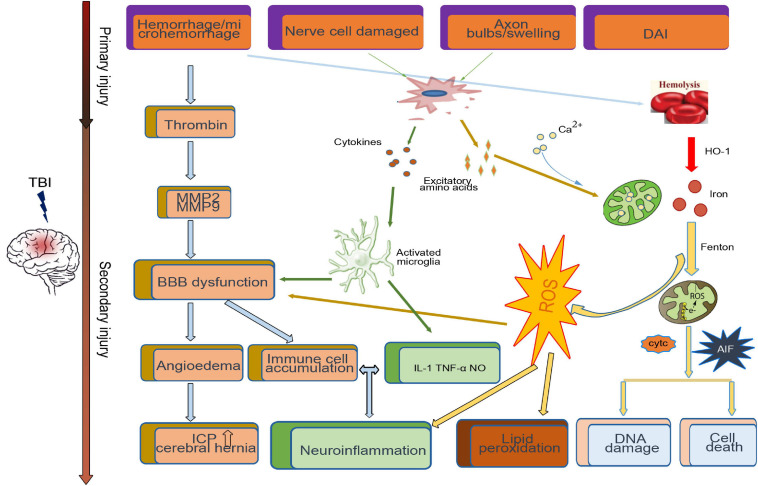
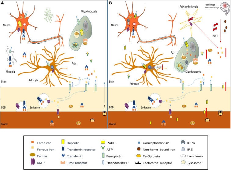
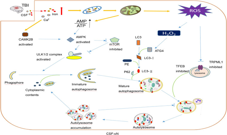
Traumatic brain injury (TBI) is one of the most harmful forms of acute brain injury and predicted to be one of the three major neurological diseases that cause neurological disabilities by 2030. A series of secondary injury cascades often cause cognitive dysfunction of TBI patients leading to poor prognosis. However, there are still no effective intervention measures, which drive us to explore new therapeutic targets. In this process, the most part of mild traumatic brain injury (mTBI) is ignored because its initial symptoms seemed not serious. Unfortunately, the ignored mTBI accounts for 80% of the total TBI, and a large part of the patients have long-term cognitive dysfunction. Iron deposition has been observed in mTBI patients and accompanies the whole pathological process. Iron accumulation may affect long-term cognitive dysfunction from three pathways: local injury, iron deposition induces tau phosphorylation, the formation of neurofibrillary tangles; neural cells death; and neural network damage, iron deposition leads to axonal injury by utilizing the iron sensibility of oligodendrocytes. Thus, iron overload and metabolism dysfunction was thought to play a pivotal role in mTBI pathophysiology. Cerebrospinal fluid-contacting neurons (CSF-cNs) located in the ependyma have bidirectional communication function between cerebral-spinal fluid and brain parenchyma, and may participate in the pathway of iron-induced cognitive dysfunction through projected nerve fibers and transmitted factor, such as 5-hydroxytryptamine, etc. The present review provides an overview of the metabolism and function of iron in mTBI, and to seek a potential new treatment target for mTBI with a novel perspective through combined iron and CSF-cNs.

Keywords: autophagy; cerebrospinal-fluid contacting neuron; cognitive dysfunction; iron metabolism; traumatic brain injury.


Conflict of interest statement

The authors declare that the research was conducted in the absence of any commercial or financial relationships that could be construed as a potential conflict of interest.

Figures

FIGURE 1
FIGURE 1
Primary and secondary pathophysiology in traumatic brain injury. Immediately at the time of impact, the brain suffers a direct mechanical trauma, which induced TBI primary injury including hemorrhage/microhemorrhage, cell death, diffuse axonal injury, etc. Hours to days after impact, the secondary injury initiated by the primary injury, such as the microglia activated by cytokines and excitatory amino acids from damaged cell, cooperates with the dysfunctional blood–brain barrier (BBB)-induced immune cell accumulation and neuroinflammation. Excessive iron and calcium-mediated mitochondrial dysfunction increase the generation of free radical and oxidative species, which induce lipid peroxidation, DNA damage, and cell death.
FIGURE 2
FIGURE 2
Iron metabolism disorder in traumatic brain injury. (A) In normal brain, iron transport across the luminal membrane of the BBB mainly relies on the transferrin/transferrin receptor (Tf/TfR) pathway, Tf-TfR-bounding iron complex under the help of divalent metal transporter 1 (DMT1) released from the endosome then transported across the abluminal membrane by ferroportin 1/hephaestin (Fpn1/Heph) and/or Fpn1/ceruloplasmin (CP). Non-transferrin-bound iron can be transported across the BBB by DMT1, etc. After release from the microvascular endothelial cells through Fpn1, iron mainly in the forms of Tf-fe3+, ferritin-Fe3+, and ATP-Fe2+, circulates in the cerebral spinal fluid (CSF), which is very convenient to be utilized by nerve cells. Astrocytes take iron via DMT1, and oligodendrocytes take iron in the form of ferritin via the Tim-2 receptor. Neurons and microglias expressing TfR can take iron from the CSF through TfR and DMT1. Cytoplasm iron: (1) Participates in cell metabolic activities, such as the synthesis of heme in mitochondria. (2) Contained in poly(rC)-binding protein (PCBP), ferritin, lysosome to prevent free iron from generating ROS. (B) In traumatic brain injury, iron accumulation occurs in the situation of hemorrhage/microhemorrhage. Excessive iron suppresses iron regulatory proteins (IRPS) combined with TfR and Fpn1, ferritin iron regulatory elements (IRES) induce TfR decrease and Fpn1, and ferritin increased to prevent neuron iron overload, but brain hepcidin expression is increased including local and peripheral hepcidin, which is transported into the brain through the dysfunctional BBB. Hepcidin internalizes Fpn1 to suppress the output of iron. At the same time, increased DMT1 enhances iron uptake in all nerve cells except oligodenrocytes, but increased ferritin promotes oligodenrocytes and takes iron through the Tim2 receptor. A large amount of increased cytoplasm iron is transported into the mitochondria generating ROS-induced organelle damage including lysosome and cell death.
FIGURE 3
FIGURE 3
A hypothesis of cerebrospinal fluid-contacting neuron (CSF-cN) autophagy abnormality in traumatic brain injury (TBI). After traumatic brain injury, both iron and calcium are increased in cerebral spinal fluid, which are largely taken by CSF-cNs. Excessive Ca2+ activates calcium/calmodulin-dependent protein kinase IIβ (CAMK2B) and increases the permeability of mitochondria resulting in respiratory chain destruction and AMP/ATP ratio increase. Both activated CAMK2B and increased AMP/ATP ratio could activate AMPK to promote the formation of immature autophagosomes by directly activating ULK1 or inhibiting mTOR. Excessive iron in the mitochondria increases the generation of ROS including H2O2. H2O2 could combine with cysteine 81 sites of ATG4 to promote lipidation of LC3 to LC3-II, which participates in mature autophagosome formation. In addition, excessive ROS may inhibit lysosomal ion channel TRPML1, which may be one of the mechanisms that mediate the abnormality of CSF-cN autophagy after TBI and transcription factor EB (TFEB) to affect lysosome biogenesis and suppress the hydrolysis ability of the lysosome. At last, autophagosome formation increases and autolysosome hydrolysis is abnormal, both together leading to autophagy flux abnormality and cytoplasmic content accumulation including damaged mitochondria, misfolded protein, etc.

Similar articles

Cited by

References

    1. Abdalkader M., Lampinen R., Kanninen K. M., Malm T. M., Liddell J. R. (2018). Targeting Nrf2 to suppress ferroptosis and mitochondrial dysfunction in neurodegeneration. Front. Neurosci. 12:466. 10.3389/fnins.2018.00466. - DOI - PMC - PubMed
    1. Af Geijerstam J. L., Britton M. (2003). Mild head injury - mortality and complication rate: meta-analysis of findings in a systematic literature review. Acta Neurochir (Wien) 145 843–850 10.1007/s00701-003-0115-1 - DOI - PubMed
    1. Agoston D. V., Risling M., Bellander B. -M. (2012). Bench-To-Bedside and bedside back to the bench; coordinating clinical and experimental traumatic brain injury studies. Front. Neurol. 3:3. 10.3389/fneur.2012.00003. - DOI - PMC - PubMed
    1. Andersen H. H., Johnsen K. B., Moos T. (2013). Iron deposits in the chronically inflamed central nervous system and contributes to neurodegeneration. Cell. Mol. Life Sci. 71 1607–1622. 10.1007/s00018-013-1509-8 - DOI - PMC - PubMed
    1. Arboix A., Comes E., García-Eroles L., Massons J., Oliveres M., Balcells M., et al. (2002). Site of bleeding and early outcome in primary intracerebral hemorrhage. Acta Neurol. Scand 105 282–288. 10.1034/j.1600-0404.2002.1o170.x - DOI - PubMed

LinkOut - more resources