. 2021 Mar 16:15:642211.
doi: 10.3389/fncel.2021.642211. eCollection 2021.

Spike Generators and Cell Signaling in the Human Auditory Nerve: An Ultrastructural, Super-Resolution, and Gene Hybridization Study

Affiliations

Spike Generators and Cell Signaling in the Human Auditory Nerve: An Ultrastructural, Super-Resolution, and Gene Hybridization Study

Wei Liu et al. Front Cell Neurosci. .

Abstract

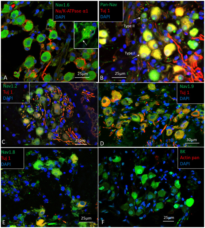
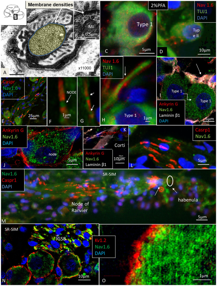
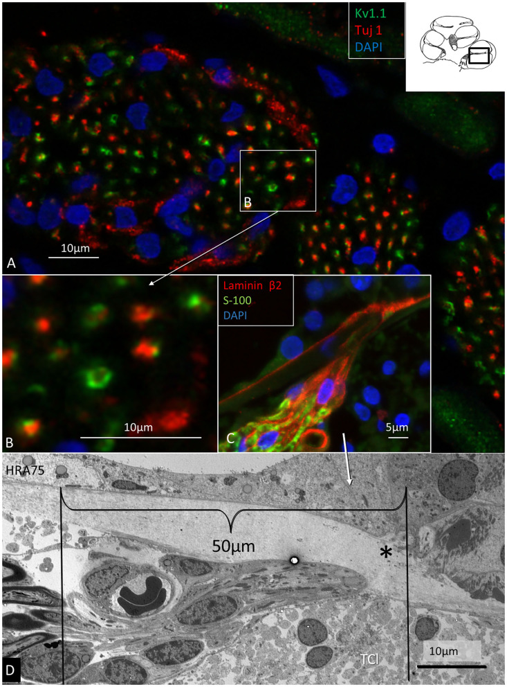
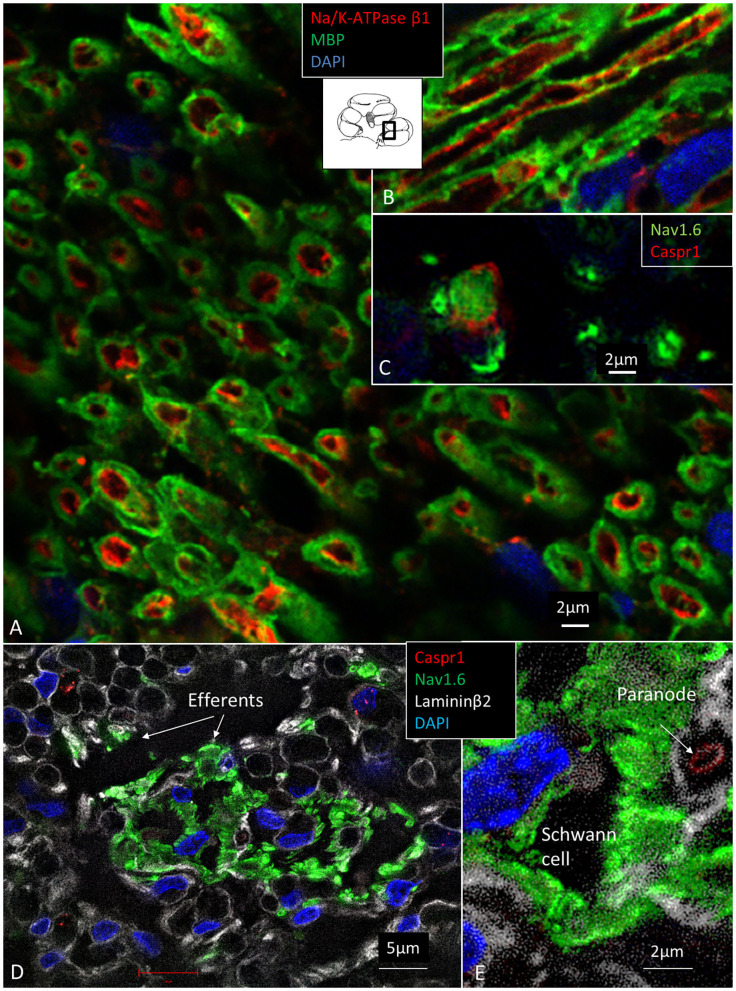
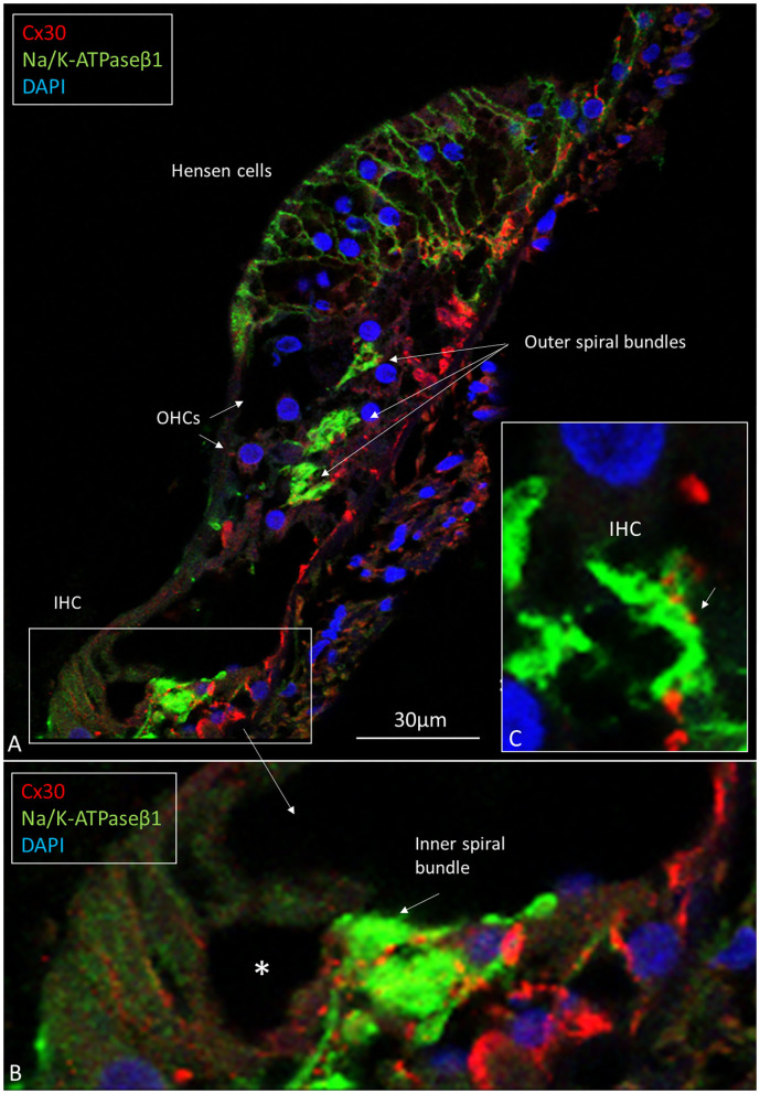
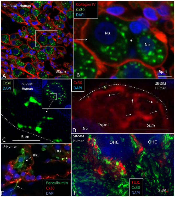
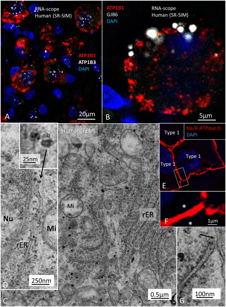
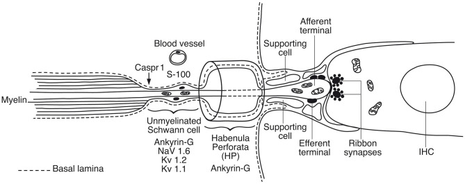
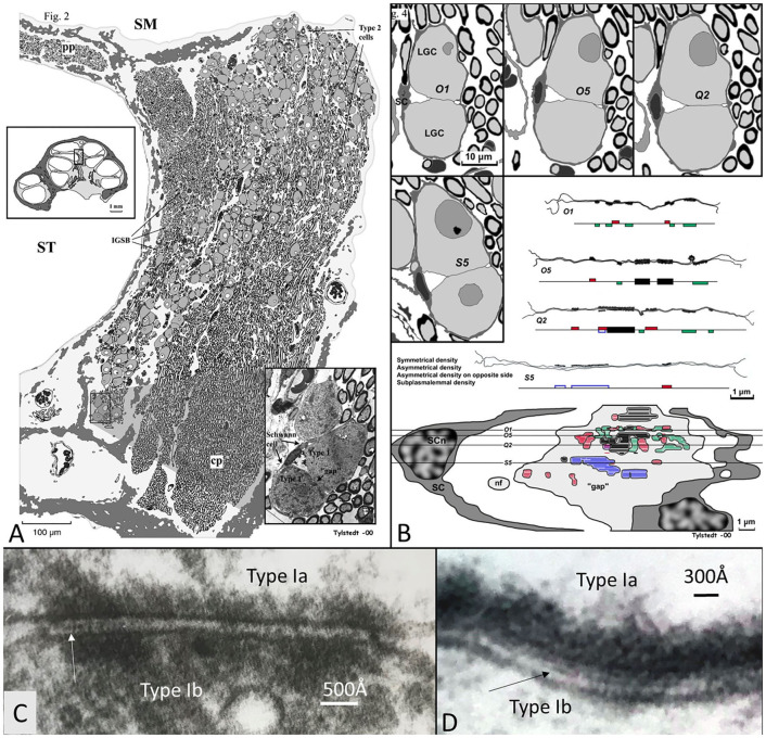
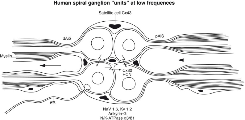
Background: The human auditory nerve contains 30,000 nerve fibers (NFs) that relay complex speech information to the brain with spectacular acuity. How speech is coded and influenced by various conditions is not known. It is also uncertain whether human nerve signaling involves exclusive proteins and gene manifestations compared with that of other species. Such information is difficult to determine due to the vulnerable, "esoteric," and encapsulated human ear surrounded by the hardest bone in the body. We collected human inner ear material for nanoscale visualization combining transmission electron microscopy (TEM), super-resolution structured illumination microscopy (SR-SIM), and RNA-scope analysis for the first time. Our aim was to gain information about the molecular instruments in human auditory nerve processing and deviations, and ways to perform electric modeling of prosthetic devices. Material and Methods: Human tissue was collected during trans-cochlear procedures to remove petro-clival meningioma after ethical permission. Cochlear neurons were processed for electron microscopy, confocal microscopy (CM), SR-SIM, and high-sensitive in situ hybridization for labeling single mRNA transcripts to detect ion channel and transporter proteins associated with nerve signal initiation and conductance. Results: Transport proteins and RNA transcripts were localized at the subcellular level. Hemi-nodal proteins were identified beneath the inner hair cells (IHCs). Voltage-gated ion channels (VGICs) were expressed in the spiral ganglion (SG) and axonal initial segments (AISs). Nodes of Ranvier (NR) expressed Nav1.6 proteins, and encoding genes critical for inter-cellular coupling were disclosed. Discussion: Our results suggest that initial spike generators are located beneath the IHCs in humans. The first NRs appear at different places. Additional spike generators and transcellular communication may boost, sharpen, and synchronize afferent signals by cell clusters at different frequency bands. These instruments may be essential for the filtering of complex sounds and may be challenged by various pathological conditions.

Keywords: auditory nerve; gene expression; human; spike generation; structured illumination microscopy.


Conflict of interest statement

MED-EL Medical Electronics, R&D, GmbH, and Innsbruck, Austria provided salary support for one research group member (WL) in accordance with the contract agreement with Uppsala University, Sweden during 2018. The funder had no role in study design, data collection and analysis, decision to publish, or preparation of the manuscript. The remaining authors declare that the research was conducted in the absence of any commercial or financial relationships that could be construed as a potential conflict of interest.

Figures

Figure 1
Figure 1
(A) SEM of a decalcified hemi-sectioned human cochlea. Framed area shows the OC. The SG (yellow) contains two types of afferent neurons: one innervating outer (5%) and one IHCs (95%). In total, there are about 30,000 nerve fibers (NFs). Efferent NFs (red) also reach and interact with SG cell bodies, IHC nerve terminals, and outer hair cells OHCs. Printed with permission from Hearing, Balance, and Communication 2020, https://doi.org/10.1080/21695717.2020.1807259. (B) SEM of a human OC. There are four rows of OHCs and one row of IHCs. Efferent NFs are colored green. (C) Higher magnification of framed area in (B). A basal afferent tunnel fiber runs in the inner pillar cell foot. It is an afferent fiber innervating OHCs. IHC afferent terminals are swollen. A similar, but not identical, image was earlier published in Anatomical Record 2012 (Rask-Andersen et al., 2012).
Figure 2
Figure 2
Human efferent innervation. MicroCT, 3D reconstruction and modeling of soft tissue in a right human IAC (anterior-medial view, broken line represents cochlear nerve at fundus). For clarity, some nerves are semi-transparent. An efferent cochlear nerve supply is mediated via the vestibular-cochlear anastomosis of Oort (blue). NFs exit from the inferior vestibular and saccular nerves and reach the cochlea and SG ~3–4 mm from its basal end. Their role in signal modulation, protection, and spatial hearing is still unclear.
Figure 3
Figure 3
Cross-sectioned human auditory nerve at different levels shown in (A). (B) The nerve near the fundus. (C,D) A transitional zone with central glia and astrocyte tissue project into the nerve (lucent part). It is surrounded by the peripheral component containing Schwann cells. (E) Higher magnification of framed area shown in (D). Inset shows TEM image at the transitional zone with astrocyte tissue (asterisk). Tissue was fixed immediately in 3% buffered glutaraldehyde and post-stained in 1% osmium tetroxide.
Figure 4
Figure 4
(A) Immune staining of a cross-sectioned human auditory nerve corresponding to the level shown in Figure 3B. (A) Nerve fibers express the myelin marker MBP and neuron marker TUJ1. Few fibers are unmyelinated (arrow) and may represent NRs. (B) Cochlear nerve at the transitional zone (corresponding to level shown in C). Astrocytes stain positive for GFAP (green) and Cx43 (inset). MBP, myelin basic protein; TUJ1, tubulin-1; GFAP, glia-fibrillary acidic protein; Cx43, connexin43.
Figure 5
Figure 5
(A–E) Expression of Nav, and TUJ1 in the human SG. The large cell bodies show various expression of Nav channels, mostly restricted to the cell bodies and the AIS. (F) Spiral ganglion cell bodies also expressed calcium-activated potassium channel (BK channel). Fixation in 4% PFA.
Figure 6
Figure 6
(A,B) TEM of a cross-sectioned node/paranode in the basal RC. Axoplasm is stained yellow. Radially oriented arrays of Schwann cell microvilli contact the axolemma (B). Mi, mitochondria. (C) Co-expression of Nav1.6 and TUJ1 in a large type I cell after fixation in 2% PFA. (D) Small type II cell with adjoining Nav1.6-positive fibers fixed in 2% PFA. (E,F) Nav1.6 expression in NRs. (G) A double NR (arrows). (H) Type I AIS (arrow) expresses Nav1.6. (I,J) Ankyrin G expression in Type I cell axon hillock (arrow) and plasmalemma. (K) Ankyrin G expressed in neurons at the HP. (L) Caspr1 is expressed in neurons beneath the HP. (M) Radial NFs express Caspr1 beneath the HP (arrows) and at NRs. (N) Expression of Kv1.2 and Nav1.6 in type I SGNs. Efferent fibers in the IGSB also express Nav1.6. (O) Higher magnification shows expression of Kv1.2 in the type I SGN plasmalemma.
Figure 7
Figure 7
Immunohistochemistry (A–C) and TEM (D) of spiral lamina NFs in a human cochlea. (A) Double-labeling of TUJ1 and Kv1.1. Framed area is magnified in (B). (C) Radial section at the HP with a fiber bundle expressing lamininβ2 and S-100. (D) TEM of radial NFs beneath the HP (asterisk). The length of the unmyelinated region is around 50 μm. The unmyelinated NFs are rich in mitochondria. A blood capillary is located typically near the nerve bundle. The basal lamina is folded at the habenular opening. TCL, tympanic covering layer.
Figure 8
Figure 8
(A) Spiral lamina NFs cross-sectioned and double-labeled with antibodies against MBP and Na/K-ATPase β1. (B) Longitudinal section. Axolemma expresses Na/K-ATPase β1 all the way. (C) Cross-sectioned axons labeled with antibodies against Nav1.6 and Caspr1 at and near a NR. (D) The spiral lamina contains bundles of thin NFs expressing Nav1.6 believed to represent efferent nerve fibers. The larger myelinated NFs are Nav1.6-negative. (E) Higher magnification of Nav1.6-positive NFs shown in (D). A fourth channel (white) shows lamininβ2 expression in the basal lamina. MBP, myelin basic protein; Nav1.6, voltage-gated sodium channel 1.6; Caspr1, contactin-associated protein 1.
Figure 9
Figure 9
(A) CM of the human OC double-labeled with antibodies against Na/K-ATPase β1 (green) and Cx30 (red). Inner and outer spiral bundles express Na/K-ATPase β1 with mostly separate Cx30 puncta (B,C). *IHC nerve terminals are swollen.
Figure 10
Figure 10
(A) TEM image of a human IHC. A myriad of afferent (*) and efferent nerve terminals are located at the basal pole. (B,C) Afferent boutons (aff) have different morphology with multiple synaptic plaques. There are large numbers of mitochondria (Mi) in the IHC synaptic region. (D–F) An efferent axo-synaptic contact is shown with multiple synaptic vesicles, mitochondria, and large dense-core vesicles (D). Some terminals face more than one ribbon synapse (G). Fixation was done directly in the surgical room in oxygenated 3% buffered glutaraldehyde and then post-stained in 1% osmium tetroxide.
Figure 11
Figure 11
CM and SR-SIM of human SG and OC. (A) Collagen IV and Cx30 co-staining shows a basal lamina “honeycomb” layer in the apical portion of the cochlea richly expressing Cx30. Framed area is magnified in (B). (B) A SGC with no basal lamina is seen between the two large SG cell bodies (*). (C,D) SR-SIM shows labeled Cx30 near the plasmalemma (broken lines). (E) Inner hair cell with afferent (arrow) shows expression of parvalbumin but no Cx30. Surrounding cells heavily express Cx30. (F) SR-SIM at lower poles of OHCs. Neurons express TUJ1 but there is no co-expression with Cx30. Nu: nucleus.
Figure 12
Figure 12
(A) Localization of ATP1B1 and ATP1B3 in human large, type I SG cell bodies encoding Na/K-ATPaseβ1 and β3. ATPB1 gene expression is most concentrated at the periphery near the cell membrane while ATP1B3 is mostly expressed in the cell nuclei. (B) GJB6 encoding Cx30 protein is expressed near the plasmalemma of a type I ganglion cell. (C) TEM image of a type I ganglion cell shows many mitochondria (Mi) and extensive rough endoplasmic reticulum (rER) and free ribosomes in the cytoplasm. (D) Several ribosomes are located at the nuclear envelope. (D) shows ribosome (rib) in higher magnification. (E) A rER is closely associated with the plasmalemma (arrow). (F) SR-SIM shows expression of the Na/K-ATPaseβ1 protein in the plasmalemma of three large ganglion cells. The cell membranes lie close to each other (asterisks). Framed area is magnified in (G). Nu; cell nucleus.
Figure 13
Figure 13
Graphic illustration of the principal organization of the human inner hair cell receptor-neural junction. A basal lamina margins the neural pathway that coalesces with the organ of Corti. Action potentials are believed to be generated in the sub-receptor zone beneath the habenular canal. In rodents Nav1.6 is localized in a hemi-node consistent with the location of spike generation (Hossain et al., 2005). There are also low- and high-voltage activated potassium channels essential for adaptation and regulation of AP activity (Smith et al., ; Kim and Rutherford, 2016).
Figure 14
Figure 14
(A) 3D model of membrane specializations between two human type I SG cell bodies in the apical turn of a human cochlea (voice fundamental frequency F0 ~ 100–250 Hz, 630–730°, normal subjective hearing). Two type I cells were serially sectioned and graphically reconstructed. Surrounding satellite cells (SCs) show a “gap” in the SC interface between the two cells with several membrane specializations. These are both symmetrical (black) and asymmetrical with different polarity (red and green). Some areas show sub-plasmalemmal densities (blue). (C,D) High-power TEM of somato-somal membrane densities shown in (B). Magnification ×160,000. Occasionally, a thin precipitous lamina is seen in the intercellular cleft (arrows). Fixed in oxygenated fluorocarbon containing 2% glutaraldehyde solution and 0.05 M sodium phosphate buffer (Tylstedt and Rask-Andersen, 2001). Published with permission from Kluwer Academic Publishers, License Number 4877550695963.
Figure 15
Figure 15
Principal model of large or type I spiral ganglion cell “units” in the human modiolus. Cell bodies and distal (dAIS) and proximal (pAIS) axonal initial segments are unmyelinated in human and surrounded by satellite glial cells. The close proximity between the perikarya may allow inter-cellular communication suggesting an electric filtering at this level. HCN channels were identified in the perikarya with particular intense staining of HCN 1, 2, and 4 at adjoining cell membranes which may boost coupling and synchronize AP firing (Luque et al., 2021). Cx30 was so far not identified in the plasmalemma. Eff., efferent.

Similar articles

Cited by

References

    1. Adamson C. L., Reid M. A., Mo Z. L., Bowne-English J., Davis R. L. (2002). Firing features and potassium channel content of murine spiral ganglion neurons vary with cochlear location. J. Comp. Neurol. 447, 331–350. 10.1002/cne.10244 - DOI - PubMed
    1. Amir R., Devor M. (1996). Chemically mediated cross-excitation in rat dorsal root ganglia. J. Neurosci. 16, 4733–4741. 10.1523/jneurosci.16-15-04733.1996 - DOI - PMC - PubMed
    1. Ariyasu L., Galey F. R., Hilsinger R., Byl F. M. (1989). Computer-generated three-dimensional reconstruction of the cochlea. Otolaryngol. Neck Surg. 100, 87–91. 10.1177/019459988910000201 - DOI - PubMed
    1. Arnold W. (1987). Myelination of the human spiral ganglion. Acta Otolaryngol. 436, 76–84. 10.3109/00016488709124979 - DOI - PubMed
    1. Arnold W., Wang J. B., Linnenkohl S. (1980). [Anatomical observations in the spiral ganglion of human newborns (author's transl)]. Arch. Otorhinolaryngol. 228, 69–84. - PubMed