. 2021 Mar 16:12:653035.
doi: 10.3389/fphar.2021.653035. eCollection 2021.

Positive Effect of Andrographolide Induced Autophagy on Random-Pattern Skin Flaps Survival

Affiliations

Positive Effect of Andrographolide Induced Autophagy on Random-Pattern Skin Flaps Survival

Jingtao Jiang et al. Front Pharmacol. .

Abstract

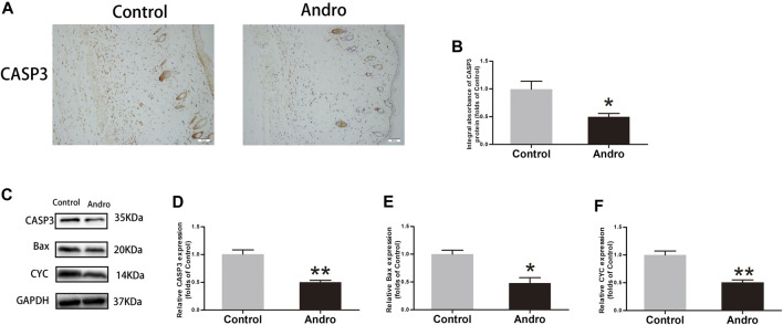
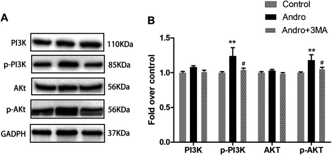
Random-pattern skin flap replantation is generally used in the reconstruction of surgical tissues and covering a series of skin flap defects. However, ischemia often occurs at the flap distal parts, which lead to flap necrosis. Previous studies have shown that andrographolide (Andro) protects against ischemic cardiovascular diseases, but little is known about the effect of Andro on flap viability. Thus, our study aimed to building a model of random-pattern skin flap to understand the mechanism of Andro-induced effects on flap survival. In this study, fifty-four mice were randomly categorized into the control, Andro group, and the Andro+3-methyladenine group. The skin flap samples were obtained on postoperative day 7. Subsequently, the tissue samples were underwent a series of evaluations such as changes in the appearance of flap tissue, the intensity of blood flow, and neovascularization density of skin flap. In our study, the results revealed that Andro enhanced the viability of random skin flaps by enhancing angiogenesis, inhibiting apoptosis, and reducing oxidative stress. Furthermore, our results have also demonstrated that the administration of Andro caused an elevation in the autophagy, and these remarkable impact of Andro were reversed by 3-methyladenine (3-MA), the most common autophagy inhibitor. Together, our data proves novel evidence that Andro is a potent modulator of autophagy capable of significantly increasing random-pattern skin flap survival.

Keywords: PI3K/Akt signaling pathway; andrographolide; angiogenesis; apoptosis; autophagy; oxidative stress; random-pattern flap.


Conflict of interest statement

The authors declare that the research was conducted in the absence of any commercial or financial relationships that could be construed as a potential conflict of interest.

Figures

FIGURE 1
FIGURE 1
Dose-dependent effect of Andro on random pattern flap survival (A) Chemical structure of Andrographolide. (B) Effects of different concentrations of Andrographolide on the survival/necrosis area after operation (7th day). (C) The histogram of survival area percentage on postoperative day seventh. The obtained data were presented as means ± SEM. Significance: *p-value < 0.05 and **p-value < 0.01, vs. control group (n = 6 per group).
FIGURE 2
FIGURE 2
Andro enhances the random-pattern skin flap survival (A) The digital images of survival/necrosis area in both groups after the operation (on the third day and seventh day). (B) The histogram of survival area percentage on postoperative day seventh. (C) The image of tissue edema and necrosis in the Control and Andro group on day seventh of the post-surgery. (D) The histogram reveals the percentage of water content in tissues. (E) The blood supply and vascular flow in both groups. (F) The histogram shows the signal intensities of the blood flow in flaps. (G) The H&E staining between two groups showing the vessels (original magnification ×200; scan bar, 50 μm). (H) The histogram depicts the MVD percentage. (I) The IHC 0f CD34 to mark vessels in vascular endothelial cells in the skin flap (original magnification ×200; scale bar, 50 µm). (J) The histogram depicts the CD34-positive vessel density percentage. The obtained data were presented as means ± SEM. Significance: *p-value < 0.05 and **p-value < 0.01, vs. control group (n = 6 per group).
FIGURE 3
FIGURE 3
The upregulation of angiogenesis in the skin flaps via Andro (A,C) IHC of Cadherin5 and VEGF in both groups of ischemic skin flaps (original magnification, ×200; scan bar, 50 μm). (B,D) The total absorbance of Cadherin5 and VEGF in IHC. (E) The results of immunoblotting i.e., the expressions of cadherin 5, MMP9, VEGF in the control as well as Andro groups. (F–H) The quantification of Cadherin5, MMP9, and VEGF expressions in the flaps by measuring their optical densities. The obtained data were presented as means ± SEM. Significance: *p-value < 0.05 and **p-value < 0.01, vs. control group (n = 6 per group).
FIGURE 4
FIGURE 4
Apoptosis inhibition in the skin flaps via Andro (A) The CASP3 expression evaluated via IHC in flaps (original magnification ×200; scale bar, 50 µm). (B) The histogram representing the CASP3 total absorbance in IHC. (C) The immunoblotting of CASP3, Bax, and CYC expressions in each group. (D–F) The quantification of CASP3, Bax, and CYC expressions in the flaps by measuring their optical densities. The obtained data were presented as means ± SEM. Significance: *p-value < 0.05 and **p-value < 0.01, vs. control group (n = 6 per group).
FIGURE 5
FIGURE 5
The alleviation of oxidative stress in the skin flaps via Andro (A) The expression of SOD1 evaluated via IHC in flaps (original magnification ×200; scale bar, 50 µm). (B) The histogram representing the total SOD1 absorbance in IHC. (C) The immunoblotting of SOD1, HO1and eNOS expressions in each group. (D–F) The quantification of SOD1, HO1and eNOS expressions in the flaps by measuring their optical densities. The obtained data were presented as means ± SEM. Significance: *p-value < 0.05 and **p-value < 0.01, vs. control group (n = 6 per group).
FIGURE 6
FIGURE 6
The enhancement of autophagy in the skin flaps via Andro (A) The expression of CTSD evaluated via IHC in flaps (original magnification ×200; scale bar, 50 µm). (B) The histogram representing the total CTSD absorbance in IHC. (C) The immunofluorescence results of LC3II in the ischemic skin flap (scan bar, 50 μm). (D) The histogram depicts the percentage of the LC3II-positive cells. (E) The immunoblotting of Beclin 1, LC3II, CTSD, VPS34, and p62 expressions in each group. (F) The quantification of Beclin 1, LC3II, CTSD, VPS34, and p62 expressions in the flaps by measuring their optical densities. The obtained data were presented as means ± SEM. Significance: *p-value < 0.05 and **p-value < 0.01, vs. control group (n = 6 per group).
FIGURE 7
FIGURE 7
3MA reversed the effects of Andro on random-pattern skin flap viability (A) The immunofluorescence data of LC3II in the ischemic skin flap (scan bar, 50 μm). (B) The histogram representing the percentage of the LC3II-positive cells. (C,E) The immunoblotting of the autophagy associated proteins VPS34, p62, LC3II, Beclin 1, and CTSD; angiogenesis associated proteins VEGF, cadherin5, and MMP9; the oxidative stress-related proteins i.e., SOD1, HO1, and eNOS and the apoptosis-associated proteins i.e., Bax, CYC, and CASP3. (D,F) The quantification of autophagy-, angiogenesis, and oxidative stress-related proteins by measuring their optical densities, as previously mentioned. The obtained data were presented as means ± SEM. Significance: *p-value < 0.05 and **p-value < 0.01, vs. control group (n = 6 per group).
FIGURE 8
FIGURE 8
Suppression of autophagy reversed the effects of Andro on flap vitality (A) The digital images of survival/necrosis area in Andro and Andro+ 3-methyladenine (3MA) groups after the operation (POD3 and POD7). (B) The histogram of survival area percentage on postoperative day seventh. (C) The image of tissue edema and necrosis in the Andro group and Andro+3MA group on the seventh day after surgery. (D) The histogram reveals the percentage of water content in tissue. (E) The blood supply and vascular flow in both groups. (F) The histogram shows the signal intensities of the blood flow in flaps. (G) The staining of H&E (original magnification ×200; scan bar, 50 μm). (H) The histogram depicts the MVD percentage. (I) The IHC 0f CD34 to mark vessels in vascular endothelial cells in the skin flap (original magnification ×200; scale bar, 50 µm). (J) The histogram depicts the percentage of CD34-positive vessel density. The obtained data were presented as means ± SEM. Si/gnificance: *p-value < 0.05 and **p-value < 0.01, vs. control group (n = 6 per group).
FIGURE 9
FIGURE 9
Andro Attenuates Apoptosis by Regulating the PI3K/Akt Signaling Pathway (A) The immunoblotting expressions of PI3K, p-PI3K, Akt, and p-Akt in the control, Andro, and Andro+3MA groups. (B) The quantification of PI3K,p-PI3K,Akt, and p-Akt expressions in the each group by measuring their optical densities. *p < 0.05 and **p < 0.01, vs. control group; #p < 0.05, ##p < 0.01, vs. Andro group (n = 6 per group).

References

    1. Ahrens T., Lambert M., Pertz O., Sasaki T., Schulthess T., Mège R.-M., et al. (2003). Homoassociation of VE-cadherin follows a mechanism common to “classical” cadherins. J. Mol. Biol. 325 (4), 733–742. 10.1016/s0022-2836(02)01286-x - DOI - PubMed
    1. Alawi K. M., Aubdool A. A., Liang L., Wilde E., Vepa A., Psefteli M. P., et al. (2015). The sympathetic nervous system is controlled by transient receptor potential vanilloid 1 in the regulation of body temperature. FASEB J. 29 (10), 4285–4298. 10.1096/fj.15-272526 - DOI - PMC - PubMed
    1. Chehelcheraghi F., Eimani H., Homayoonsadraie S., Torkaman G., Amini A., Alavi Majd H., et al. (2016). Effects of acellular amniotic membrane matrix and bone marrow-derived mesenchymal stem cells in improving random skin flap survival in rats. Iran Red Crescent Med. J. 18 (6), e25588. 10.5812/ircmj.25588 - DOI - PMC - PubMed
    1. Chen G., Shen H., Zang L., Su Z., Huang J., Sun Y., et al. (2018). Protective effect of luteolin on skin ischemia-reperfusion injury through an AKT-dependent mechanism. Int. J. Mol. Med. 42 (6), 3073–3082. 10.3892/ijmm.2018.3915 - DOI - PMC - PubMed
    1. Duan M.-X., Zhou H., Wu Q.-Q., Liu C., Xiao Y., Deng W., et al. (2019). Andrographolide protects against HG-induced inflammation, apoptosis, migration, and impairment of angiogenesis via PI3K/AKT-eNOS signalling in HUVECs. Mediators Inflamm. 2019, 6168340. 10.1155/2019/6168340 - DOI - PMC - PubMed

LinkOut - more resources