. 2021 Mar 16:12:642867.
doi: 10.3389/fimmu.2021.642867. eCollection 2021.

Activation of Neutrophil Granulocytes by Platelet-Activating Factor Is Impaired During Experimental Sepsis

Affiliations

Activation of Neutrophil Granulocytes by Platelet-Activating Factor Is Impaired During Experimental Sepsis

Stefan Hug et al. Front Immunol. .

Abstract

Platelet-activating factor (PAF) is an important mediator of the systemic inflammatory response. In the case of sepsis, proper activation and function of neutrophils as the first line of cellular defense are based on a well-balanced physiological response. However, little is known about the role of PAF in cellular changes of neutrophils during sepsis. Therefore, this study investigates the reaction patterns of neutrophils induced by PAF with a focus on membrane potential (MP), intracellular pH, and cellular swelling under physiological and pathophysiological conditions and hypothesizes that the PAF-mediated response of granulocytes is altered during sepsis. The cellular response of granulocytes including MP, intracellular pH, cellular swelling, and other activation markers were analyzed by multiparametric flow cytometry. In addition, the chemotactic activity and the formation of platelet-neutrophil complexes after exposure to PAF were investigated. The changes of the (electro-)physiological response features were translationally verified in a human ex vivo whole blood model of endotoxemia as well as during polymicrobial porcine sepsis. In neutrophils from healthy human donors, PAF elicited a rapid depolarization, an intracellular alkalization, and an increase in cell size in a time- and dose-dependent manner. Mechanistically, the alkalization was dependent on sodium-proton exchanger 1 (NHE1) activity, while the change in cellular shape was sodium flux- but only partially NHE1-dependent. In a pathophysiological altered environment, the PAF-induced response of neutrophils was modulated. Acidifying the extracellular pH in vitro enhanced PAF-mediated depolarization, whereas the increases in cell size and intracellular pH were largely unaffected. Ex vivo exposure of human whole blood to lipopolysaccharide diminished the PAF-induced intracellular alkalization and the change in neutrophil size. During experimental porcine sepsis, depolarization of the MP was significantly impaired. Additionally, there was a trend for increased cellular swelling, whereas intracellular alkalization remained stable. Overall, an impaired (electro-)physiological response of neutrophils to PAF stimulation represents a cellular hallmark of those cells challenged during systemic inflammation. Furthermore, this altered response may be indicative of and causative for the development of neutrophil dysfunction during sepsis.

Keywords: flow cytometry; intracellular pH; membrane potential; neutrophil granulocytes; platelet-activating factor; sepsis.


Conflict of interest statement

The authors declare that the research was conducted in the absence of any commercial or financial relationships that could be construed as a potential conflict of interest.

Figures

Figure 1
Figure 1
PAF elicited a rapid increase in size of human neutrophils. (A) FSC-A (forward scatter area) of neutrophils stimulated by PAF (1 μM, black) or PBS (CTRL, gray), measured by continuous acquisition by flow cytometry for 5 min, x indicating baseline measurement, n = 5. (B) FSC-A of neutrophils stimulated with PAF or PBS for 60 min, n = 15. For comparison, neutrophils exposed to C5a (green) are shown. (C) Calculated diameter of neutrophils using counting beads (10, 15, and 20 μm) from one representative donor with or without PAF exposure. (D) Mean diameter and volume of neutrophils stimulated by PAF or PBS measured by Coulter counter, n = 8. Data are mean ± SD. **p < 0.01, Wilcoxon matched-pairs signed rank test comparing PAF-stimulated neutrophils with unstimulated control cells.
Figure 2
Figure 2
PAF depolarized the membrane potential and increased the intracellular pH of human neutrophils within seconds. (A) Changes in membrane potential of neutrophils stimulated with PAF (1 μM) or C5a (green, 10 nM), n = 15, unstimulated cells = 0 mV. (B) Intracellular pH of neutrophils upon stimulation with PAF, C5a, or PBS (CTRL), n = 15. (C) Changes in membrane potential and intracellular pH of neutrophils exposed to PAF measured by continuous acquisition on flow cytometry. x represents the baseline measurement prior to stimulation. Depicted is one representative donor out of five independent experiments. (D) PAF-induced effects on neutrophils were concentration-dependent (ROS, reactive oxygen species; SSC-A, side scatter area; FSC-A, forward scatter area; pHi, intracellular pH; CD11b, integrin alpha M; MP, membrane potential; CD62L, L-selectin) and respective EC50 ± interquartile range were calculated (n = 5–10). (A,B) data are mean ± SD. *p < 0.05, **p < 0.01, ***p < 0.001, Wilcoxon signed-rank test (A) and Wilcoxon matched-pairs signed rank test (B) comparing PAF-stimulated neutrophils with unstimulated control cells.
Figure 3
Figure 3
PAF mediated the formation of platelet–neutrophil complexes (PNCs). (A,B) Flow cytometric measurements of PNCs using APC-labeled anti-CD41 and PerCP-labeled anti-CD61 antibodies. Shown is one representative donor for blood stimulated with PBS (CTRL, A) or PAF 1 μM (B). Blue indicates CD41- and CD61-negative cells, yellow indicates either CD41- or CD61-positive neutrophils, red refers to neutrophils positive for CD41 and CD61. (C) Light microscopy of an exemplary PNC. (D) Quantification of PAF-induced (1 μM) PNC formation by flow cytometry (n = 7). (E) PAF mediated PNC formation as detected by blood smear and manual counting (n = 6). Data are mean ± SD. *p < 0.05, Wilcoxon matched-pairs signed rank test.
Figure 4
Figure 4
(A) Amiloride (200 μM), (B) BIX (5 μM), (C) NPPB (100 μM), and (D) ebselen (10 nM) modulated the PAF-mediated response of neutrophils. While 0% indicates the regular response induced by 1 μM PAF, −100% indicates full inhibition of the PAF-mediated response = control level. For the calculation, see Supplement 3. Data are mean ± SD (n = 5–12). *p < 0.05, **p < 0.01, ***p < 0.001, Wilcoxon signed-rank test.
Figure 5
Figure 5
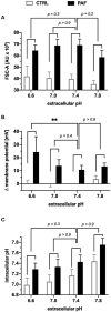
Extracellular acidosis increased the PAF-mediated depolarization in human neutrophils in vitro. Neutrophils were incubated in buffers with different extracellular pH and subsequently stimulated by 1 μM PAF. (A) The increase in the FSC-A induced by PAF in comparison with unstimulated neutrophils in an acidotic or alkaline environment did not significantly alter the cellular response in comparison to an extracellular pH of 7.4. (B) Depolarization of the membrane potential upon PAF stimulation was enhanced in an acidic environment (0 mV = CTRL extracellular pH 7.4). (C) Intracellular pH of neutrophils stimulated with PAF (black) or PBS (CTRL, white). Data are mean ± SD (n = 6). **p < 0.01, Kruskal Wallis and Dunn's post-hoc test comparing the PAF-induced response for each extracellular pH.
Figure 6
Figure 6
LPS-driven inflammation altered the PAF-induced response pattern of human neutrophils in an ex vivo whole blood model. Blood was incubated for 1 h with or without 100 ng/ml LPS. Subsequently, blood (A,B,F) or isolated neutrophils (C–E) were stimulated with 1 μM PAF in vitro. (A,B) Surface expression of L-selectin (CD62L, (A)) and integrin alpha M (CD11b, (B)) on neutrophils. (C) Cellular size of neutrophils as indicated by the FSC-A. (D) PAF-induced depolarization after exposure to LPS. (E) Intracellular pH of neutrophils stimulated with or without PAF. (F) PAF-mediated ROS generation. Data are mean ± SD of the PAF-induced response (n = 6). *p < 0.05, **p < 0.01, Wilcoxon matched-pairs signed rank test comparing the response induced by PAF of neutrophils with (LPS) or without (PBS as CTRL) stimulation in the whole blood model. MFI, mean fluorescence intensity.
Figure 7
Figure 7
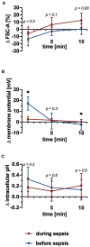
PAF-mediated depolarization was significantly impaired and there was a trend for increased cellular swelling during experimental polymicrobial sepsis, whereas alkalization remained stable. (A) Changes of the FSC-A upon stimulation with 1 μM PAF vs. unstimulated cells before (blue) and during (red) sepsis (n = 9). (B) PAF-induced alteration in membrane potential (n = 7). (C) Alterations in intracellular pH of PAF-exposed neutrophils (n = 9). Data are mean ± SD. *p < 0.05, Wilcoxon matched-pairs signed rank test.
Figure 8
Figure 8
Graphical summary of the neutrophil response upon exposure to platelet-activating factor (PAF), including depolarization, NHE1-dependent alkalization, and cellular swelling and functional thromboinflammatory activity. The (electro-)physiological response mediated by PAF is modulated in inflammatory environments. Endotoxemia increases intracellular pH and cell size of neutrophils while in parallel diminishing an additional change inducible by PAF. Depolarization is enhanced by extracellular acidosis, while being significantly disrupted during experimental sepsis. ROS, reactive oxygen species; PNC, platelet–neutrophil complex; NOX, NADPH oxidase; NHE1, sodium-proton exchanger 1.

References

    1. Lordan R, Tsoupras A, Zabetakis I, Demopoulos CA. Forty years since the structural elucidation of Platelet-Activating Factor (PAF): historical, current, and future research perspectives. Molecules. (2019) 24:4414. 10.3390/molecules24234414 - DOI - PMC - PubMed
    1. Gill P, Jindal NL, Jagdis A, Vadas P. Platelets in the immune response: revisiting platelet-activating factor in anaphylaxis. J Allergy Clin Immunol. (2015) 135:1424–32. 10.1016/j.jaci.2015.04.019 - DOI - PubMed
    1. Ramakrishnan AVKP, Varghese TP, Vanapalli S, Nair NK, Mingate MD. Platelet activating factor: a potential biomarker in acute coronary syndrome? Cardiovasc Ther. (2017) 35:64–70. 10.1111/1755-5922.12233 - DOI - PubMed
    1. Yost CC, Weyrich AS, Zimmerman GA. The platelet activating factor (PAF) signaling cascade in systemic inflammatory responses. Biochimie. (2010) 92:692–7. 10.1016/j.biochi.2010.02.011 - DOI - PMC - PubMed
    1. Singer M, Deutschman CS, Seymour CW, Shankar-Hari M, Annane D, Bauer M, et al. . The Third International Consensus Definitions for Sepsis and Septic Shock (Sepsis-3). JAMA. (2016) 315:801–10. 10.1001/jama.2016.0287 - DOI - PMC - PubMed

Publication types