Review
. 2021 Mar 16:12:652846.
doi: 10.3389/fimmu.2021.652846. eCollection 2021.

IL-1 Family Antagonists in Mouse and Human Skin Inflammation

Affiliations
Review

IL-1 Family Antagonists in Mouse and Human Skin Inflammation

Praxedis Martin et al. Front Immunol. .

Abstract

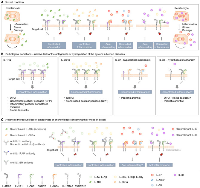
Interleukin (IL)-1 family cytokines initiate inflammatory responses, and shape innate and adaptive immunity. They play important roles in host defense, but excessive immune activation can also lead to the development of chronic inflammatory diseases. Dysregulated IL-1 family signaling is observed in a variety of skin disorders. In particular, IL-1 family cytokines have been linked to the pathogenesis of psoriasis and atopic dermatitis. The biological activity of pro-inflammatory IL-1 family agonists is controlled by the natural receptor antagonists IL-1Ra and IL-36Ra, as well as by the regulatory cytokines IL-37 and IL-38. These four anti-inflammatory IL-1 family members are constitutively and highly expressed at steady state in the epidermis, where keratinocytes are a major producing cell type. In this review, we provide an overview of the current knowledge concerning their regulatory roles in skin biology and inflammation and their therapeutic potential in human inflammatory skin diseases. We further highlight some common misunderstandings and less well-known observations, which persist in the field despite recent extensive interest for these cytokines.

Keywords: atopic dermatitis; cytokine; inflammation; interleukin-1; psoriasis; skin.


Conflict of interest statement

The authors declare that the research was conducted in the absence of any commercial or financial relationships that could be construed as a potential conflict of interest.

Figures

Figure 1
Figure 1
Structure of human skin. HE stained section of normal human skin at ×10 (left panel) and ×40 (right panel) original magnification. Epidermis, papillary (upper), and reticular (lower) dermis are shown (left panel). The epidermis contains keratinocytes arranged from bottom to top in four typical layers: basal layer, spinous layer, granular layer, and cornified layer (right panel). Photomicrography image credit: Lutz Slomianka 1998–2009, Blue Histology (http://www.lab.anhb.uwa.edu.au/mb140/).
Figure 2
Figure 2
Inflammatory IL-1, IL-18, IL-33, and IL-36 signaling is controlled by natural antagonists and regulatory molecules of the IL-1 family. (A–D) Upon binding of IL-1α or IL-1β to IL-1R1, of IL-33 to ST2, of IL-36α, IL-36β or IL-36γ to IL-36R or of IL-18 to IL-18Rα the co-receptors IL-1RAP or IL-18RAP, respectively, are recruited. Signaling is then initiated by the juxtaposition of the cytoplasmic TIR domains, which are present in both the ligand-binding and accessory protein chain. This leads to MyD88 and IRAK binding, activation of NF-κB and mitogen-activated protein kinase (MAPK) pathways and a pro-inflammatory signaling cascade. In order to control these inflammatory responses, different mechanisms exist. (A+D) The natural antagonists IL-1Ra or IL-36Ra bind to IL-1R1 or IL-36R, respectively, without recruiting the co-receptor IL-1RAP. Receptor antagonist binding competitively inhibits IL-1 cytokine- or IL-36 cytokine-mediated signaling. (A) Membrane-bound and soluble IL-1R2 (sIL-1R2), which lack the intracellular TIR domain, act as decoy receptors and bind to IL-1α or IL-1β with high affinity. In the case of membrane-bound IL-1R2, the IL-1RAP co-receptor is recruited. However, as IL-1R2 lacks the cytoplasmic TIR domain, no signaling cascade is initiated (thin red cross). Soluble IL-1RAP (sIL-1RAP) increases the affinity of sIL-1R2 binding to IL-1 and thus enhances the ability to inhibit IL-1 activity (thick red cross). In addition, IL-1R2 sequesters IL-1RAP thereby blocking IL-1R1/IL-1RAP receptor complex formation. (B) IL-18BP prevents the binding of IL-18 to its receptors IL-18Rα and IL-18RAP and acts as a soluble decoy receptor to control excessive IL-18-mediated inflammatory responses. (C) IL-33-mediated signaling can be inhibited by the soluble decoy receptor sST2 (thin red cross), with sIL-1RAP further enhancing the ability of sST2 to inhibit the effect of IL-33 (thick red cross).
Figure 3
Figure 3
SIGIRR is an inhibitory IL-1 family receptor. No specific ligand for SIGIRR has been described. However, SIGIRR can disrupt IL-1-, IL-33-, and IL-18-mediated signaling by competing for MyD88 and IRAK recruitment.
Figure 4
Figure 4
Anti-inflammatory IL-37 and IL-38 signaling. (A) IL-37 binds to IL-18Rα, but does not induce IL-18RAP recruitment. Instead, a complex of IL-18Rα and the inhibitory IL-1 family receptor SIGIRR is described to mediate anti-inflammatory effects of IL-37, such as inhibition of LPS or IL-1β-induced responses. Direct binding of IL-37 to IL-18BP and the formation of a heterotrimeric complex with IL-18RAP inhibits its association with IL-18Rα to transduce IL-18 signals. (B) IL-38 was reported to bind to IL-36R in vitro and to exert similar anti-inflammatory effects as IL-36Ra, although this has not been firmly demonstrated in in vivo studies. Furthermore, truncated IL-38 was proposed to limit inflammatory cytokine production by macrophages by acting as a ligand for TIGIRR-2.
Figure 5
Figure 5
Role and therapeutic use of anti-inflammatory IL-1 family cytokines in human inflammatory skin diseases. (A) IL-1Ra, IL-36Ra, IL-37, and IL-38 are constitutively expressed in keratinocytes as intracellular proteins. During inflammation, or in response to stress or cell damage, these cytokines are passively released by dying cells or actively secreted through leaderless pathways, and exert regulatory roles to control skin inflammation. The classical receptor antagonists IL-1Ra and IL-36Ra specifically antagonize the effects of, respectively, IL-1 or IL-36 cytokines, while IL-37 and IL-38 exert broader anti-inflammatory effects. (B) Evidence derived from individuals with genetic deficiencies and clinical trials highlights essential roles for IL-1Ra and IL-36Ra in the regulation of the inflammatory response in human skin. While genetic association and in vitro studies also suggest anti-inflammatory properties for IL-37 and IL-38 in the context of human skin diseases, the role of these two cytokines in skin homeostasis in vivo remains to be determined. (C) Therapeutic agents developed to target IL-1 and IL-36 signaling include receptor antagonists and monoclonal antibodies against pro-inflammatory cytokines or their receptors. Since both IL-1R and IL-36R bind several agonists, bispecific antibodies neutralizing two agonists or antibodies blocking the receptors conceptually represent better therapeutic agents than antibodies specifically targeting a single ligand. A recently described monoclonal antibody targeting the co-receptor IL-1RAP may also prove useful to target IL-1 and IL-36 signaling simultaneously. Finally, it remains to be determined if treatment with recombinant IL-37 or IL-38 might be of therapeutic interest in specific inflammatory skin diseases.

Similar articles

Cited by

References

    1. Eyerich S, Eyerich K, Traidl-Hoffmann C, Biedermann T. Cutaneous barriers and skin immunity: differentiating a connected network. Trends Immunol. (2018) 39:315–27. 10.1016/j.it.2018.02.004 - DOI - PubMed
    1. Goleva E, Berdyshev E, Leung DY. Epithelial barrier repair and prevention of allergy. J Clin Invest. (2019) 129:1463–74. 10.1172/JCI124608 - DOI - PMC - PubMed
    1. Proksch E, Brandner JM, Jensen JM. The skin: an indispensable barrier. Exp Dermatol. (2008) 17:1063–72. 10.1111/j.1600-0625.2008.00786.x - DOI - PubMed
    1. Doebel T, Voisin B, Nagao K. Langerhans cells - the macrophage in dendritic cell clothing. Trends Immunol. (2017) 38:817–28. 10.1016/j.it.2017.06.008 - DOI - PubMed
    1. Clayton K, Vallejo AF, Davies J, Sirvent S, Polak ME. Langerhans cells-programmed by the epidermis. Front Immunol. (2017) 8:1676. 10.3389/fimmu.2017.01676 - DOI - PMC - PubMed

Publication types