. 2021 Mar 16:11:637710.
doi: 10.3389/fcimb.2021.637710. eCollection 2021.

RIPK3-Dependent Necroptosis Is Induced and Restricts Viral Replication in Human Astrocytes Infected With Zika Virus

Affiliations

RIPK3-Dependent Necroptosis Is Induced and Restricts Viral Replication in Human Astrocytes Infected With Zika Virus

Chunxia Wen et al. Front Cell Infect Microbiol. .

Abstract

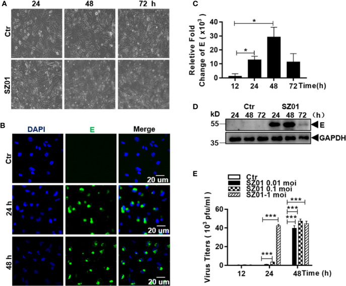
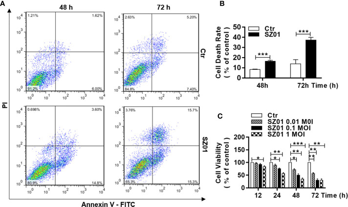
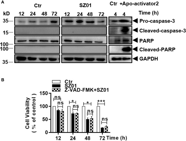
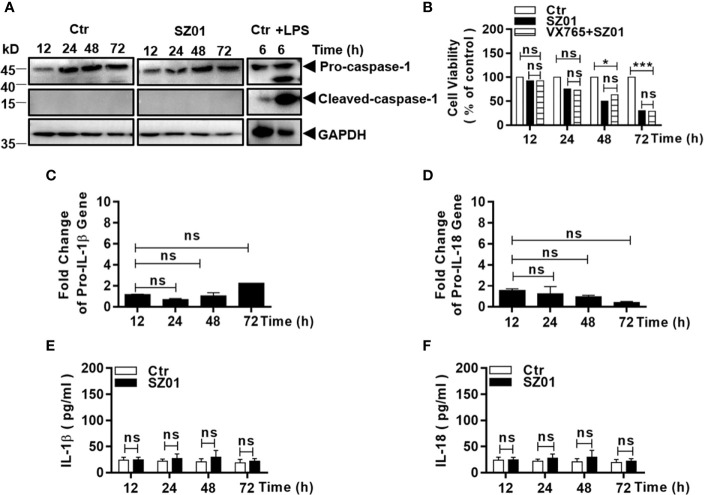
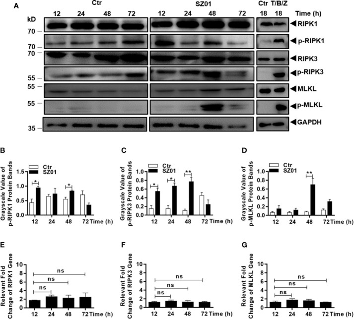
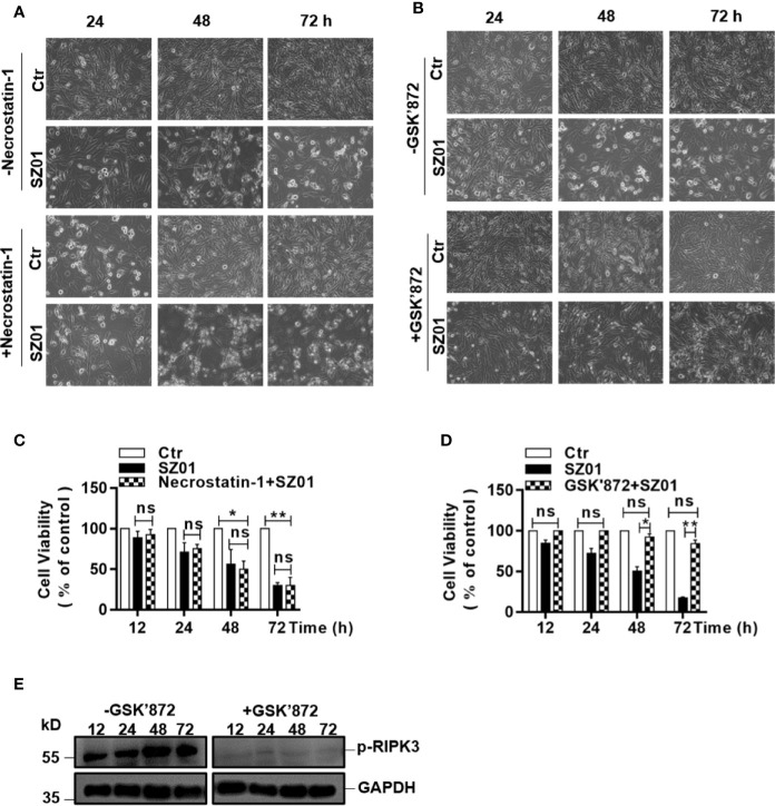
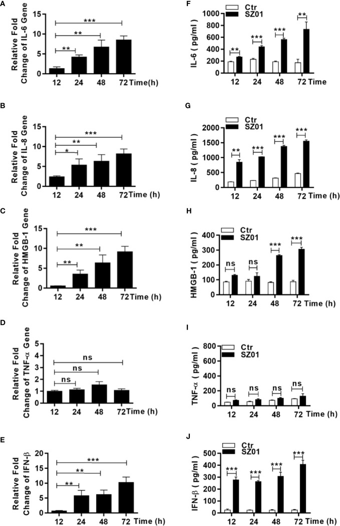
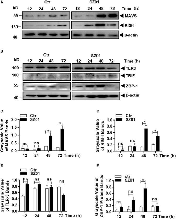
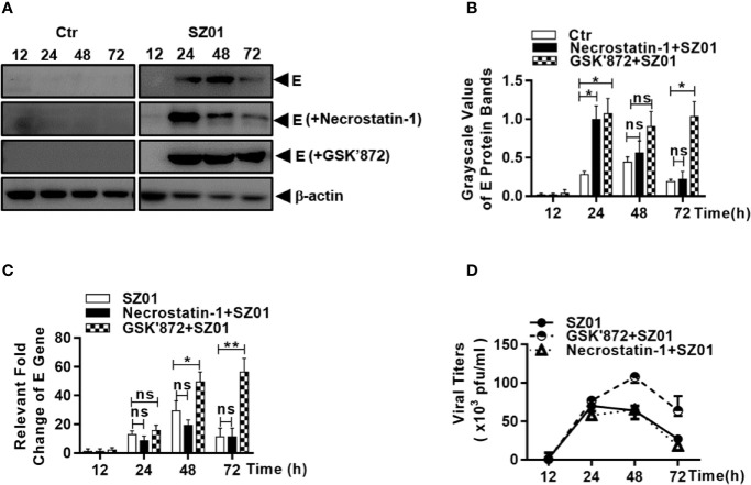
Apoptosis, pyroptosis and necroptosis are regulated processes of cell death which can be crucial for viral disease outcomes in hosts because of their effects on viral pathogenicity and host resistance. Zika virus (ZIKV) is a mosquito-borne flavivirus, which infects humans and can cause neurological disorders. Neural developmental disorders and microcephaly could occur in infected fetuses. Several types of nervous cells have been reported to be susceptible to ZIKV infection. Human astrocytes play important roles in the nutritional support and defense of neurons. In this study, we show that human astrocytes are susceptible to ZIKV infection and undergo progressive cell death after infection. In infected astrocytes we detected no cleavage or activation of pro-caspase-3 and pro-caspase-1. Apoptotic substrates and increased secretion of interleukin (IL)-1β or IL-18 were not detected, either. These ruled out the occurrence of apoptosis or pyroptosis in ZIKV-infected astrocytes. We detected, however, an increase of phosphorylated receptor-interacting serine/threonine-protein kinase (RIPK)1, RIPK3, and mixed lineage kinase domain-like (MLKL) protein, indicating that programmed necrosis, or necroptosis, was induced in infected astrocytes. The phosphorylation and cell death were inhibited in cells pre-treated with GSK'872, an inhibitor of RIPK3, while inhibition of RIPK1 with an inhibitor, Necrostatin-1, had no effect, suggesting that ZIKV-induced necroptosis was RIPK1-independent in astrocytes. Consistent with this finding, the inhibition of RIPK1 had no effect on the phosphorylation of MLKL. We showed evidence that MLKL phosphorylation was RIPK3-dependent and ZBP-1, which could stimulate RIPK3, was upregulated in ZIKV-infected astrocytes. Finally, we demonstrated that in GSK'872-pre-treated astrocytes, viral replication increased significantly, which indicates that necroptosis may be protective against viral replication in astrocytes. Our finding that astrocytes uniquely underwent necroptosis in response to ZIKV infection provides insight and helps us better understand the viral pathogenesis in the ZIKV-infected central nervous system.

Keywords: RIPK1; RIPK3; Zika virus; astrocytes; necroptosis.


Conflict of interest statement

The authors declare that the research was conducted in the absence of any commercial or financial relationships that could be construed as a potential conflict of interest.

Figures

Figure 1
Figure 1
Susceptibility of human astrocytes to ZIKV and its pathologic effects on infected cells. (A) U251 cells were infected with ZIKV SZ01 at MOI of 1 and cytopathic effects (CPE) were observed at 24, 48, and 72 h p.i. (Magnification x40). (B) ZIKV E protein was expressed and detected by immunofluorescence assay with a specific antibody for E protein using confocal microscopy. (C) Replication of ZIKV genome in the infected cells. ZIKV E gene copies were quantified at various time points p.i. by real-time RT-PCR with specific primers for the viral E gene. Data were representative of three experiments and each experiment was performed in triplicate. Unpaired Student’s T-test was used to analyze the differences of data and error bars represent standard errors of the means. (D) Expressed viral E protein was detected in the infected cells by western blot with specific antibody (1:1000) for the E protein. (E) Viral titers in U251 cells infected with ZIKV at various MOI. Culture supernatants from the infected cells was collected at indicated time point p.i. and the virus titrated in BHK21 cells by a standard plaque assay (*p < 0.05; ***p < 0.001).
Figure 2
Figure 2
Cell death and reduced viability observed in human astrocytes infected with ZIKV. (A) U251 cells with or without ZIKV infection (MOI of 1) co-stained with Annexin V and PI at 48 and 72 h p.i. for flowcytometric analyses. (B) Quantitative analyses of cell death at various time points p.i. in U251 cells infected with ZIKV. ***p < 0.001. (C) Cell viability of U251 cells infected with ZIKV at various MOI was assessed with an MTT assay. Triplicate cultures were analyzed at the indicated time points (*p < 0.05; **p < 0.01; ***p < 0.001).
Figure 3
Figure 3
ZIKV did not induce apoptosis in human astrocytes. (A) U251 cells were infected with ZIKV (MOI of 1) and cell lysates prepared and subjected to western blot analyses with specific antibodies (1:1000) as indicated. HeLa cells were treated with or without Apoptosis activator2 (Apoa2, 10μM) and the lysates analyzed as a positive control for apoptosis. (B) Cell viability analyses in pre-treated U251 cells infected with ZIKV(MOI of 1). The cells were pre-treated with or without Z-VAD-FMK, a pan inhibitor of caspases, prior to ZIKV infection and analyzed for viability at various times p.i. with the MTT assay. The experiments were repeated at least three times (*p < 0.05; ***p < 0.001; ns, no significance).
Figure 4
Figure 4
ZIKV did not activate pyroptosis in human astrocytes. (A) U251 cells were infected with 1 MOI of ZIKV and cell lysates were prepared at various time points p.i. and subjected to western blot analyses with specific antibodies (1:1000) as indicated. Monocytic THP-1 cells, were treated with or without lipopolysaccharide (LPS, 1μg/ml), and the lysates analyzed as a positive control for pyroptosis. (B) Cell viability was analyzed in pre-treated U251 cells infected with 1 MOI of ZIKV. The cells were pre-treated with or without VX765, an inhibitor of pro-caspase-1, prior to ZIKV infection and analyzed for viability at various times p.i. with the MTT assay. The experiments were repeated at least three times (ns p>0.05; *p < 0.05; ***p < 0.001). (C, D) No transcriptional changes occurred to pro-IL-1β and pro-IL-18 in infected cells. Total RNA was prepared from the U251 cells infected with ZIKV to measure mRNA transcript copies using quantitative real time PCR with specific primers for genes of IL-1β (C) and IL-18 (D), respectively. (E, F) No change in secreted IL-1β and IL-18 levels in the cell cultures after ZIKV infection. Culture media was collected at various time points p.i. for measurement of IL-1β and IL-18 by ELISA. Each data point represents the mean values from triplicate cultures performed at least three times (ns p > 0.05).
Figure 5
Figure 5
Necroptosis was induced by ZIKV-infected human astrocytes. (A) Increased phosphorylation of necroptosis-associated proteins was detected in ZIKV-infected cells. Cell lysates were prepared at various time points p.i. and subjected to SDS-PAGE and western blot analyses with specific antibodies (1:1000) for the proteins, either non-phosphorylated or phosphorylated, as indicated. The efficacies of the antibodies were confirmed with the lysates prepared from HT-29 cells treated with or without TNF-α (20 ng/ml), BV-6 (100 nM) and Z-VAD-FMK (20 µM) for induction of necroptosis. (B–D) Quantitative analyses of the gray scale values of the phosphorylated RIPK1 (B), RIPK3 (C), and MLKL (D) between ZIKV infected and non-infected cells at various time points p.i. (E–G). Relative level of each protein was normalized to GAPDH at indicated time points. No changes of RIPK1, RIPK3 and MLKL at the transcriptional level in U251 cells after infection with ZIKV. Total RNA was prepared from infected cells at several time points p.i. for measuring mRNA transcript copy numbers by real time PCR with specific primers for RIPK1 (E), RIPK3 (F), and MLKL (G), respectively. The experiments were repeated three times and the data are shown as means ± SEM (*p < 0.05; **p < 0.01; ns p > 0.05).
Figure 6
Figure 6
Effect of inhibiting the phosphorylation of RIPK1 or RIPK3 on necroptosis in ZIKV-infected human astrocytes. (A, B) U251 cells were pre-treated without (top) or with (bottom) necrostatin-1, an inhibitor of RIPK1 (A) or GSK’872, an inhibitor of RIPK3 (B), prior to infection with ZIKV. Cell death was observed at 24, 48, and 72 h p.i. by light microscopy (magnification x 200). (C, D) Quantitative analyses of cell viabilities at 24, 48, and 72 h p.i. in U251 cells, untreated or pre-treated with necrostatin-1 (C) or GSK’872 (D), prior to infection with ZIKV. The experiments were repeated three times and the values presented are means ± SEM (*p < 0.05; **p < 0.01; ns p > 0.05). (E) Inhibition of the RIPK3 phosphorylation. U251 cells were untreated or pre-treated with the inhibitor, followed by ZIKV infection. Cell lysates were prepared for SDS-PAGE and western blot analysis with indicated antibodies.
Figure 7
Figure 7
Upregulation and increased release of proinflammatory cytokines and DAMP from human astrocytes infected with ZIKV. (A–E) U251 cells were infected with ZIKV at an MOI of 1 and total RNA was prepared at various time points p.i. for measurement of mRNA transcript levels of selected cytokines and DAMP by real time RT-PCR with specific primers for IL-6 (A), IL-8 (B), HMGB-1 (C), TNF-α (D), and IFN-β (E). (F–I, J) U251 cells were infected with ZIKV at an MOI of -1 and the culture media was collected at various time points p.i. for measurement of secreted protein levels of selected cytokines and DAMP by ELISA. IL-6 (F), IL-8 (G), HMGB-1 (H), TNF-α (I), and IFN-β (J) were tested with respective reagents. Values represent means ± SEM obtained from triplicate cultures and each test was repeated for at least three times (*p < 0.05; **p < 0.01; ***p < 0.001; ns p > 0.05).
Figure 8
Figure 8
Upregulation of cellular sensors of viral nucleic acids in ZIKV-infected human astrocytes. U251 cells were infected with 1 MOI of ZIKV and cell lysates were prepared for SDS-PAGE analyses with specific antibodies (1:1000) as indicated for MAVS, RIG-1 (A), or TLR3, TRIF, and ZBP-1 (B). Grayscale values were analyzed for quantitative comparison of the protein expressions of MAVS (C), RIG-1 (D), TLR3 (E), and ZBP-1 (F) in ZIKV-infected cells. Relative grayscale of each protein was normalized to β-actin at indicated time point. The experiments were repeated at least three times and the values presented were means ± SEM (*p < 0.05; ns p > 0.05).
Figure 9
Figure 9
Necroptosis restricted viral replication in human astrocytes infected with ZIKV. U251 cells were untreated (left) or pre-treated with (right) necrostatin-1 or GSK’872 prior to infection with ZIKV. (A) Cell lysates were prepared from the infected cells at 12, 24, 48, and 72 h p.i. for SDS-PAGE and western blot analyses with anti-ZIKV E protein for levels of the viral protein E. (B) Quantitative analyses of the viral E protein levels in ZIKV-infected U251 cells pre-treated without or with necrostatin-1 or GSK’872. The E protein grayscale was normalized by β-actin at indicated time point. (C) Total RNA were prepared from the infected cells at 12, 24, 48, and 72 h p.i. for real time RT-PCR with primers specific for the viral E gene to measure the E RNA copy numbers in ZIKV-infected cells pre-treated with or without inhibitors. (D) Culture media was collected at various time points p.i. from ZIKV-infected cells, untreated or pre-treated with the inhibitors, for titration in Vero cells to determine infectious viral titers. The experiments were repeated for at least three times and the values presented were means ± SEM (*p < 0.05; **p < 0.01; ns p > 0.05).

References

    1. Bell B. P., Boyle C. A., Petersen L. R. (2016). Preventing Zika Virus Infections in Pregnant Women: An Urgent Public Health Priority. Am. J. Public Health 106, 589–590. 10.2105/AJPH.2016.303124 - DOI - PMC - PubMed
    1. Bian P., Zheng X., Wei L., Ye C., Fan H., Cai Y., et al. . (2017). MLKL Mediated Necroptosis Accelerates JEV-Induced Neuroinflammation in Mice. Front. Microbiol. 8, 303. 10.3389/fmicb.2017.00303 - DOI - PMC - PubMed
    1. Blish C. (2017). Zika virus infection induces cranial neural crest cells to produce cytokines at levels detrimental for neurogenesis. J. Neuroimmune Pharmacol. 12, S9–S10. 10.1016/j.chom.2016.09.006 - DOI - PMC - PubMed
    1. Brasil P., Pereira J. P., Moreira M. E., Ribeiro Nogueira R. M., Damasceno L., Wakimoto M., et al. . (2016). Zika Virus Infection in Pregnant Women in Rio de Janeiro. N Engl. J. Med. 375, 2321–2334. 10.1056/NEJMoa1602412 - DOI - PMC - PubMed
    1. Cho Y. S., Challa S., Moquin D., Genga R., Ray T. D., Guildford M., et al. . (2009). Phosphorylation-driven assembly of the RIP1-RIP3 complex regulates programmed necrosis and virus-induced inflammation. Cell 137, 1112–1123. 10.1016/j.cell.2009.05.037 - DOI - PMC - PubMed

Publication types

Substances