Case Reports
. 2021 Mar;7(1):17-25.
doi: 10.1159/000508517. Epub 2020 Aug 25.

Intratumoral Heterogeneity in Uveal Melanoma

Affiliations
Case Reports

Intratumoral Heterogeneity in Uveal Melanoma

Cristina Fonseca et al. Ocul Oncol Pathol. 2021 Mar.

Abstract

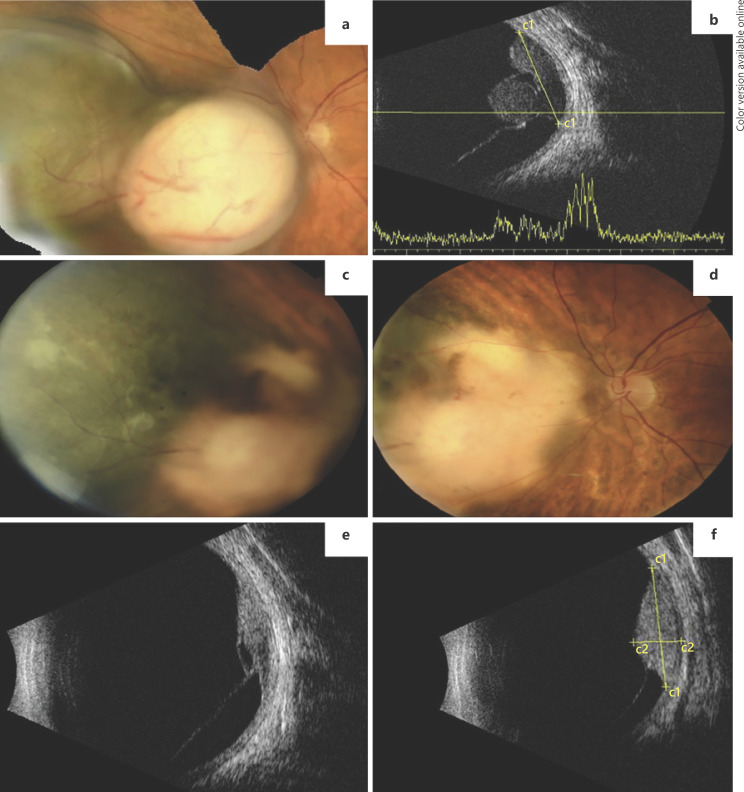
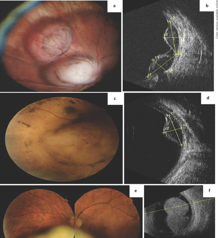
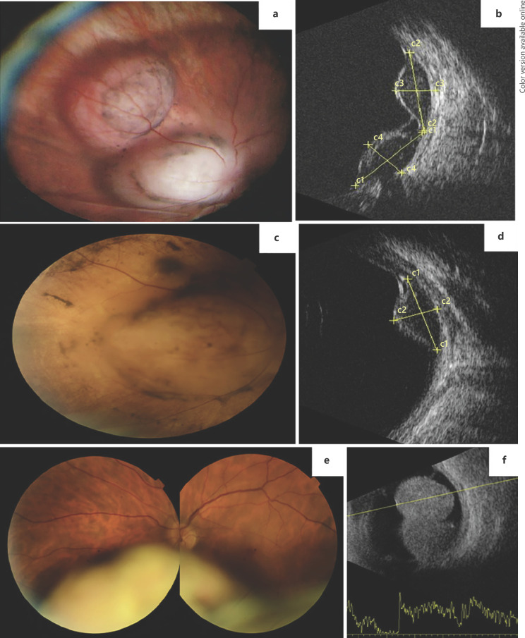
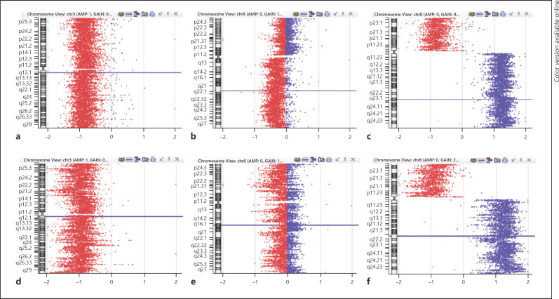
Tumor biopsies in uveal melanoma (UM) serve mainly the purpose of prognostication and assessment of individual metastatic risk, but can be used for diagnosis in selected cases. The importance of precise information is paramount for selecting adequate surveillance protocols, patient counseling, and optimization of treatment strategies. However, intratumoral heterogeneity and sample representativity are major concerns and can interfere with the correct prediction of the patient's prognosis. We report a series of cases of UM with distinct morphologically identifiable areas, highlighting the differences in clinical behavior, as well as histopathological and genetic features.

Keywords: Biopsy; Prognosis; Tumor heterogeneity; Uveal melanoma.


Conflict of interest statement

The authors have no conflict of interest to declare.

Figures

Fig. 1
Fig. 1
Case 1. a Fundus photograph of a bilobed uveal melanoma nasal to the optic disc, composed of 2 distinct portions. b Ultrasound (US) examination showing the 2 lobes of the tumor and inferior retinal detachment. c, d Fundus photographs 1 year after EBT treatment clearly showing regression of the melanotic part but stability of the pigmented lobe. e, f US examination showing regression of the amelanotic lobe and stability of the pigmented one.
Fig. 2
Fig. 2
Case 2. a Wide-field fundus photograph of a bilobed amelanotic uveal melanoma nasal to the optic disc. b US examination showing the 2 lobes of the tumor. c Fundus photograph 6 months after PBR clearly showing total regression of one of the lobes but stability of the other. d US examination showing only the remaining lobe of the tumor after PBR. Case 3. e Fundus photograph of a bilobed inferior uveal melanoma showing an amelanotic portion and a more anterior pigmented area. f US examination showing a large uveal melanoma occupying a great portion of the vitreous cavity.
Fig. 3
Fig. 3
Case 3. a Macroscopy of the ocular globe showing a small non-pigmented area in the base of the tumor (arrow). b Enucleated globe showing the inferior choroidal melanoma infiltrating the ciliary body (hematoxylin and eosin). c 10× magnification of the pigmented portion (inset with 40× magnification view of both epithelioid and spindle cells). d 10× magnification of the amelanotic region (inset with 40× magnification view of epithelioid cells). e PAS-stained 10× magnification view of the pigmented portion displaying vascular loops (arrow) in contrast to the same power view of the amelanotic area, without vascular loops (f).
Fig. 4
Fig. 4
Case 3. a IHC stain of the transition area between morphologically different regions of the tumor (arrow and arrowhead), showing differences in tumor lymphocytic infiltration (anti-CD3 staining). b 20× magnification of the amelanotic portion, showing absence of lymphocytic infiltration. c 20× magnification of the pigmented portion showing the presence of lymphocytes (anti-CD3 staining) infiltrating the tumor. d IHC stain of the transition area between morphologically different regions of the tumor (arrow and arrowhead), showing differences in macrophage infiltration (anti-CD68 staining). e 20× magnification of the amelanotic portion, showing absence of macrophage infiltration. f 20× magnification of the pigmented portion showing the presence of macrophages (anti-CD68 staining) infiltrating the tumor.
Fig. 5
Fig. 5
a–c Chromosome view of oligoarray-CGH profile of the pigmented area. a Monosomy 3. b 6q deletion. c 8p loss combined with 8q gain. d–f Chromosome view of oligoarray-CGH profile of the amelanotic portion. d Monosomy 3. e 6q without deletion. f 8p loss combined with 8q gain.

References

    1. Kaliki S, Shields CL. Uveal melanoma: relatively rare but deadly cancer. Eye (Lond) 2016 Feb;31((2)):241–257. - PMC - PubMed
    1. Collaborative Ocular Melanoma Study Group The COMS randomized trial of iodine 125 brachytherapy for choroidal melanoma: V. Twelve-year mortality rates and prognostic factors: COMS report No. 28. Arch Ophthalmol. 2006 Dec;124((12)):1684–93. - PubMed
    1. Frizziero L, Midena E, Trainiti S, Londei D, Bonaldi L, Bini S, et al. Uveal Melanoma Biopsy: A Review. Cancers (Basel) 2019 Jul;11((8)):1075. - PMC - PubMed
    1. Lim LA, Miyamoto C, Blanco P, Bakalian S, Burnier MN., Jr Case report: an atypical peripapillary uveal melanoma. BMC Ophthalmol. 2014 Feb;14((1)):13. - PMC - PubMed
    1. Mensink HW, Vaarwater J, Kiliç E, Naus NC, Mooy N, Luyten G, et al. Chromosome 3 intratumor heterogeneity in uveal melanoma. Invest Ophthalmol Vis Sci. 2009 Feb;50((2)):500–4. - PubMed

Publication types