. 2021 Mar 5;11(5):e3937.
doi: 10.21769/BioProtoc.3937.

Live Intravital Intestine with Blood Flow Visualization in Neonatal Mice Using Two-photon Laser Scanning Microscopy

Affiliations

Live Intravital Intestine with Blood Flow Visualization in Neonatal Mice Using Two-photon Laser Scanning Microscopy

Yuhki Koike et al. Bio Protoc. .

Abstract

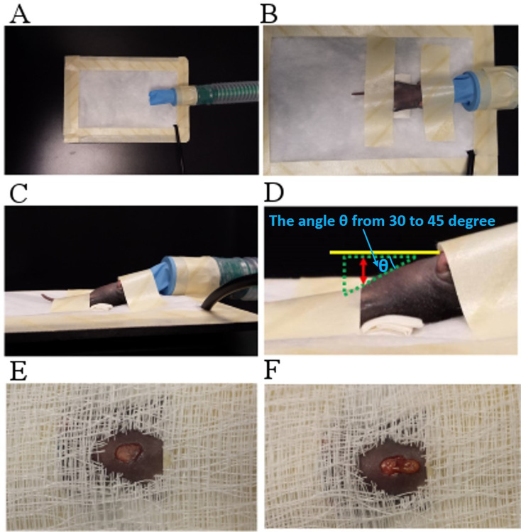
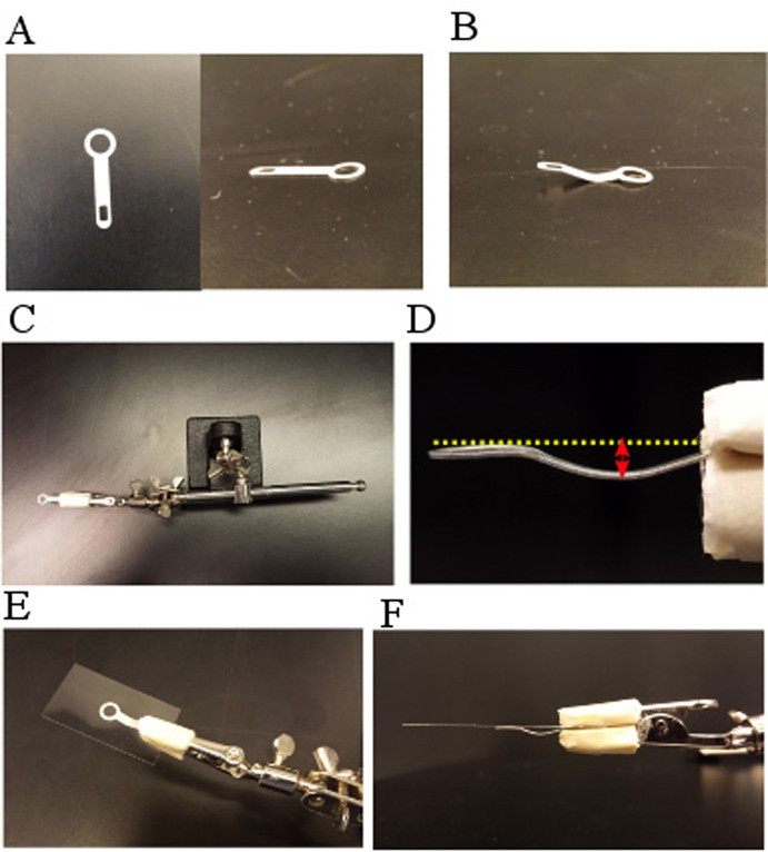
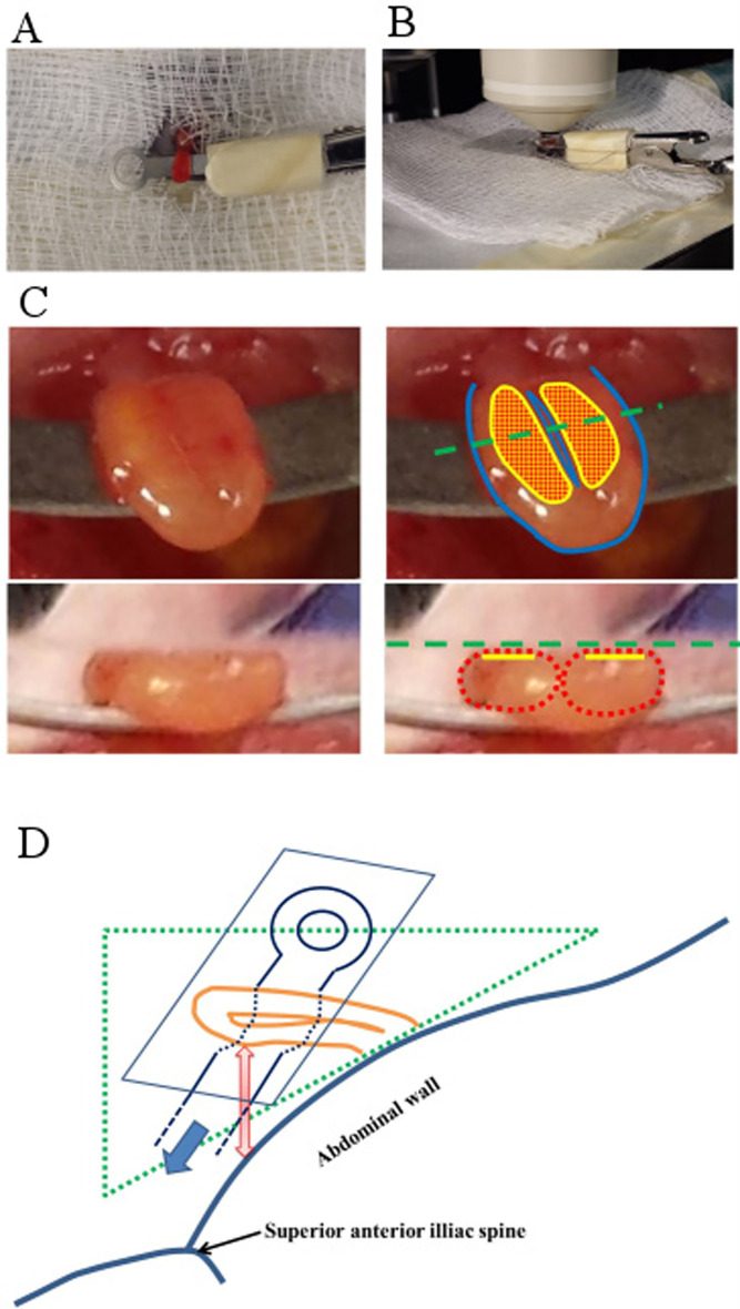
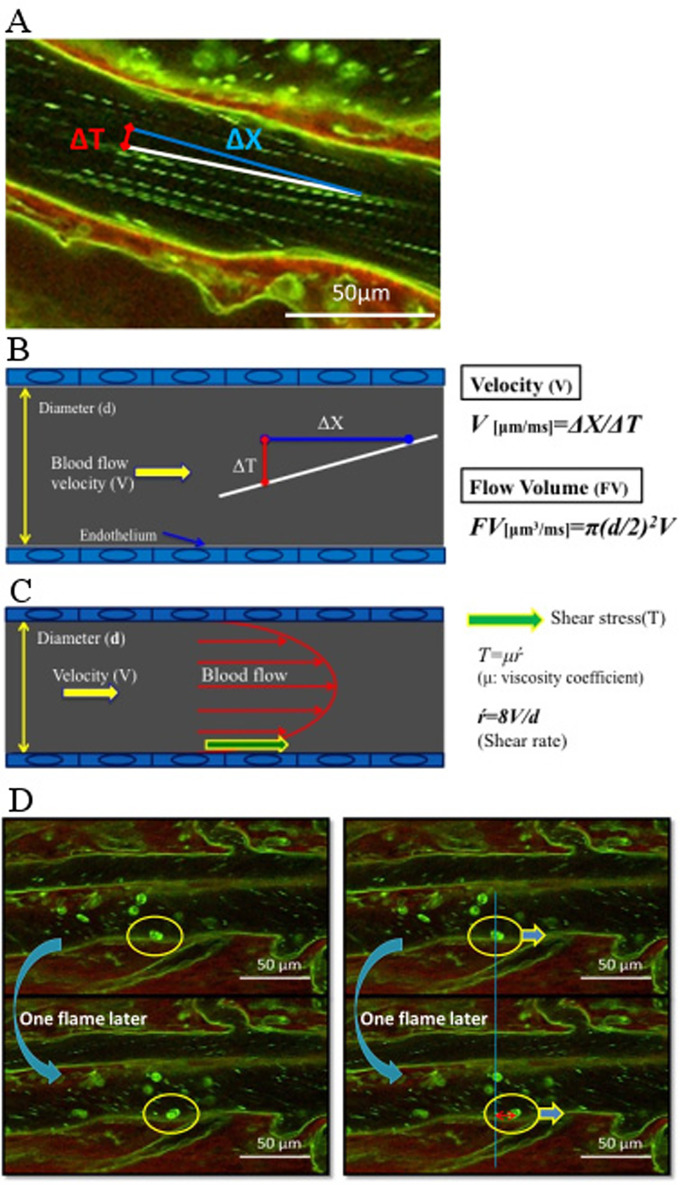
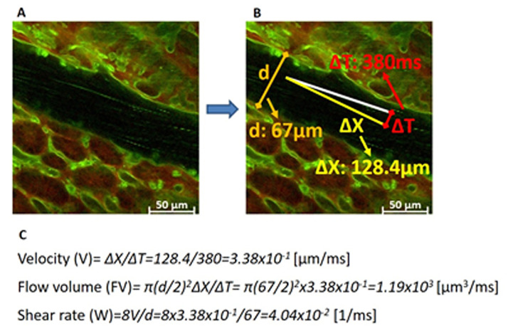
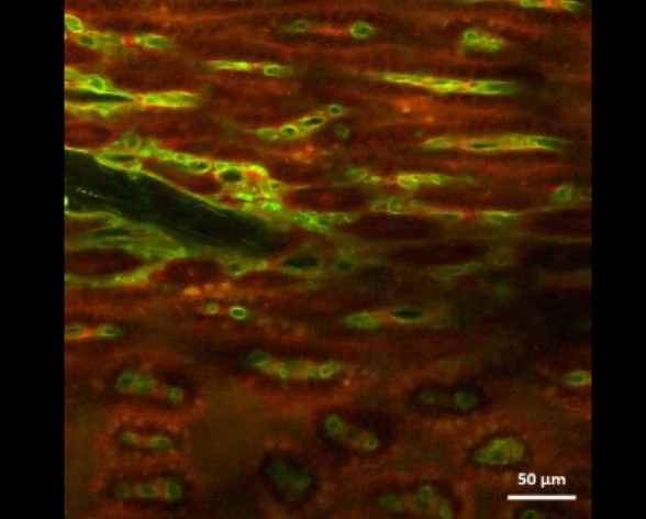
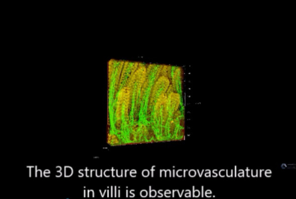
This protocol describes a novel technique to investigate the microcirculation dynamics underlying the pathology in the small intestine of neonatal mice using two-photon laser-scanning microscopy (TPLSM). Recent technological advances in multi-photon microscopy allow intravital analysis of different organs such as the liver, brain and intestine. Despite these advances, live visualization and analysis of the small intestine in neonatal rodents remain technically challenging. We herein provide a detailed description of a novel method to capture high resolution and stable images of the small intestine in neonatal mice as early as postnatal day 0. This imaging technique allows a comprehensive understanding of the development and blood flow dynamics in small intestine microcirculation.

Keywords: In vivo imaging; Intravital imaging; Necrotizing enterocolitis (NEC); Neonatal mouse imaging; Two-photon laser scanning microscopy (TPLSM).


Conflict of interest statement

Competing interestsThe authors have no conflict of interest to declare.

Figures

Figure 1.
Figure 1.. Neonatal mouse preparation for the TPLSM analysis.
(A-D) Mouse anesthesia and appropriate positioning. (E, F) Intestine exposure.
Figure 2.
Figure 2.. Fixing device setup for intestinal microscopic observation.
(A, B) Solder lug. (C, D). Solder lug attached to holding device with appropriate spacing, the spacing should be the same as the diameter of the intestine being used (generally between 1 and 3 mm). (E, F) Cover glass attached on top of the solder lug and clipped with the holding device.
Figure 3.
Figure 3.. Neonatal mouse intestine exposed for TPLSM observation.
(A-C) Mouse intestine preparation and areas of observation. The yellow marking or alternatively in the red dotted area shows the contact lesion area between small intestinal wall and the cover glass. The green dashed line (under image) shows the cover glass level (the top picture in C: view from above the cover glass, the bottom picture in C: view from the frontal view. The observable area is between the green dotted line and the yellow line from the frontal view. The red dashed circle represents the outer intestinal wall line from the frontal view. (D) Illustration of mouse intestine for microscopic observation.
Figure 4.
Figure 4.. Dynamic intestinal microcirculation in neonatal mice.
A. Intestinal microcirculation image obtained by TPLSM. B. Blood flow volume calculation. C. Blood vessel wall shear rate calculation. D. Leukocyte rolling speed calculation.
Figure 5.
Figure 5.. Example of experimental result and data analysis.
A. One still/flame image cutting from the recorded video (one flame scan speed: 380 ms). From this find a straight line of the platelet. B. Measure the tangent length of the straight line that is parallel to the blood flow direction (ΔX), scan time (ΔT), and blood vessel maximum diameter (d). C. Calculate the velocity, flow volume, and shear rate using the specific formula.
Video 1.
Video 1.. Microcirculation of the neonatal small intestine
Video 2.
Video 2.. 3-D image of the whole villi in a control neonatal mouse and the necrotizing enterocolitis mouse model

Similar articles

Cited by

References

    1. Barlow B., Santulli T. V., Heird W. C., Pitt J., Blanc W. A. and Schullinger J. N.(1974). An experimental study of acute neonatal enterocolitis--the importance of breast milk. J Pediatr Surg 9(5): 587-595. - PubMed
    1. Cao L., Kobayakawa S., Yoshiki A. and Abe K.(2012). High resolution intravital imaging of subcellular structures of mouse abdominal organs using a microstage device. PLoS One 7(3): e33876. - PMC - PubMed
    1. Coppieters K., Martinic M. M., Kiosses W. B., Amirian N. and von Herrath M.(2010). A novel technique for the in vivo imaging of autoimmune diabetes development in the pancreas by two-photon microscopy . PLoS One 5(12): e15732. - PMC - PubMed
    1. Devi S., Li A., Westhorpe C. L., Lo C. Y., Abeynaike L. D., Snelgrove S. L., Hall P., Ooi J. D., Sobey C. G., Kitching A. R. and Hickey M. J.(2013). Multiphoton imaging reveals a new leukocyte recruitment paradigm in the glomerulus. Nat Med 19(1): 107-112. - PubMed
    1. Ferrer M., Martin-Jaular L., Calvo M. and del Portillo H. A.(2012). Intravital microscopy of the spleen: quantitative analysis of parasite mobility and blood flow. J Vis Exp (59). - PMC - PubMed

LinkOut - more resources