. 2021 Jul;185(7):1972-1980.
doi: 10.1002/ajmg.a.62192. Epub 2021 Apr 2.

A novel homozygous SLC13A5 whole-gene deletion generated by Alu/Alu-mediated rearrangement in an Iraqi family with epileptic encephalopathy

Affiliations

A novel homozygous SLC13A5 whole-gene deletion generated by Alu/Alu-mediated rearrangement in an Iraqi family with epileptic encephalopathy

Ruizhi Duan et al. Am J Med Genet A. 2021 Jul.

Abstract

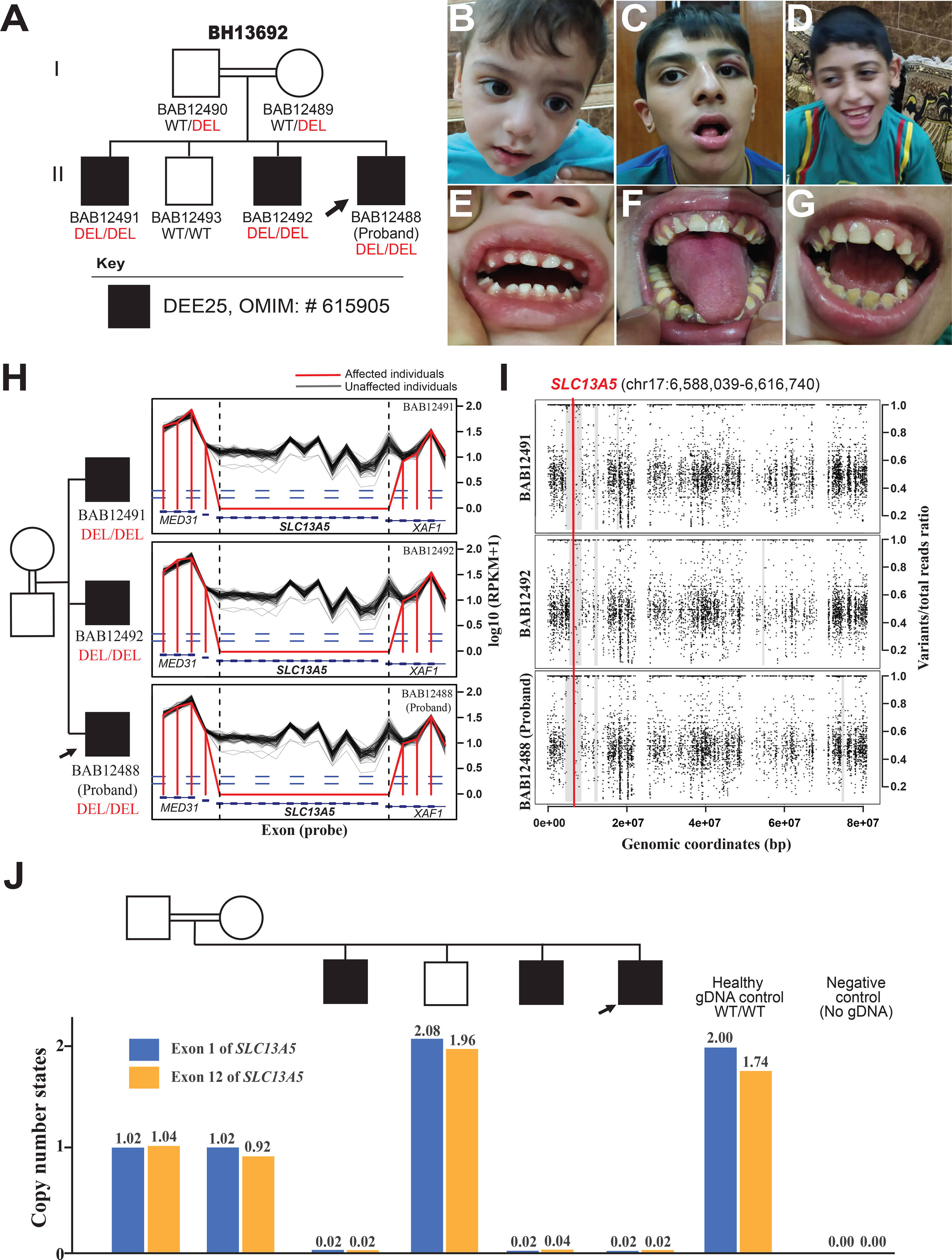
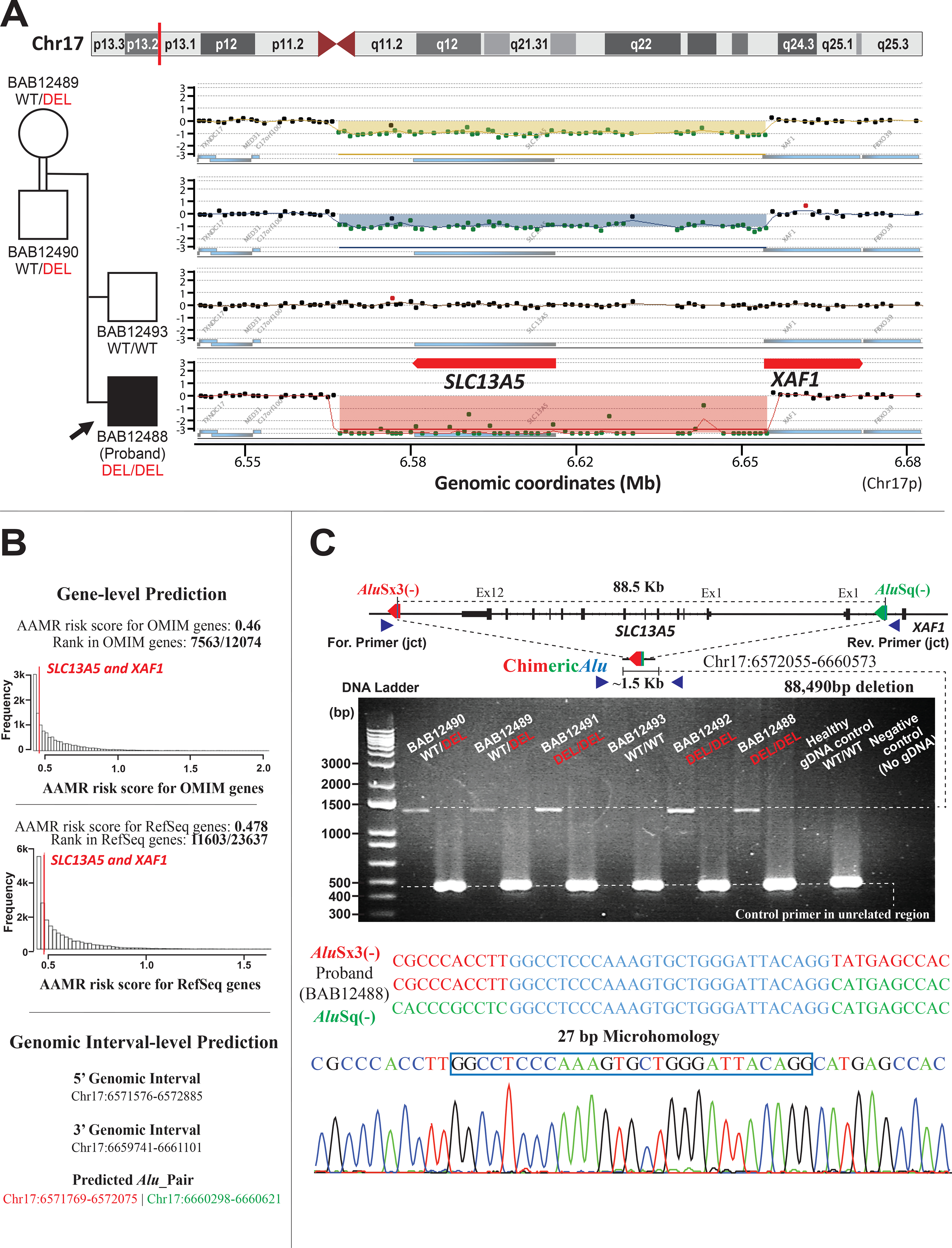
Biallelic loss-of-function (LoF) of SLC13A5 (solute carrier family 13, member 5) induced deficiency in sodium/citrate transporter (NaCT) causes autosomal recessive developmental epileptic encephalopathy 25 with hypoplastic amelogenesis imperfecta (DEE25; MIM #615905). Many pathogenic SLC13A5 single nucleotide variants (SNVs) and small indels have been described; however, no cases with copy number variants (CNVs) have been sufficiently investigated. We describe a consanguineous Iraqi family harboring an 88.5 kb homozygous deletion including SLC13A5 in Chr17p13.1. The three affected male siblings exhibit neonatal-onset epilepsy with fever-sensitivity, recurrent status epilepticus, global developmental delay/intellectual disability (GDD/ID), and other variable neurological findings as shared phenotypical features of DEE25. Two of the three affected subjects exhibit hypoplastic amelogenesis imperfecta (AI), while the proband shows no evidence of dental abnormalities or AI at 2 years of age with apparently unaffected primary dentition. Characterization of the genomic architecture at this locus revealed evidence for genomic instability generated by an Alu/Alu-mediated rearrangement; confirmed by break-point junction Sanger sequencing. This multiplex family from a distinct population elucidates the phenotypic consequence of complete LoF of SLC13A5 and illustrates the importance of read-depth-based CNV detection in comprehensive exome sequencing analysis to solve cases that otherwise remain molecularly unsolved.

Keywords: Alu/Alu-mediated rearrangement; CNV alleles; SLC13A5; developmental and epileptic encephalopathy; genomic instability; homozygous deletion.


Conflict of interest statement

Conflict of interest

J.R.L. has stock ownership in 23andMe, is a paid consultant for Regeneron Pharmaceuticals, and is a co-inventor on multiple United States and European patents related to molecular diagnostics for inherited neuropathies, eye diseases, and bacterial genomic fingerprinting. The Department of Molecular and Human Genetics at Baylor College of Medicine receives revenue from clinical genetic testing conducted at Baylor Genetics (BG) Laboratories; JRL participates in the BG scientific advisory board. Other authors have no potential conflicts to report.

Figures

Figure 1.
Figure 1.. Family pedigree, facial features, dentition, and bioinformatic characteristics of whole-gene deletion of SLC13A5 and exonic deletion of XAF1.
(A) Pedigree of the family showing the affected males as black shaded squares with the proband demarcated (arrow). The parents are second-degree cousins; extended multigenerational family pedigree analyses (Supplementary) reveals historical evidence for consanguineous relationships in previous generations. WT: Wild Type, DEL: Deletion. (B-D) Facial photographs of the proband (B), the 15-year-old affected sibling (C), and the 11-year-old affected sibling (D) with no distinct dysmorphic features. (E) A dental photograph of the proband (B) shows no evidence of dental abnormalities or AI at 2 years of age with appeared unaffected primary dentition. (F) A dental photograph of the 15-year-old affected sibling (C) shows hypoplastic enamel, dark yellow color with severely worn enamel surfaces, generalized gingival inflammation, and marginal gingival overgrowth. AI type 1 (hypoplastic enamel) is highly suspected. (G) A dental photograph of the 11-year-old affected sibling (D) shows defective and severely worn enamel surfaces, accumulation of higher calculus and plaque deposits, and dark color of the underlying dentin. Incipient caries at the lower left canine, and generalized gingival inflammation are also noted. These findings are consistent with AI type I (hypoplastic enamel). (H) The log10 RPKM profile of targeted region from HMZDelFinder raw data; Designed exon capture region is marked as blue rectangles; RPKM values of the three affected siblings are marked as red lines, while black lines delineate RPKM value from individual ES samples from an internal database with similar experimental conditions. (I) B-allele frequency of chromosome 17 for the proband and affected siblings showing a shared 2.5–2.7 Mb interval of Absence of heterozygosity (AOH) surrounding the homozygous SLC13A5 deletion and marked with a thick gray line. Few small unshared AOH blocks are also seen shaded with thin grey lines. The location of the CNV is marked with a red line across the shared AOH region. (J) Copy number state of exon 1 and exon 12 of SLC13A5, determined by relative positive droplet ratios, are independently examined by digital droplet PCR (ddPCR) on all family members. According to relative positive droplet ratios, three affected individuals manifest zero-copy of both exons; both parents present as heterozygous carriers (single copy), while the unaffected brother shows two copies of both exons.
Figure 2.
Figure 2.. Molecular genomic architecture of SLC13A5 whole-gene deletion and XAF1 exonic deletion in the family mediated by AAMR.
(A) A high-resolution array-based comparative genomic hybridization (aCGH) over Chr17p13.1 of the parents and unaffected brother, and proband, from top to bottom respectively, performed to evaluate for copy number status of SLC13A5 and XAF1. aCGH confirmed the homozygous deletion in the proband and the heterozygous deletion (carrier status) in both parents, while the unaffected brother was homozygous for the wild type (WT) allele (copy number neutral state). (B) Gene-level prediction from AluAluCNVPredictor shows SLC13A5 and XAF1 have the same AAMR risk score in OMIM genes (0.46) and RefSeq genes (0.478), respectively. Genomic interval-level prediction successfully addresses this one CNV-Alu pair (AluSx3/AluSq), generating the deletion in this family with the information of genomic intervals acquired experimentally. (C) A schematic of the genomic profile of AluSx3/AluSq pair mediated rearrangement and the primer design along with the gel electrophoresis of the PCR products. The junction primer pair was designed to produce an amplicon size of ~1.5 Kb for deleted alleles resulting from AAMR with the formation of a recombinant or chimeric Alu. Control primers were selected from a 450 bp unrelated genomic locus. Below the schematic is the 1% agarose gel electrophoresis of PCR products. From left to right of the panel: parents with lighter bands representing a heterozygous ~1.5 Kb deleted alleles; affected brother (BAB12491) with a thick band of ~1.5 Kb, consistent with the homozygous deleted allele; unaffected brother (BAB12493) showing no deleted band; affected brother (BAB12492) and proband with thick bands of ~1.5 Kb, consistent of homozygous deleted alleles; Healthy gDNA control with no deleted allele, and negative control (no template). Below the gel figure is an illustration of breakpoint junction analysis by Sanger sequencing revealed an 88,490 bp deletion resulting from recombination of AluSx3/AluSq pair in the same orientation (red and green arrow, respectively) encompassing a region of microhomology (blue line with each Alu). This results in a chimeric Alu with a 27 bp of microhomology (blue vertical line within chimeric Alu). The three sequences below the schematic from top to bottom represent the reference AluSx3 sequence (colored in red), the proband “chimeric” sequence, and the AluSq sequence (colored in green). The region of microhomology is colored in blue. The bottom image represents Sanger tracing of the breakpoint junction with the 27 bp of microhomology marked with a blue box.

References

    1. Alkuraya FS (2015). Natural human knockouts and the era of genotype to phenotype. Genome Medicine, 7(1), 48. 10.1186/s13073-015-0173-z - DOI - PMC - PubMed
    1. Boone PM, Yuan B, Campbell IM, Scull JC, Withers MA, Baggett BC, Beck CR, Shaw CJ, Stankiewicz P, Moretti P, Goodwin WE, Hein N, Fink JK, Seong MW, Seo SH, Park SS, Karbassi ID, Batish SD, Ordóñez-Ugalde A, … Lupski JR (2014). The Alu-rich genomic architecture of SPAST predisposes to diverse and functionally distinct disease-associated CNV alleles. American Journal of Human Genetics, 95(2), 143–161. 10.1016/j.ajhg.2014.06.014 - DOI - PMC - PubMed
    1. Carvalho CMB, Vasanth S, Shinawi M, Russell C, Ramocki MB, Brown CW, Graakjaer J, Skytte AB, Vianna-Morgante AM, Krepischi ACV, Patel GS, Immken LD, Aleck K, Lim C, Cheung SW, Rosenberg C, Katsanis N & Lupski JR (2014). Dosage changes of a segment at 17p13.1 lead to intellectual disability and microcephaly as a result of complex genetic interaction of multiple genes. American Journal of Human Genetics, 95(5), 565–578. 10.1016/j.ajhg.2014.10.006 - DOI - PMC - PubMed
    1. Dharmadhikari AV, Ghosh R, Yuan B, Liu P, Dai H, Al Masri S, Scull J, Posey JE, Jiang AH, He W, Vetrini F, Braxton AA, Ward P, Chiang T, Qu C, Gu S, Shaw CA, Smith JL, Lalani S, … Bi W (2019). Copy number variant and runs of homozygosity detection by microarrays enabled more precise molecular diagnoses in 11,020 clinical exome cases. Genome Medicine, 11(1), 1–17. 10.1186/s13073-019-0639-5 - DOI - PMC - PubMed
    1. Fromer M & Purcell SM (2014). Using XHMM Software to Detect Copy Number Variation in Whole-Exome Sequencing Data. Current Protocols in Human Genetics, 81, 7.23.1–21. 10.1002/0471142905.hg0723s81 - DOI - PMC - PubMed

Publication types

LinkOut - more resources