

Clinical performance of long axial field of view PET/CT: a head-to-head intra-individual comparison of the Biograph Vision Quadra with the Biograph Vision PET/CT
- PMID: 33797596
- PMCID: PMC8241747
- DOI: 10.1007/s00259-021-05282-7
Clinical performance of long axial field of view PET/CT: a head-to-head intra-individual comparison of the Biograph Vision Quadra with the Biograph Vision PET/CT
Abstract
Purpose: To investigate the performance of the new long axial field-of-view (LAFOV) Biograph Vision Quadra PET/CT and a standard axial field-of-view (SAFOV) Biograph Vision 600 PET/CT (both: Siemens Healthineers) system using an intra-patient comparison.
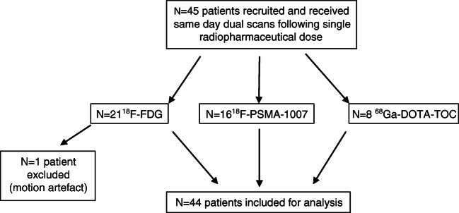
Methods: Forty-four patients undergoing routine oncological PET/CT were prospectively included and underwent a same-day dual-scanning protocol following a single administration of either 18F-FDG (n = 20), 18F-PSMA-1007 (n = 16) or 68Ga-DOTA-TOC (n = 8). Half the patients first received a clinically routine examination on the SAFOV (FOVaxial 26.3 cm) in continuous bed motion and then immediately afterwards on the LAFOV system (10-min acquisition in list mode, FOVaxial 106 cm); the second half underwent scanning in the reverse order. Comparisons between the LAFOV at different emulated scan times (by rebinning list mode data) and the SAFOV were made for target lesion integral activity, signal to noise (SNR), target lesion to background ratio (TBR) and visual image quality.
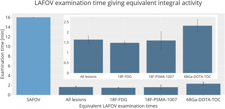
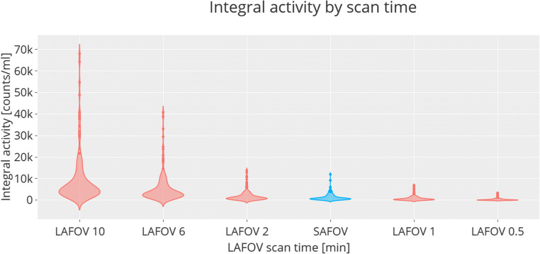
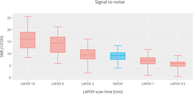
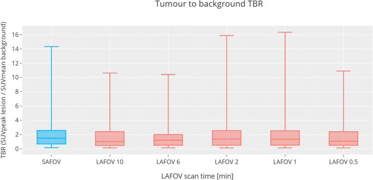
Results: Equivalent target lesion integral activity to the SAFOV acquisitions (16-min duration for a 106 cm FOV) were obtained on the LAFOV in 1.63 ± 0.19 min (mean ± standard error). Equivalent SNR was obtained by 1.82 ± 1.00 min LAFOV acquisitions. No statistically significant differences (p > 0.05) in TBR were observed even for 0.5 min LAFOV examinations. Subjective image quality rated by two physicians confirmed the 10 min LAFOV to be of the highest quality, with equivalence between the LAFOV and the SAFOV at 1.8 ± 0.85 min. By analogy, if the LAFOV scans were maintained at 10 min, proportional reductions in applied radiopharmaceutical could obtain equivalent lesion integral activity for activities under 40 MBq and equivalent doses for the PET component of <1 mSv.
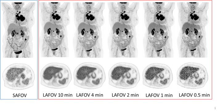
Conclusion: Improved image quality, lesion quantification and SNR resulting from higher sensitivity were demonstrated for an LAFOV system in a head-to-head comparison under clinical conditions. The LAFOV system could deliver images of comparable quality and lesion quantification in under 2 min, compared to routine SAFOV acquisition (16 min for equivalent FOV coverage). Alternatively, the LAFOV system could allow for low-dose examination protocols. Shorter LAFOV acquisitions (0.5 min), while of lower visual quality and SNR, were of adequate quality with respect to target lesion identification, suggesting that ultra-fast or low-dose acquisitions can be acceptable in selected settings.
Keywords: Digital PET; PET/CT; Positron-emission-tomography; Total-body; Ultra-long FOV PET; Whole-body.
Conflict of interest statement
HS is a full-time employee of Siemens Healthcare AG, Switzerland. AR has received research support and speaker honoraria from Siemens. All other authors have no conflicts of interest to report.
Figures
Comment in
-
First clinical experience of 106 cm, long axial field-of-view (LAFOV) PET/CT: an elegant balance between standard axial (23 cm) and total-body (194 cm) systems.Eur J Nucl Med Mol Imaging. 2021 Nov;48(12):3755-3759. doi: 10.1007/s00259-021-05505-x. Eur J Nucl Med Mol Imaging. 2021. PMID: 34424375 Free PMC article. No abstract available.
References
-
- Beyer T, Townsend DW, Brun T, Kinahan PE, Charron M, Roddy R, et al. A combined PET/CT scanner for clinical oncology. J Nucl Med. 2000;41:1369–1379. - PubMed
-
- van Sluis JJ, de Jong J, Schaar J, Noordzij W, van Snick P, Dierckx R, et al. Performance characteristics of the digital biograph vision PET/CT system. J Nucl Med. 2019. 10.2967/jnumed.118.215418. - PubMed
-
- Nguyen NC, Vercher-Conejero JL, Sattar A, Miller MA, Maniawski PJ, Jordan DW, et al. Image quality and diagnostic performance of a digital PET prototype in patients with oncologic diseases: initial experience and comparison with analog PET. J Nucl Med. 2015;56:1378–1385. doi: 10.2967/jnumed.114.148338. - DOI - PubMed
Publication types
MeSH terms
Substances
LinkOut - more resources
Full Text Sources
Other Literature Sources
Miscellaneous
