Distinct and temporally associated neural mechanisms underlying concurrent, postsuccess, and posterror cognitive controls: Evidence from a stop-signal task
- PMID: 33797816
- PMCID: PMC8127156
- DOI: 10.1002/hbm.25347
Distinct and temporally associated neural mechanisms underlying concurrent, postsuccess, and posterror cognitive controls: Evidence from a stop-signal task
Abstract
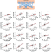
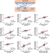
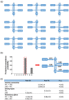
Cognitive control is built upon the interactions of multiple brain regions. It is currently unclear whether the involved regions are temporally separable in relation to different cognitive processes and how these regions are temporally associated in relation to different task performances. Here, using stop-signal task data acquired from 119 healthy participants, we showed that concurrent and poststop cognitive controls were associated with temporally distinct but interrelated neural mechanisms. Specifically, concurrent cognitive control activated regions in the cingulo-opercular network (including the dorsal anterior cingulate cortex [dACC], insula, and thalamus), together with superior temporal gyrus, secondary motor areas, and visual cortex; while regions in the fronto-parietal network (including the lateral prefrontal cortex [lPFC] and inferior parietal lobule) and cerebellum were only activated during poststop cognitive control. The associations of activities between concurrent and poststop regions were dependent on task performance, with the most notable difference in the cerebellum. Importantly, while concurrent and poststop signals were significantly correlated during successful cognitive control, concurrent activations during erroneous trials were only correlated with posterror activations in the fronto-parietal network but not cerebellum. Instead, the cerebellar activation during posterror cognitive control was likely to be driven secondarily by posterror activation in the lPFC. Further, a dynamic causal modeling analysis demonstrated that postsuccess cognitive control was associated with inhibitory connectivity from the lPFC to cerebellum, while excitatory connectivity from the lPFC to cerebellum was present during posterror cognitive control. Overall, these findings suggest dissociable but temporally related neural mechanisms underlying concurrent, postsuccess, and posterror cognitive control processes in healthy individuals.
Keywords: cerebellum; cingulo-opercular network; cognitive control; fronto-parietal network; posterror; poststop.
© 2021 The Authors. Human Brain Mapping published by Wiley Periodicals LLC.
Conflict of interest statement
Dr. Cannon has served as a consultant for Boehringer‐Ingelheim Pharmaceuticals and Biogen. Dr. Cao reports no conflicts of interest.
Figures


Similar articles
-
Measuring Cognitive Flexibility with the Flexible Item Selection Task: From fMRI Adaptation to Individual Connectome Mapping.J Cogn Neurosci. 2020 Jun;32(6):1026-1045. doi: 10.1162/jocn_a_01536. Epub 2020 Feb 4. J Cogn Neurosci. 2020. PMID: 32013686
-
Large-scale reconfiguration of connectivity patterns among attentional networks during context-dependent adjustment of cognitive control.Hum Brain Mapp. 2021 Aug 15;42(12):3821-3832. doi: 10.1002/hbm.25467. Epub 2021 May 14. Hum Brain Mapp. 2021. PMID: 33987911 Free PMC article.
-
Dissociable roles of right inferior frontal cortex and anterior insula in inhibitory control: evidence from intrinsic and task-related functional parcellation, connectivity, and response profile analyses across multiple datasets.J Neurosci. 2014 Oct 29;34(44):14652-67. doi: 10.1523/JNEUROSCI.3048-14.2014. J Neurosci. 2014. PMID: 25355218 Free PMC article.
-
Cerebellar Contributions to Proactive and Reactive Control in the Stop Signal Task: A Systematic Review and Meta-Analysis of Functional Magnetic Resonance Imaging Studies.Neuropsychol Rev. 2020 Sep;30(3):362-385. doi: 10.1007/s11065-020-09432-w. Epub 2020 Mar 18. Neuropsychol Rev. 2020. PMID: 32189178
-
The cerebellar connectome.Behav Brain Res. 2025 Mar 28;482:115457. doi: 10.1016/j.bbr.2025.115457. Epub 2025 Jan 28. Behav Brain Res. 2025. PMID: 39884319 Review.
Cited by
-
The brain selectively allocates energy to functional brain networks under cognitive control.Sci Rep. 2024 Dec 30;14(1):32032. doi: 10.1038/s41598-024-83696-7. Sci Rep. 2024. PMID: 39738735 Free PMC article.
-
Intrinsic network changes associated with cognitive impairment in patients with hearing loss and tinnitus: a resting-state functional magnetic resonance imaging study.Ann Transl Med. 2022 Jun;10(12):690. doi: 10.21037/atm-22-2135. Ann Transl Med. 2022. PMID: 35845541 Free PMC article.
-
Cerebellar Functional Dysconnectivity in Drug-Naïve Patients With First-Episode Schizophrenia.Schizophr Bull. 2023 Mar 15;49(2):417-427. doi: 10.1093/schbul/sbac121. Schizophr Bull. 2023. PMID: 36200880 Free PMC article.
-
Mapping Cerebellar Connectivity to Cognition in Psychosis: Convergent Evidence From Functional Magnetic Resonance Imaging and Transcranial Magnetic Stimulation.Biol Psychiatry. 2025 Jul 4:S0006-3223(25)01297-1. doi: 10.1016/j.biopsych.2025.06.023. Online ahead of print. Biol Psychiatry. 2025. PMID: 40617381
References
-
- Anticevic, A. , Hu, S. , Zhang, S. , Savic, A. , Billingslea, E. , Wasylink, S. , … Pittenger, C. (2014). Global resting‐state functional magnetic resonance imaging analysis identifies frontal cortex, striatal, and cerebellar dysconnectivity in obsessive‐compulsive disorder. Biological Psychiatry, 75(8), 595–605. 10.1016/j.biopsych.2013.10.021 - DOI - PMC - PubMed
Publication types
MeSH terms
LinkOut - more resources
Full Text Sources
Other Literature Sources

