. 2021 Mar 23;118(12):e2007194118.
doi: 10.1073/pnas.2007194118.

JNK signaling prevents biliary cyst formation through a CASPASE-8-dependent function of RIPK1 during aging

Affiliations

JNK signaling prevents biliary cyst formation through a CASPASE-8-dependent function of RIPK1 during aging

Katrin Müller et al. Proc Natl Acad Sci U S A. .

Abstract

The c-Jun N-terminal kinase (JNK) signaling pathway mediates adaptation to stress signals and has been associated with cell death, cell proliferation, and malignant transformation in the liver. However, up to now, its function was experimentally studied mainly in young mice. By generating mice with combined conditional ablation of Jnk1 and Jnk2 in liver parenchymal cells (LPCs) (JNK1/2LPC-KO mice; KO, knockout), we unraveled a function of the JNK pathway in the regulation of liver homeostasis during aging. Aging JNK1/2LPC-KO mice spontaneously developed large biliary cysts that originated from the biliary cell compartment. Mechanistically, we could show that cyst formation in livers of JNK1/2LPC-KO mice was dependent on receptor-interacting protein kinase 1 (RIPK1), a known regulator of cell survival, apoptosis, and necroptosis. In line with this, we showed that RIPK1 was overexpressed in the human cyst epithelium of a subset of patients with polycystic liver disease. Collectively, these data reveal a functional interaction between JNK signaling and RIPK1 in age-related progressive cyst development. Thus, they provide a functional linkage between stress adaptation and programmed cell death (PCD) in the maintenance of liver homeostasis during aging.

Keywords: MK2; cholangiocytes; liver; liver cysts; programmed cell death.


Conflict of interest statement

The authors declare no competing interest.

Figures

Fig. 1.
Fig. 1.
Combined deletion of Jnk1/2 in LPCs induces massive liver cyst formation in old mice. (A) Representative macroscopic liver pictures (Left), hematoxylin and eosin (H&E) staining (Middle), and pan-CK staining (Right) of 6-wk-old WT and JNK1/2LPC-KO animals. (B) Liver weight-to-body weight ratio of 6-wk-old WT and JNK1/2LPC-KO animals. n = 10. (C) Macroscopic picture (Left), H&E staining (Middle), and pan-CK staining (Right) of 52-wk-old WT and JNK1/2LPC-KO animals. (D) Liver weight-to-body weight ratio of 52-wk-old JNK1/2LPC-KO and WT animals. n = 5. (E) Macroscopic picture (Left), H&E staining (Middle), and pan-CK staining (Right) of 52-wk-old JNK1/2Δhep and WT animals. (F) Liver weight-to-body weight ratio of 52-wk-old JNK1/2Δhep and WT animals. n = 10 to 20. n.s., not significant; *P < 0.05, ***P < 0.001.
Fig. 2.
Fig. 2.
JNK1/2LPC-KO mice show elevated markers for liver injury. (A) Serum-level analysis of ALT, GLDH, and AP in 6-wk-old WT and JNK1/2LPC-KO mice. n = 6. (B) Serum-level analysis of ALT, GLDH, and AP in 52-wk-old WT and JNK1/2LPC-KO mice. n = 6. (C) Serum-level analysis of total bile acids in 52-wk-old WT and JNK1/2LPC-KO mice. n = 5. (D) Western blot analysis of liver protein extracts of 6-wk-old WT and JNK1/2LPC-KO animals. *P < 0.05, **P < 0.01.
Fig. 3.
Fig. 3.
Additional deletion of Caspase-8 or Mlkl does not significantly reduce cyst formation in JNK1/2LPC-KO mice. (A) Representative macroscopic liver pictures of WT (Left), JNK1/2LPC-KO (Middle Left), JNK1/2/CASP-8LPC-KO (Middle Right), and JNK1/2/MLKLLPC-KO (Right) animals at the age of 6 wk (Top) and 52 wk (Bottom). (B) Representative H&E-stained liver sections of WT (Left), JNK1/2LPC-KO (Middle Left), JNK1/2/CASP-8LPC-KO (Middle Right), and JNK1/2/MLKLLPC-KO (Right) animals at the age of 6 wk (Top) and 52 wk (Bottom). (C) Representative pan-CK–stained liver sections of WT (Left), JNK1/2LPC-KO (Middle Left), JNK1/2/CASP-8LPC-KO (Middle Right), and JNK1/2/MLKLLPC-KO (Right) animals at the age of 6 wk (Top) and 52 wk (Bottom). (D) Liver weight-to-body weight ratio of 52-wk-old WT, JNK1/2LPC-KO, JNK1/2/CASP-8LPC-KO, and JNK1/2/MLKLLPC-KO animals. n = 5 to 8. (E) Quantification of fluid-filled tissue cavities, including liver cysts, biliary ducts, and blood vessels, of 52-wk-old WT, JNK1/2LPC-KO, JNK1/2/CASP-8LPC-KO, and JNK1/2/MLKLLPC-KO mice. n = 6 to 8. (F) Serum analysis of ALT, GLDH, and AP in 52-wk-old WT, JNK1/2LPC-KO, JNK1/2/CASP-8LPC-KO, and JNK1/2/MLKLLPC-KO mice. n = 6. *P < 0.05.
Fig. 4.
Fig. 4.
Ripk1 deletion rescues JNK1/2LPC-KO mice from macroscopic visible cyst formation, which is restored by additional deletion of Caspase-8. (A) Representative macroscopic pictures of 6-wk- (Top), 52-wk- (Middle), and 66-wk- (Bottom) old WT (Left), JNK1/2LPC-KO (Middle Left), JNK1/2/RIPK1LPC-KO (Middle Right), and JNK1/2/RIPK1/CASP-8LPC-KO (Right) livers. (B) Representative H&E stainings of liver sections of 6-wk- (Top), 52-wk- (Middle), and 66-wk- (Bottom) old WT (Left), JNK1/2LPC-KO (Middle Left), JNK1/2/RIPK1LPC-KO (Middle Right), and JNK1/2/RIPK1/CASP-8LPC-KO (Right) mice. (C) Representative pan-CK stainings of liver sections of 6-wk- (Top), 52-wk- (Middle), and 66-wk- (Bottom) old WT (Left), JNK1/2LPC-KO (Middle Left), JNK1/2/RIPK1LPC-KO (Middle Right), and JNK1/2/RIPK1/CASP-8LPC-KO (Right) mice. (D) Liver weight-to-body weight ratios of 66-wk-old WT, JNK1/2LPC-KO, JNK1/2/RIPK1LPC-KO, and JNK1/2/RIPK1/CASP-8LPC-KO mice. n = 4. (E) Quantification of fluid-filled tissue cavities, including liver cysts, biliary ducts, and blood vessels, of 66-wk-old WT, JNK1/2LPC-KO, JNK1/2/RIPK1LPC-KO, and JNK1/2/RIPK1/CASP-8LPC-KO mice. n = 4. (F) Serum analysis of ALT, GLDH, and AP in 66-wk-old WT, JNK1/2LPC-KO, JNK1/2/RIPK1LPC-KO, and JNK1/2/RIPK1/CASP-8LPC-KO mice. n = 4. *P < 0.05, **P < 0.005, ***P < 0.001.
Fig. 5.
Fig. 5.
Additional deletion of Ripk1 in JNK1/2LPC-KO does not influence proliferation and DNA-damage response in cystic liver tissue. (A) Representative Ki67 (brown) and pan-CK (red) double stainings of liver sections of 52-wk-old WT, JNK1/2LPC-KO, JNK1/2/RIPK1LPC-KO, and JNK1/2/RIPK1/CASP-8LPC-KO mice and quantification of double-positive cells. n = 6. Arrowheads indicate double-positive cells. (B) Representative Ki67 stainings of liver sections of 52-wk-old WT, JNK1/2LPC-KO, JNK1/2/RIPK1LPC-KO, and JNK1/2/RIPK1/CASP-8LPC-KO mice and quantification. n = 5 or 6. (C) Representative ɣH2A.X stainings of liver sections of 52-wk-old WT, JNK1/2LPC-KO, JNK1/2/RIPK1LPC-KO, and JNK1/2/RIPK1/CASP-8LPC-KO mice and quantification. n = 5 to 7. *P < 0.05.
Fig. 6.
Fig. 6.
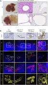
Liver cysts in JNK1/2 ablated livers originate from bile duct cells. (A) Representative macroscopic liver picture (Left), H&E staining (Middle), and pan-CK staining (Right) of 52-wk-old WT and JNK1/2Sox9-cre/ERT2 animals 48 wk after tamoxifen injection. (B) Stepwise cystic transformation of intrahepatic bile ducts in JNK1/2LPC-KO animals. Representative CK19 immunohistochemistry stainings (first row) and three-dimensional (3D) analysis of pan-CK/DAPI–immunostained intrahepatic bile ducts/ductules showing progressive stepwise cystic transformation of intrahepatic bile duct/ductules in JNK1/2LPC-KO animals at the indicated age (low-magnification 3D maximum-intensity projection, second row; corresponding optical 2D view and 3D maximum-intensity projection of enlarged insets, third and fourth rows; corresponding 3D pan-CK + surface/interconnectivity analysis using the IMARIS surface protocol where different colors represent individual interconnected panCK+ segments, fifth row). White dashed lines, portal tract; white dashed circles, portal vein; BD, bile duct; PV, portal vein. (Scale bars, 50 µm.)
Fig. 7.
Fig. 7.
Human liver samples of Caroli disease show expression of RIPK1 in biliary cystic epithelium. (A) Cystic bile duct in Caroli disease showing strong patchy RIPK1 staining in biliary epithelium. (B) Another case of Caroli disease with weak RIPK1 expression in some biliary epithelial cells lining the cystically dilated bile duct. (C) Focal RIPK1 staining in Caroli syndrome. Note the lack of RIPK1 staining in a corresponding ductal plate malformation of this patient (F). (D) Barely visible RIPK1 immunosignal in this example of autosomal dominant polycystic liver disease. (E) No expression in this example of autosomal recessive polycystic liver disease.

Similar articles

Cited by

References

    1. Schwabe R. F., Luedde T., Apoptosis and necroptosis in the liver: A matter of life and death. Nat. Rev. Gastroenterol. Hepatol. 15, 738–752 (2018). - PMC - PubMed
    1. Sabio G., Davis R. J., TNF and MAP kinase signalling pathways. Semin. Immunol. 26, 237–245 (2014). - PMC - PubMed
    1. Seki E., Brenner D. A., Karin M., A liver full of JNK: Signaling in regulation of cell function and disease pathogenesis, and clinical approaches. Gastroenterology 143, 307–320 (2012). - PMC - PubMed
    1. Kuan C. Y., et al. ., The Jnk1 and Jnk2 protein kinases are required for regional specific apoptosis during early brain development. Neuron 22, 667–676 (1999). - PubMed
    1. Vernia S., et al. ., The PPARα-FGF21 hormone axis contributes to metabolic regulation by the hepatic JNK signaling pathway. Cell Metab. 20, 512–525 (2014). - PMC - PubMed

Publication types

MeSH terms

Substances

Supplementary concepts