. 2021 Apr 2;17(4):e1009479.
doi: 10.1371/journal.ppat.1009479. eCollection 2021 Apr.

FOXO regulates the expression of antimicrobial peptides and promotes phagocytosis of hemocytes in shrimp antibacterial immunity

Affiliations

FOXO regulates the expression of antimicrobial peptides and promotes phagocytosis of hemocytes in shrimp antibacterial immunity

Cang Li et al. PLoS Pathog. .

Abstract

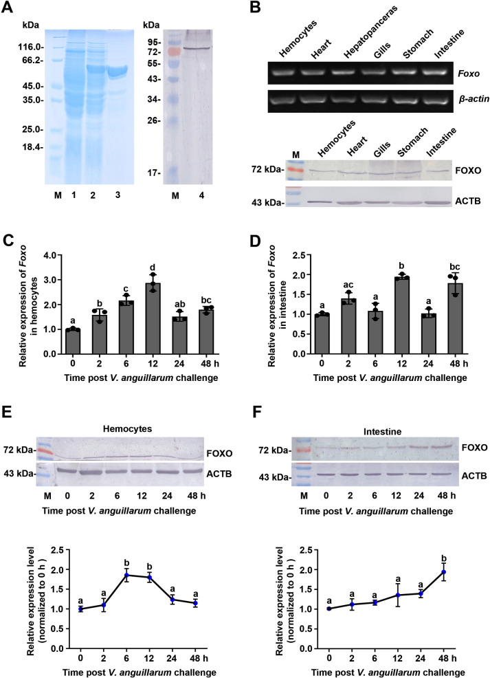
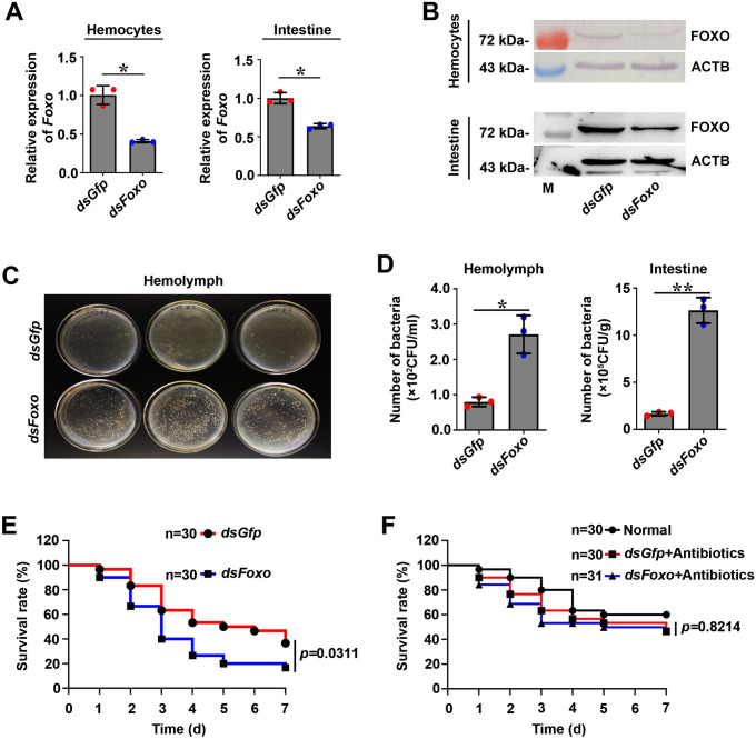
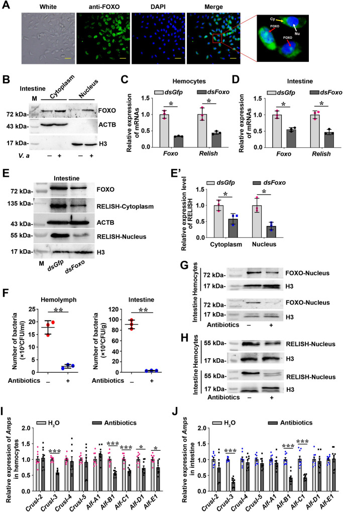
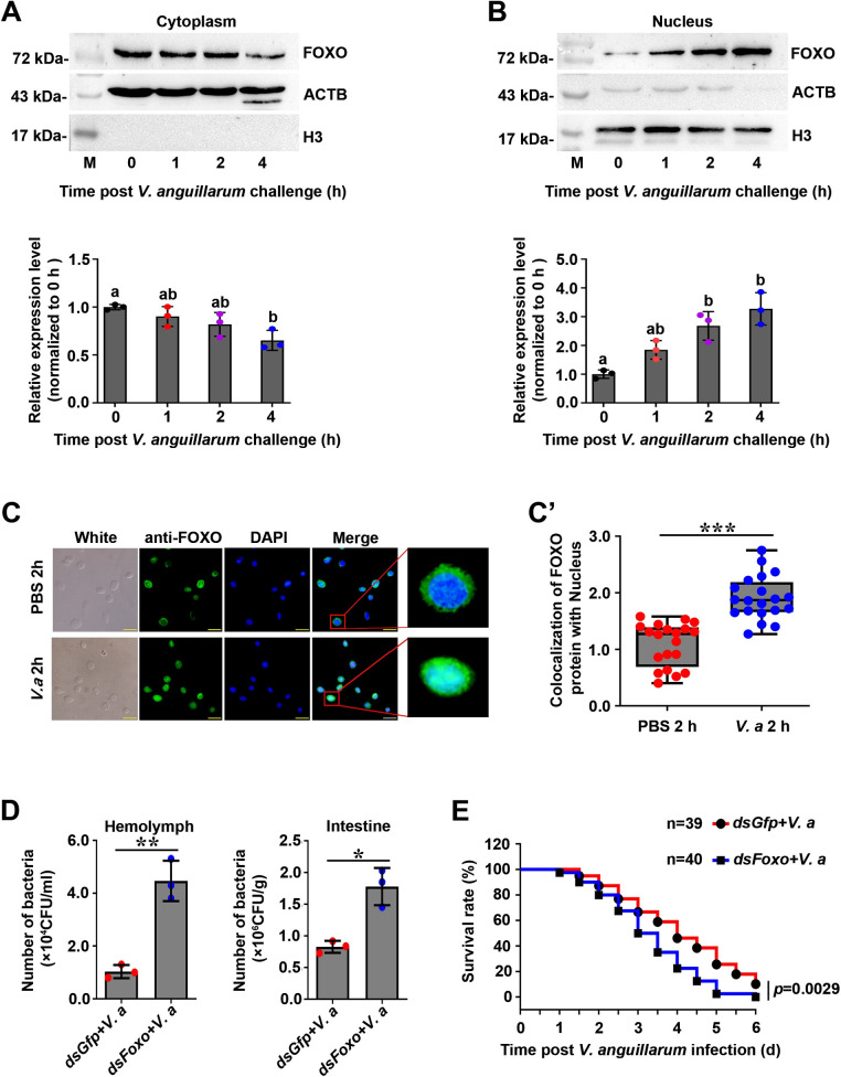
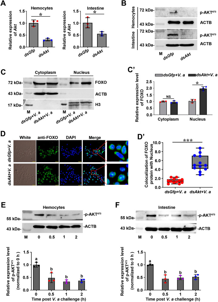
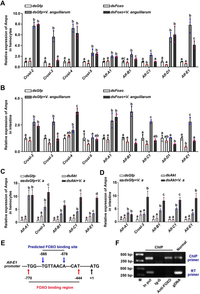
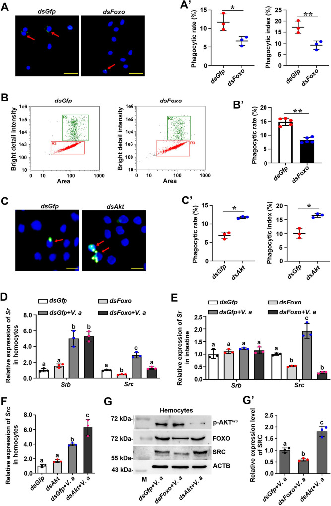
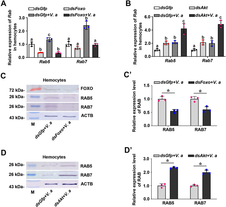
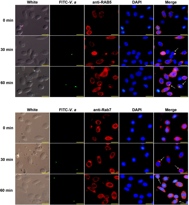
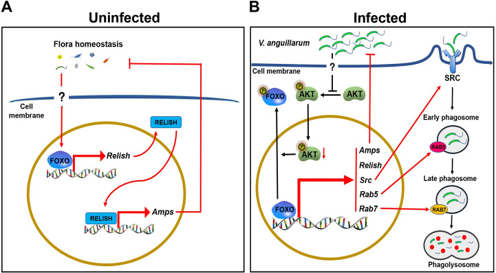
Invertebrates rely on innate immunity, including humoral and cellular immunity, to resist pathogenic infection. Previous studies showed that forkhead box transcription factor O (FOXO) participates in mucosal immune responses of mammals and the gut humoral immune regulation of invertebrates. However, whether FOXO is involved in systemic and cellular immunity regulation in invertebrates remains unknown. In the present study, we identified a FOXO from shrimp (Marsupenaeus japonicus) and found that it was expressed at relatively basal levels in normal shrimp, but was upregulated significantly in shrimp challenged by Vibrio anguillarum. FOXO played a critical role in maintaining hemolymph and intestinal microbiota homeostasis by promoting the expression of Relish, the transcription factor of the immune deficiency (IMD) pathway for expression of antimicrobial peptides (AMPs) in shrimp. We also found that pathogen infection activated FOXO and induced its nuclear translocation by reducing serine/threonine kinase AKT activity. In the nucleus, activated FOXO directly regulated the expression of its target Amp and Relish genes against bacterial infection. Furthermore, FOXO was identified as being involved in cellular immunity by promoting the phagocytosis of hemocytes through upregulating the expression of the phagocytotic receptor scavenger receptor C (Src), and two small GTPases, Rab5 and Rab7, which are related to phagosome trafficking to the lysosome in the cytoplasm. Taken together, our results indicated that FOXO exerts its effects on homeostasis of hemolymph and the enteric microbiota by activating the IMD pathway in normal shrimp, and directly or indirectly promoting AMP expression and enhancing phagocytosis of hemocytes against pathogens in bacteria-infected shrimp. This study revealed the different functions of FOXO in the mucosal (local) and systemic antibacterial immunity of invertebrates.


Conflict of interest statement

The authors have declared that no competing interests exist.

Figures

Fig 1
Fig 1. FOXO was upregulated in shrimp challenged by V. anguillarum.
(A) Recombinant expression of the FH domain of FOXO in E. coli and western blotting to detect FOXO using polyclonal antibodies against FOXO prepared in rabbits. Lane 1, the total proteins of E. coli with Foxo-pGEX4T-1. Lane 2, the total proteins of the bacteria after IPTG induction. Lane 3, purified recombinant FOXO. Lane 4, FOXO in the intestines of untreated shrimp detected using western blotting. M, Protein molecular mass markers. (B) The tissue distribution of Foxo at mRNA (upper panel) and protein (lower panel) levels detected by RT-PCR and western blotting, respectively. ACTB is the abbreviation of β-actin. (C, D) The mRNA expression patterns of Foxo in hemocytes (C) and intestines (D) analyzed by qPCR using geometric mean expression of β-actin and Ef-1α as endogenous control genes. (E, F) The protein expression patterns of FOXO in hemocytes (E) and intestines (F) detected by western blotting. The results of statistical analysis of three replicates were shown in the lower panels. The bands on the western blot were digitalized using ImageJ software by scanning the bands from three independent repeats. Relative expression levels of FOXO/β-actin were expressed as the mean ± SD, and the value of the control shrimp was set as one. Error bars in the figure indicate SDs (three replicates).
Fig 2
Fig 2. FOXO participates in the intestinal (local) and systemic antibacterial immune responses.
(A, B) Efficiency of Foxo-RNAi in hemocytes (A) and intestines (B) of shrimp, as determined using qPCR (A) and western blotting (B). The qPCR date analysis using geometric mean expression of β-actin and Ef-1α as endogenous control genes. Error bars show SDs. (C) Detection of hemolymph bacteria of Foxo-knockdown shrimp by solid LB culture (three repeats); dsGfp injection was used as the control. The dilution ratio was 1:10. (D). The number of bacteria in the hemolymph and intestines in Foxo-knockdown shrimp and dsGfp-injection shrimp without bacterial challenge. (E). The survival rate of Foxo-RNAi shrimp without bacterial challenge. dsGfp injection was used as the control. (F). The survival rate of Foxo-RNAi shrimp treated with antibiotics. dsGfp injection was used as control. Normal: wild-type shrimp not treated with antibiotics.
Fig 3
Fig 3. A basal amount of FOXO translocates into the nuclei of hemocytes and intestine cells and promotes Relish expression and subsequently activates the IMD pathway to regulate hemolymph and intestinal microbiota homeostasis under normal conditions.
(A) Immunocytochemistry was used to detect the subcellular distribution of FOXO in hemocytes of normal shrimp. Scale bar = 20 μm. Cy: Cytoplasm; Nu: Nucleus. (B) Western blotting was used to detect the subcellular distribution of FOXO in intestine cells from normal shrimp and V. anguillarum infected shrimp. (C) The expression of Relish at mRNA level was detected in hemocytes of shrimp after knockdown of Foxo by qPCR using geometric mean expression of β-actin and Ef-1α as endogenous control genes. (D) The expression of Relish at the mRNA level in the intestines of shrimp after knockdown of Foxo was detected by qPCR using geometric mean expression of β-actin and Ef-1α as endogenous control genes. (E) The RELISH level was detected using western blotting in shrimp after knockdown of Foxo. (E’) Three replicates of panel E were digitalized using ImageJ software. (F) The bacterial load of hemolymph and intestines in shrimp 24 h post antibiotics treatment. (G) FOXO in nucleus was detected using western blotting in hemocytes and intestines of shrimp 24 h post treatment with antibiotics. (H) RELISH in nucleus was detected using western blotting in shrimp hemocytes and intestines at 24 h post treatment with antibiotics. (I, J) The expression of Amps in hemocytes (I) and intestines (J) of germ-free shrimp determined by qPCR using β-actin and Ef-1α as an internal reference gene. Normal shrimp (H2O injection) was used as the control. The data were analyzed statistically using the Mann-Whitney U test. All the error bars in the figure indicate SDs.
Fig 4
Fig 4. FOXO translocated into nucleus in shrimp challenged by V. anguillarum.
(A, B) The FOXO distribution patterns in the cytoplasm (A) and nucleus (B) of intestine cells of shrimp challenged by bacteria, as analyzed using western blotting (lower panels of A and B). The western blotting bands were digitalized using ImageJ software by scanning the bands from three repeats. The relative expression levels of FOXO/β-actin or FOXO/Histone 3 were expressed as the mean ± SD, and the value of the control shrimp was set as one. (C) The nuclear translocation of FOXO in hemocytes of shrimp 2 h post V. anguillarum challenge was detected using fluorescent immunocytochemical assays. PBS injection was used as control. Scale bar = 20 μm. (C’) Statistic analysis of panel C. the colocalization of FOXO and DAPI-stained nuclei in hemocytes was analyzed by WCIF ImageJ software. (D) The bacterial number in the hemolymph and intestines of Foxo-knockdown shrimp followed V. anguillarum injection; dsGfp-injection in shrimp following bacterial injection was used as control. (E) The survival rate of Foxo-RNAi shrimp infected by V. anguillarum. dsGfp injection was used as the control.
Fig 5
Fig 5. Pathogen infection decreased AKT activity, which promoted FOXO nuclear translocation in hemocytes and enteric cells of shrimp.
(A, B) Efficiency of Akt-RNAi in the hemocytes and intestines of shrimp, as determined by qPCR using geometric mean expression of β-actin and Ef-1α as endogenous control genes (A) and western blotting (B). (C) The amount of FOXO in the cytoplasm and nucleus of intestines from Akt-knockdown shrimp analyzed by western blotting. dsGfp was used as control. (C’) The statistical analysis of panel (C). ImageJ software was used to digitalized the bands by scanning three-repeat pictures. (D) The nuclear translocation of FOXO in hemocytes of Akt-knockdown shrimp was detected 2 h post V. anguillarum challenge using a fluorescent immunocytochemical assay. dsGfp was used as control. Scale bar = 20 μm. (D’) Statistical analysis of (D). WCIF ImageJ software was used to analyze the co-localization by fluorescence intensity ratio of anti-FOXO (green) and DAPI-stained nucleus (blue) in hemocytes after fixed the exposure value. (E, F) Content change of phosphorylated AKT (p-AKT) in hemocytes (E) and intestines (F) of shrimp challenged by V. anguillarum. The lower panels are digitalized figures of (E) and (F) based on bands of western blotting and statistical analysis, respectively.
Fig 6
Fig 6. FOXO performs its antibacterial roles in shrimp by regulating the expression of AMPs.
(A, B) The expression of Amps in hemocytes (A) and intestines (B) of Foxo-knockdown shrimp, as determined by qPCR using geometric mean expression of β-actin and Ef-1α as endogenous control genes; dsGfp injection in shrimp was used as the control. The data were analyzed statistically using the Mann-Whiteny U test. (C, D) The expression of Amps in hemocytes (C) and intestines (D) of Akt-knockdown shrimp was analyzed by qPCR using geometric mean expression of β-actin and Ef-1α as endogenous control genes; dsGfp injection in shrimp was used as the control. Significant differences were analyzed using one-way ANOVA. (E) Schematic diagram of the predicted binding sites of FOXO on the Alf-E1 promoter. (F) ChIP and RT-PCR were used to detect FOXO binding to the Alf-E1 promoter sequence using primers (Alf-E1-F and Alf-E1-R, Table 1). RT-PCR was also used to amplify encoding region of Alf-E1 with RT-PCR primers (Alf-E1-RT-F and Alf-E1-RT-R. Table 1) using the ChIP obtained DNA as template. IgG antibody was used as control, and the normal genomic DNA amplified fragment was used to confirm the target band.
Fig 7
Fig 7. FOXO enhances hemocyte phagocytosis of pathogens in shrimp by promoting SRC expression.
(A) Hemocyte phagocytosis of V. anguillarum observed using a fluorescent immunocytochemical assay under a fluorescence microscope. V. anguillarum was labeled with FITC (green) and cell nuclei were stained with DAPI (blue). The red arrow indicates the phagocytic hemocytes. Scale bar = 20 μm. (A’) Statistical analysis of the phagocytic rate and phagocytic index in Foxo-knockdown shrimp. The dsGfp injection group was used as the control. Five hundred hemocytes were counted under the fluorescence microscope in each experiment. The data were analyzed statistically using Student’s t-test. (B) Bacterial phagocytosis by hemocytes analyzed using flow cytometry. The phagocytosed hemocytes containing bacteria (R2) were separated from non-phagocytosed hemocytes (R3) based on fluorescence signal of labeled bacteria in hemocytes using IDEAS Application v6.0 software. (B’) The phagocytic rate of hemocytes based on the flow cytometry data. Three thousand hemocytes were counted for each analysis and three repetitions were carried out. The dsGfp group was used as the control. (C) Hemocyte phagocytosis of V. anguillarum in Akt-knockdown shrimp observe under the fluorescence microscope. Scale bar = 10 μm. (C’) Statistical analysis of phagocytic rate and phagocytic index based on data in panel (C). (D) The mRNA expression level of Srb and Src in hemocytes from Foxo-knockdown shrimp analyzed by qPCR using geometric mean expression of β-actin and Ef-1α as endogenous control genes. dsGfp injection was used as the control. (E) The mRNA expression of Srb and Src in intestines of Foxo-RNAi shrimp analyzed by qPCR using geometric mean expression of β-actin and Ef-1α as endogenous control genes. (F) The mRNA expression of Src in hemocytes of Akt-RNAi shrimp analyzed by qPCR using geometric mean expression of β-actin and Ef-1α as endogenous control genes. (G) The protein level of SRC in hemocytes of Foxo-RNAi or Akt-RNAi shrimp, as analyzed using western blotting. dsGfp-injection used as control. (G’) The statistical analysis based on data of three replicates of panel (G).
Fig 8
Fig 8. RAB5 and RAB7 are involved in FOXO-mediated phagocytosis of hemocytes in shrimp.
(A, B) qPCR was used to analyze the mRNA expression levels of Rab5 and Rab7 in the hemocytes of Foxo-knockdown shrimp (A) and Akt-knockdown shrimp (B) with and without bacterial infection. The qPCR used geometric mean expression of β-actin and Ef-1α as endogenous control genes. (C) The protein levels of the RAB5 and RAB7 in hemocytes of Foxo-knockdown shrimp at 2 h post V. anguillarum challenge determined using western blotting. (C’) The statistical analysis based on data of three replicates of panel (C). (D) The protein levels of the RAB5 and RAB7 in hemocytes of Akt-knockdown shrimp at 2 h post V. anguillarum challenge determined using western blotting. (D’) The statistical analysis based on data of three replicates of panel (D).
Fig 9
Fig 9. RAB5 and RAB7 co-localize with V. anguillarum.
The co-localization of RAB5 and RAB7 with FITC labeled-V. anguillarum in shrimp hemocytes, as analyzed using a fluorescent immunocytochemical assay. The bacteria were labeled with FITC (green) and injected into shrimp. Hemocytes were collected from five shrimp at 30 min and 1 h after bacterial injection and incubated with anti-RAB5 or anti-RAB7 antibodies. The secondary antibody was antirabbit IgG Alexa-546 (red). Nuclei were stained with DAPI (blue). Co-localization in hemocytes is indicated by white arrows. Scale bar = 10 μm.
Fig 10
Fig 10. Schematic representation of FOXO involvement in local and systemic innate immunity in shrimp.
(A) In uninfected conditions, FOXO maintains the homeostasis of the hemolymph and the intestinal microbiota by promoting Relish and subsequently AMP expression. (B) In infected conditions, AKT phosphorylation is decreased by pathogen infection, resulting in increasing of FOXO content in the nucleus. FOXO directly promotes the expression of Relish and AMPs to eliminate invasive pathogens; on the other hand, FOXO promotes the phagocytosis of pathogens through the SRC-mediated phagocytosis pathway.

Similar articles

Cited by

References

    1. Calnan DR, Brunet A. The FoxO code. Oncogene. 2008;27(16):2276–2288. 10.1038/onc.2008.21 . - DOI - PubMed
    1. Tran H, Brunet A, Grenier JM, Datta SR, Fornace AJ, DiStefano PS, et al.. DNA repair pathway stimulated by the forkhead transcription factor FOXO3a through the Gadd45 protein. Science. 2002;296(5567):530–534. 10.1126/science.1068712 . - DOI - PubMed
    1. Martins R, Lithgow GJ, Link W. Long live FOXO: unraveling the role of FOXO proteins in aging and longevity. Aging Cell. 2016;15(2):196–207. 10.1111/acel.12427 . - DOI - PMC - PubMed
    1. Vogt PK, Jiang H, Aoki M. Triple layer control-phosphorylation, acetylation and ubiquitination of FOXO proteins. Cell Cycle. 2005;4(7):908–913. 10.4161/cc.4.7.1796 . - DOI - PubMed
    1. Daitoku H, Sakamaki J, Fukamizu A. Regulation of FoxO transcription factors by acetylation and protein-protein interactions. Bba-Mol Cell Res. 2011;1813(11):1954–1960. 10.1016/j.bbamcr.2011.03.001 . - DOI - PubMed

Publication types

Substances

Supplementary concepts

LinkOut - more resources