

The frontier of live tissue imaging across space and time
- PMID: 33798422
- PMCID: PMC8034393
- DOI: 10.1016/j.stem.2021.02.010
The frontier of live tissue imaging across space and time
Abstract
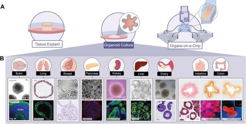
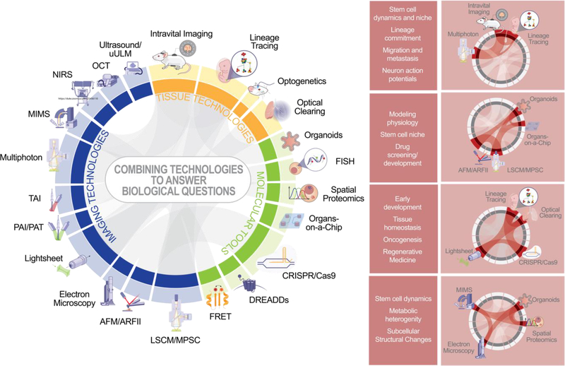
What you see is what you get-imaging techniques have long been essential for visualization and understanding of tissue development, homeostasis, and regeneration, which are driven by stem cell self-renewal and differentiation. Advances in molecular and tissue modeling techniques in the last decade are providing new imaging modalities to explore tissue heterogeneity and plasticity. Here we describe current state-of-the-art imaging modalities for tissue research at multiple scales, with a focus on explaining key tradeoffs such as spatial resolution, penetration depth, capture time/frequency, and moieties. We explore emerging tissue modeling and molecular tools that improve resolution, specificity, and throughput.
Copyright © 2021 Elsevier Inc. All rights reserved.
Conflict of interest statement
Declaration of interests The authors declare no competing interests.
Figures


References
-
- Baptista PM, Siddiqui MM, Lozier G, Rodriguez SR, Atala A, and Soker S (2011). The use of whole organ decellularization for the generation of a vascularized liver organoid. Hepatology (Baltimore, Md) 53, 604–617. - PubMed
Publication types
MeSH terms
Grants and funding
LinkOut - more resources
Full Text Sources
Other Literature Sources
