Review
. 2021 Aug 6;433(16):166968.
doi: 10.1016/j.jmb.2021.166968. Epub 2021 Mar 30.

Molecular Mechanisms for Bacterial Potassium Homeostasis

Affiliations
Review

Molecular Mechanisms for Bacterial Potassium Homeostasis

Janina Stautz et al. J Mol Biol. .

Abstract

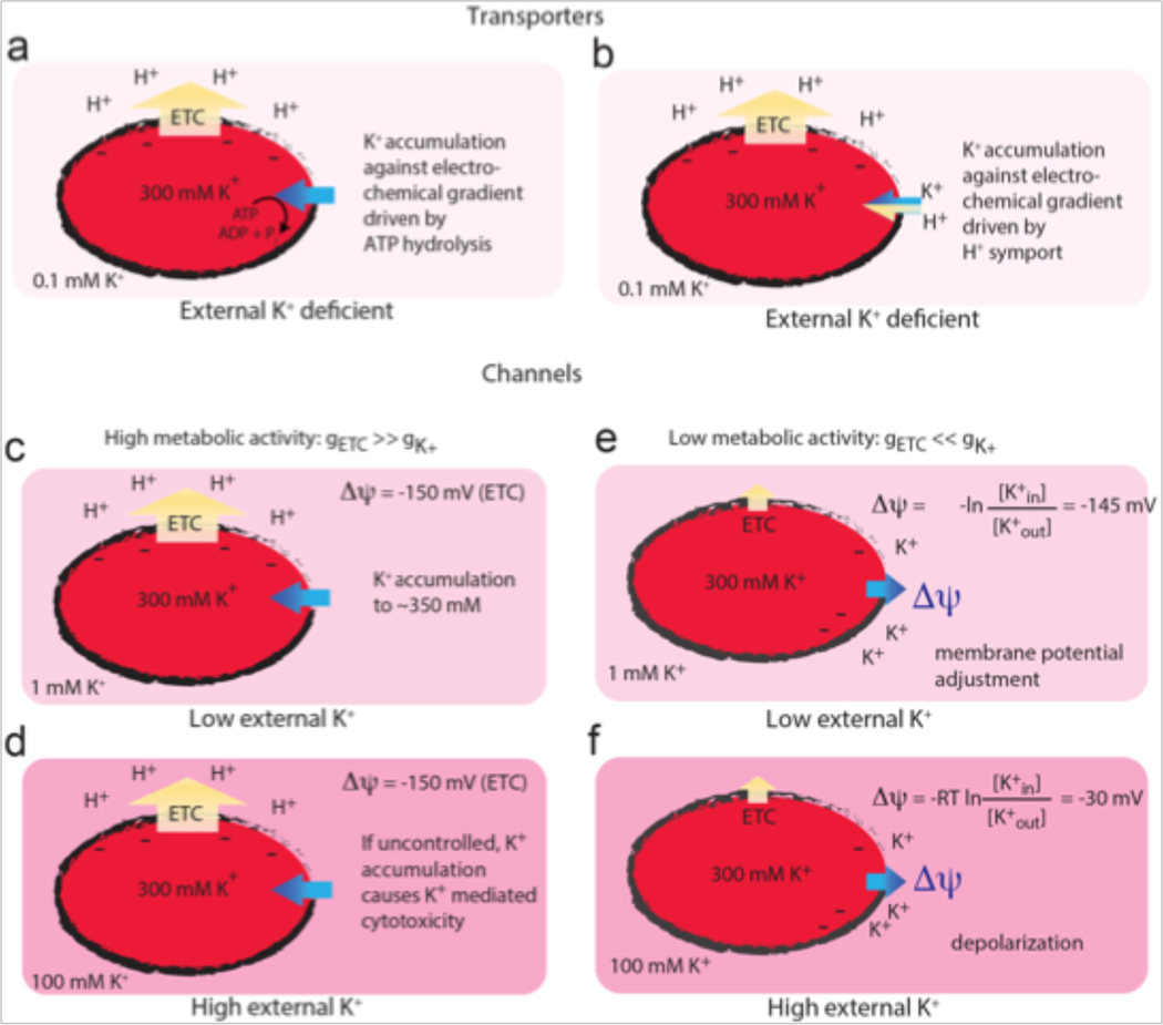
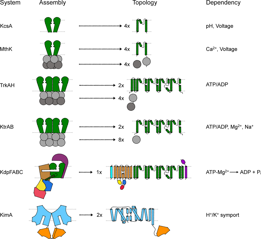
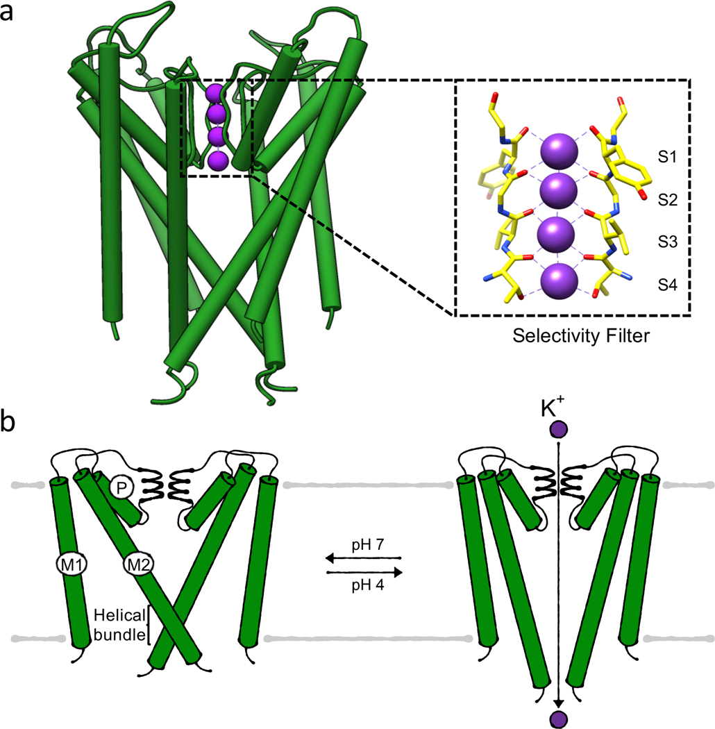
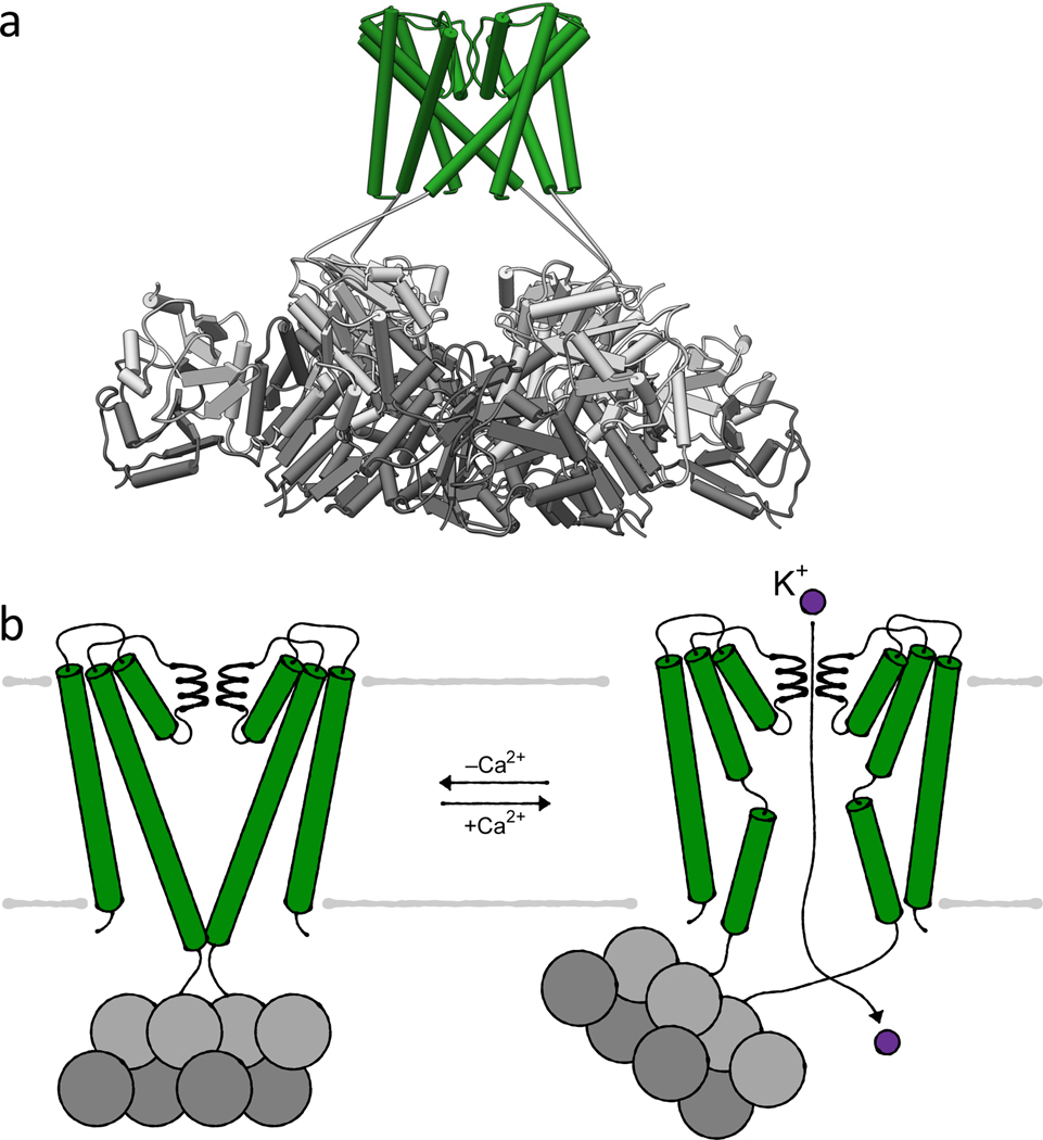
Potassium ion homeostasis is essential for bacterial survival, playing roles in osmoregulation, pH homeostasis, regulation of protein synthesis, enzyme activation, membrane potential adjustment and electrical signaling. To accomplish such diverse physiological tasks, it is not surprising that a single bacterium typically encodes several potassium uptake and release systems. To understand the role each individual protein fulfills and how these proteins work in concert, it is important to identify the molecular details of their function. One needs to understand whether the systems transport ions actively or passively, and what mechanisms or ligands lead to the activation or inactivation of individual systems. Combining mechanistic information with knowledge about the physiology under different stress situations, such as osmostress, pH stress or nutrient limitation, one can identify the task of each system and deduce how they are coordinated with each other. By reviewing the general principles of bacterial membrane physiology and describing the molecular architecture and function of several bacterial K+-transporting systems, we aim to provide a framework for microbiologists studying bacterial potassium homeostasis and the many K+-translocating systems that are still poorly understood.

Keywords: K(+) transport; bacterial physiology; membrane potential; principles of K(+) transporters and channels; structural biology.


Conflict of interest statement

Declaration of Competing Interest The authors declare that they have no known competing financial interests or personal relationships that could have appeared to influence the work reported in this paper.

Figures

Figure 1.
Figure 1.. Simplified scenarios showing the interplay between bacterial metabolism, K+ chemical gradient, and membrane potential.
Yellow arrows indicate proton movement and blue arrows indicate K+ movement. Darker shades of pink indicate higher K+ concentrations. Transporters (a, b) utilize energy derived from ATP hydrolysis (a) or the proton motive force (b) to accumulate intracellular K+ against its electrochemical gradient. Channels (c–f) facilitate the thermodynamically favorable movement of K+ down its electrochemical gradient. In the first case (c, d), an inside-negative membrane potential is established by the proton gradient due to the high relative permeability of protons from the electron transport chain (gETC). K+ can be accumulated to physiological levels in the cytoplasm even at low external concentrations (c), but the accumulation must be regulated to prevent uncontrolled K+ uptake causing K+-mediated cytotoxicity at higher external K+ concentrations (d). When the relative permeability of K+ (gK+) surpasses that of protons due to a decrease in metabolic activity or K+ channel opening (e,f), the degree of membrane depolarization is determined by the external K+ concentration.
Figure 2.
Figure 2.. Assembly and topology of bacterial K+-translocating systems.
Overview of the assembly of the different systems discussed. Channel-like subunits/domains are shown in green, RCK subunits/domains in gray, the P-type ATPase KdpB in beige with blue, red and yellow, KdpC in purple, KdpF in cyan, the LeuT-fold domain in light blue, and the adenylyltransferase-like domain in orange. The topologies provide more molecular detail of the transmembrane domains. Characteristic are the M1PM2 domains of the channel-like proteins (green), the insertion of the cytosolic N, P and A domains (red, blue, and yellow, respectively) into loops of the transmembrane domain of KdpB (beige) and the 5+5 inverted repeat of the LeuT-fold domain (light blue) of KimA.
Figure 3.
Figure 3.. Gating of potassium channel KcsA.
(a) Structure (PDB 1J95) of the membrane-inserted domains of KcsA in its conductive, closed state with the inset highlighting the selectivity filter, (b) Bundle gate mechanism. Cartoons of closed, conductive and open, conductive states, respectively. For simplicity only two M1PM2 domains are shown.
Figure 4.
Figure 4.. Ca2+-gating by K+ channel MthK.
(a) Structure (PDB 6U6H) of MthK in its closed, conductive state in the absence of Ca2+ with the inset highlighting the selectivity filter. (b) Helical bundle gating controlled by the cytosolic RCK domains (tethered RCK domains light grey, soluble RCK domains dark grey). Cartoons of closed, conductive and open, conductive state, respectively. For simplicity only two M1PM2 domains are shown.
Figure 5.
Figure 5.. Gating of nucleotide- and cation-gated two-pored K+ channels KtrAB and TrkAH.
(a) Structure (PDB 4J7C) of KtrAB in its ATP-bound (but still closed) state with the inset highlighting the selectivity filter, (b) Intramembrane loop gating of KtrAB controlled by the cytosolic RCK subunits. Cartoons of ADP-bound, closed and ATP-bound, open state, respectively, (c) Structure (PDB 4J9U) of TrkAH in its ADP-bound, closed state, (d) Intramembrane loop gating of TrkAH controlled by the cytosolic RCK subunits (RCK2 light grey, RCK1 dark grey). Cartoons of ADP-bound, closed and ATP- bound, open state, respectively.
Figure 6.
Figure 6.. Ion transport models through K+ pump KdpFABC.
(a) Structure (PDB 5MRW) of KdpFABC in its E1 state. (b) Transport cycle of the coupling helix model. (c) Transport cycle of the intersubunit transport model.
Figure 7.
Figure 7.. K+/H+ symport by KimA.
(a) Structure (PDB 6S3K) of KimA in its inward, occluded state, rainbow-colored from N terminus (blue) to C terminus (red). (b) Cartoon of the alternating access transport by KimA.

References

    1. Benarroch JM, Asally M. The Microbiologist’s Guide to Membrane Potential Dynamics. Trends Microbiol. 2020;28:304–14. - PubMed
    1. Beagle SD, Lockless SW. Unappreciated Roles for K(+) Channels in Bacterial Physiology. Trends Microbiol. 2020. - PMC - PubMed
    1. Zhou M, Morais-Cabral JH, Mann S, MacKinnon R. Potassium channel receptor site for the inactivation gate and quaternary amine inhibitors. Nature. 2001;411:657–61. - PubMed
    1. Fan C, Sukomon N, Flood E, Rheinberger J, Allen TW, Nimigean CM. Ball-and-chain inactivation in a calcium-gated potassium channel. Nature. 2020;580:288–93. - PMC - PubMed
    1. Vieira-Pires RS, Szollosi A, Morais-Cabral JH. The structure of the KtrAB potassium transporter. Nature. 2013;496:323–8. - PubMed

Publication types

LinkOut - more resources