. 2021 Jan-Jun:296:100615.
doi: 10.1016/j.jbc.2021.100615. Epub 2021 Mar 30.

IL-4Rα signaling by CD8α+ dendritic cells contributes to cerebral malaria by enhancing inflammatory, Th1, and cytotoxic CD8+ T cell responses

Affiliations

IL-4Rα signaling by CD8α+ dendritic cells contributes to cerebral malaria by enhancing inflammatory, Th1, and cytotoxic CD8+ T cell responses

Xianzhu Wu et al. J Biol Chem. 2021 Jan-Jun.

Abstract

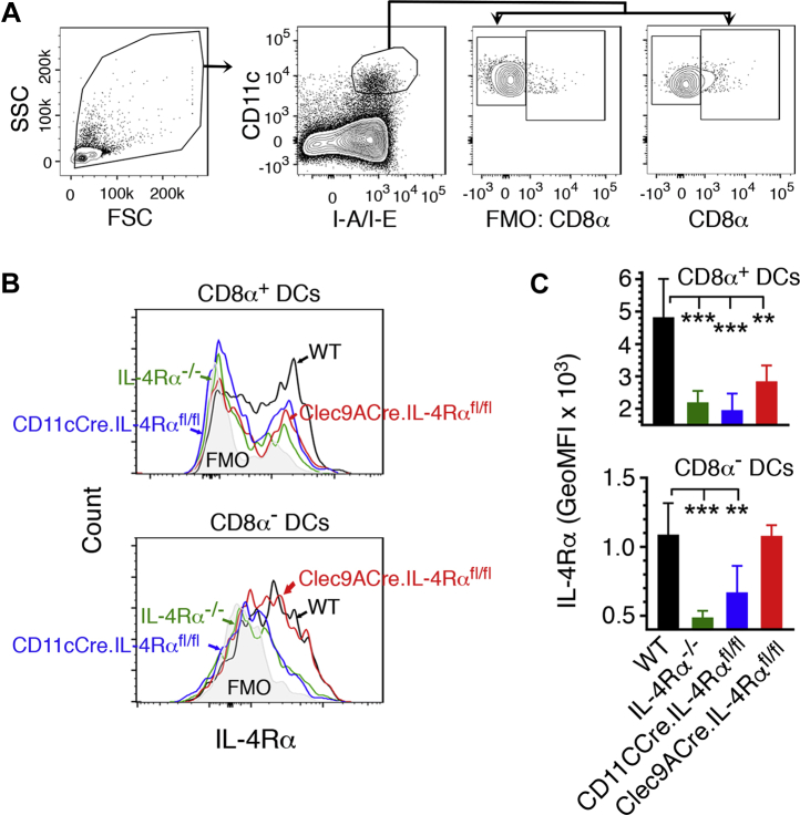
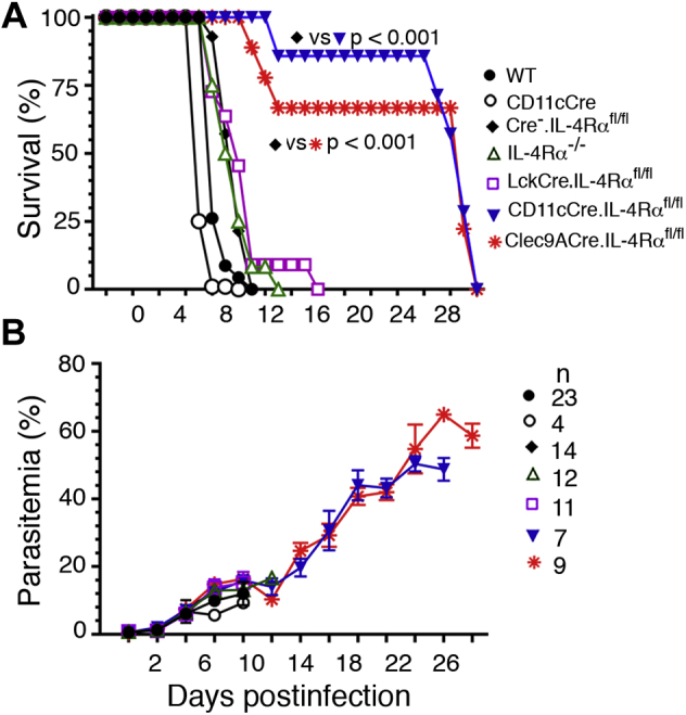
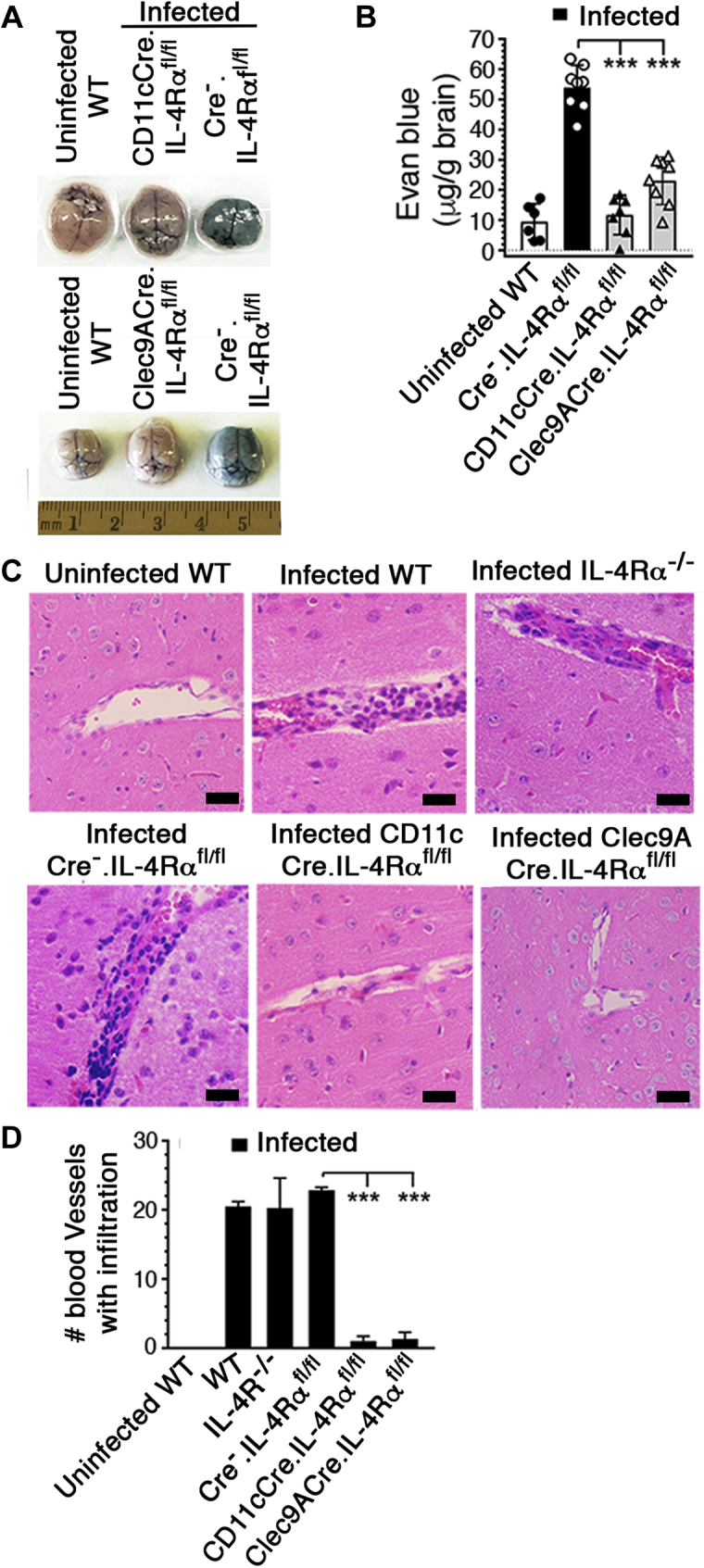
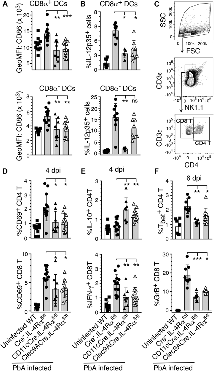
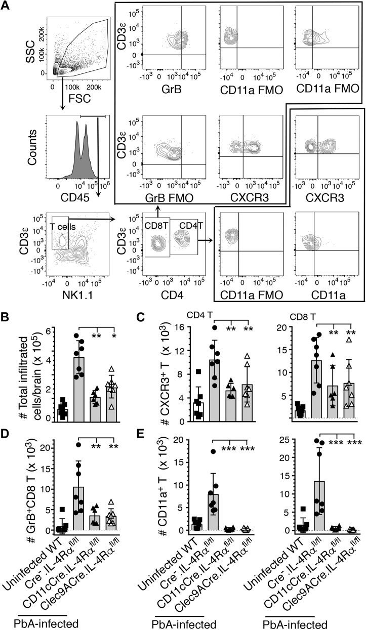
Persistent high levels of proinflammatory and Th1 responses contribute to cerebral malaria (CM). Suppression of inflammatory responses and promotion of Th2 responses prevent pathogenesis. IL-4 commonly promotes Th2 responses and inhibits inflammatory and Th1 responses. Therefore, IL-4 is widely considered as a beneficial cytokine via its Th2-promoting role that is predicted to provide protection against severe malaria by inhibiting inflammatory responses. However, IL-4 may also induce inflammatory responses, as the result of IL-4 action depends on the timing and levels of its production and the tissue environment in which it is produced. Recently, we showed that dendritic cells (DCs) produce IL-4 early during malaria infection in response to a parasite protein and that this IL-4 response may contribute to severe malaria. However, the mechanism by which IL-4 produced by DCs contributing to lethal malaria is unknown. Using Plasmodium berghei ANKA-infected C57BL/6 mice, a CM model, we show here that mice lacking IL-4Rα only in CD8α+ DCs are protected against CM pathogenesis and survive, whereas WT mice develop CM and die. Compared with WT mice, mice lacking IL-4Rα in CD11c+ or CD8α+ DCs showed reduced inflammatory responses leading to decreased Th1 and cytotoxic CD8+ T cell responses, lower infiltration of CD8+ T cells to the brain, and negligible brain pathology. The novel results presented here reveal a paradoxical role of IL-4Rα signaling in CM pathogenesis that promotes CD8α+ DC-mediated inflammatory responses that generate damaging Th1 and cytotoxic CD8+ T cell responses.

Keywords: IL-4Rα; cerebral malaria; cytotoxic T cells; endothelial damage; infiltration to brain; inflammatory cytokines.


Conflict of interest statement

Conflict of interest The authors declare that they have no conflicts of interest with the contents of this article.

Figures

Figure 1
Figure 1
Flow cytometry analysis of IL-4Rα expression in DCs of the conditional IL-4Rα knockout mice. Spleen cells of uninfected WT and IL-4Rα−/−, CD11cCre.IL-4Rαfl/fl, and Clec9ACre.IL-4Rαfl/fl mice (n = 4–5) were stained with dye-conjugated antibodies against surface markers and anti-mouse IL-4Rα antibody. A, shows gating of CD8α+ and CD8α DCs. B, histogram showing the IL-4Rα expression of a representative mouse from each group on gated CD8α+ and CD8α DCs. C, shows plot of mean of IL-4Rα expression. Error bars indicate GeoMFI ±SD. ∗∗p < 0.01, ∗∗∗p < 0.001.
Figure 2
Figure 2
IL-4Rα signaling in cDCs or CD8α+DCs contributes to the development of ECM and mortality.A and B, mice were infected with 2 × 105 PbA IRBCs. Shown are survival (A) and parasitemia (B) in CD11cCre.IL-4Rαfl/fl mice, Clec9ACre.IL-4Rαfl/fl mice, and LckCre.IL-4Rαfl/fl mice. Cohoused WT, CD11cCre, and IL-4Rα−/− mice, and the littermate WT (Cre.IL-4Rαfl/fl) mice obtained by crossing CD11cCre or Clec9ACre mice with IL-4Rαfl/fl mice were used as controls. Error bars indicate mean values of data ±SD.
Figure 3
Figure 3
IL-4Rα signaling in cDCs or CD8α+DCs contributes to brain pathology.AD, mice were infected with 2 × 105 PbA IRBCs. At 6 dpi, Evans blue was injected into mice (n = 6–8/group) via tail vein. After 2 h, brains were harvested and photographed, and the amounts of Evans blue entered into the brains were assessed as outlined under Experimental procedures. A and B, photographs of the brain of a representative mouse in each group; uninfected WT, and infected CD11cCre.IL-4Rαfl/fl, Clec9ACre.IL-4Rαfl/fl, and Cre.IL-4Rαfl/fl mice (A), and the amounts of Evans blue entered into the brains (B). Mean data ±SD plotted. Panel A, the numbers in the horizontal scale represent cm. C, at 6 dpi, 6-μm brain sections of the indicated mice (n = 3–4/group) were assessed by H&E staining and 40× images of a representative mouse in each group are shown. In each image, the black bar is 10 μm. Images of uninfected CD11cCre.IL-4Rαfl/fl and Clec9ACre.IL-4Rαfl/fl mice were similar to those of uninfected WT mice. D, 20× microscopic views of the brain sections of mice were assessed under light microscopy and the numbers of blood vessels having large numbers of immune cell filtration were counted and mean values plotted. Error bars indicate mean values of data ±SD plotted. ∗∗∗p ≤ 0.001.
Figure 4
Figure 4
IL-4Rα signaling in cDCs or CD8α+DCs contributes to inflammatory responses by DCs and T cells of PbA-infected mice. The indicated mice (n = 8–12/group) were infected with 2 × 105 PbA IRBCs. AF, spleen cells from the infected and uninfected WT control mice were analyzed by flow cytometry. A and B, the expression of costimulatory molecule CD86 at 4 dpi (A) and the percentages of IL-12p70+ at 5 dpi (B) in CD8α+ DCs and CD8α DCs. C, gating strategy of CD4+ T and CD8+ T cells. D, the percentages of CD69 in the CD4+ T and CD8+ T cells at 4 dpi. E, the percentages of IL-10+CD4+ T and IFN-γ+CD8+ T cells at 4 dpi. F, the percentages of Tbet+ CD4+ T and granzyme B+CD8+ T cells at 6 dpi. Error bars indicate mean values ±SD. ns, not significant, ∗p ≤ 0.05, ∗∗p ≤ 0.01, ∗∗∗p ≤ 0.001.
Figure 5
Figure 5
IL-4Rα signaling in cDCs or CD8α+DCs contributes to infiltration of immune cells, including pathogenic CD8+T cells, to the brain.AD, the indicated mice (n = 6–7/group) were infected with 2 × 105 PbA IRBCs. At 6 dpi, immune cells in the brains were isolated, stained with dye-conjugated antibodies, and analyzed by flow cytometry. A, cell gating strategy. BE, the numbers of immune cells infiltrated into the brains; total immune cells (B), CXCR3+ CD4+ T and CD8+ T cells (C), granzyme B-expressing CD8+ T cells (D), and CD11a/LFA-1+ CD4+ T and CD8+ T cells. (E). Error bars indicate mean values ±SD. ∗p ≤ 0.05, ∗∗p ≤ 0.01, ∗∗∗p ≤ 0.001.

References

    1. de Souza M.C., Pádua T.A., das Graças Henriques M. Multiple organ dysfunction during severe malaria: The role of the inflammatory response. In: Rodriguez-Marales A.J., editor. Current Topics in Malaria. InTech Publishing; Rijeka, Croatia: 2016.
    1. Storm J., Craig A.G. Pathogenesis of cerebral malaria-inflammation and cytoadherence. Front. Cell. Infect. Microbiol. 2014;4:1–8. - PMC - PubMed
    1. Hunt N.H., Grau G.E. Cytokines: Accelerators and brakes in the pathogenesis of cerebral malaria. Trends Immunol. 2003;24:491–499. - PubMed
    1. Amorim K.N., Chagas D.C., Sulczewski F.B., Boscardin S.B. Dendritic cells and their multiple roles during malaria infection. J. Immunol. Res. 2016;2016 - PMC - PubMed
    1. Deroost K., Pham T.-T., Opdenakker G., Van den Steen P.E. The immunological balance between host and parasite in malaria. FEMS Microbiol. Rev. 2015;40:208–257. - PubMed

Publication types

MeSH terms

LinkOut - more resources