. 2021 Mar 11;26(6):1527.
doi: 10.3390/molecules26061527.

Kaempferia parviflora Extract Alleviated Rat Arthritis, Exerted Chondroprotective Properties In Vitro, and Reduced Expression of Genes Associated with Inflammatory Arthritis

Affiliations

Kaempferia parviflora Extract Alleviated Rat Arthritis, Exerted Chondroprotective Properties In Vitro, and Reduced Expression of Genes Associated with Inflammatory Arthritis

Siriwan Ongchai et al. Molecules. .

Abstract

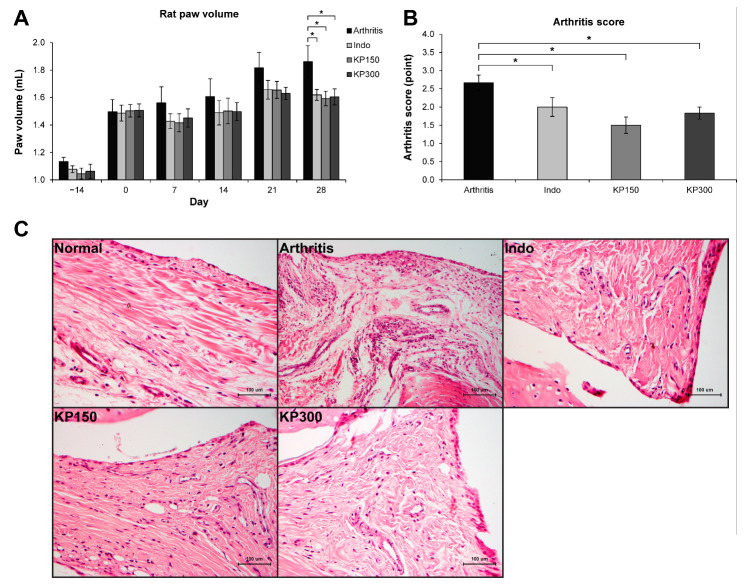
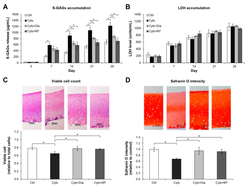
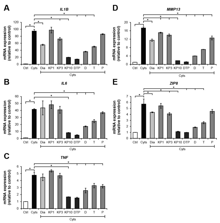
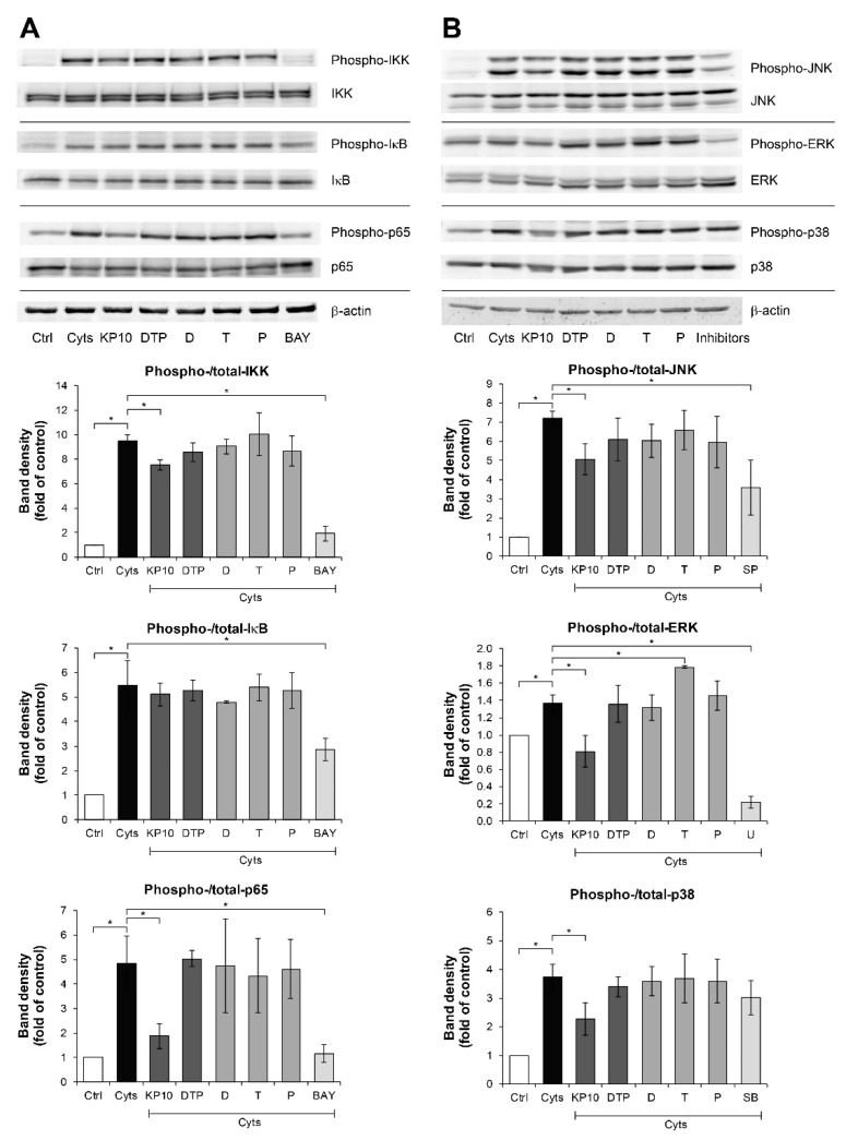
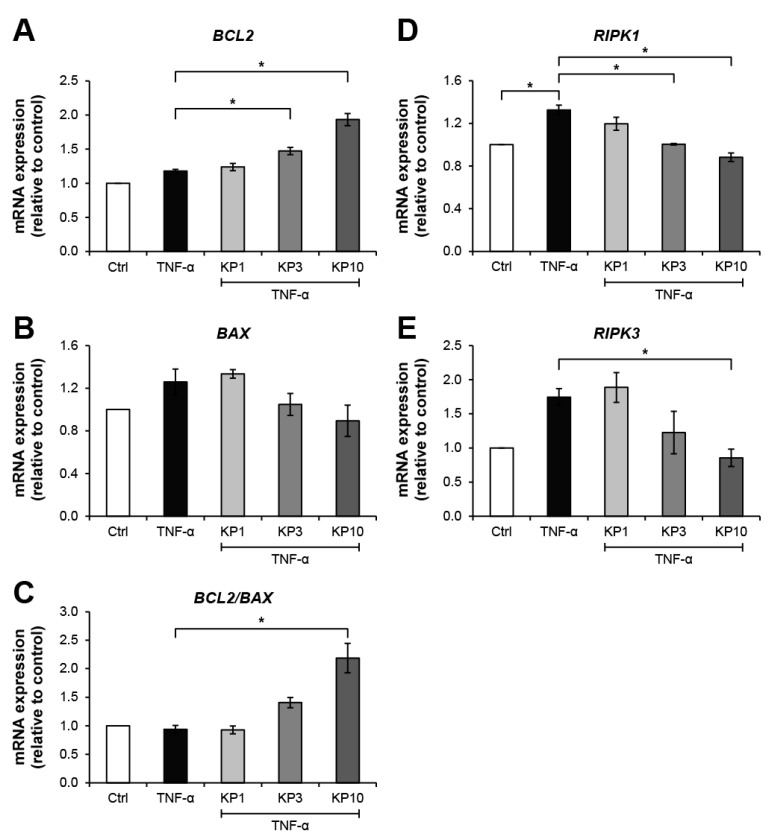
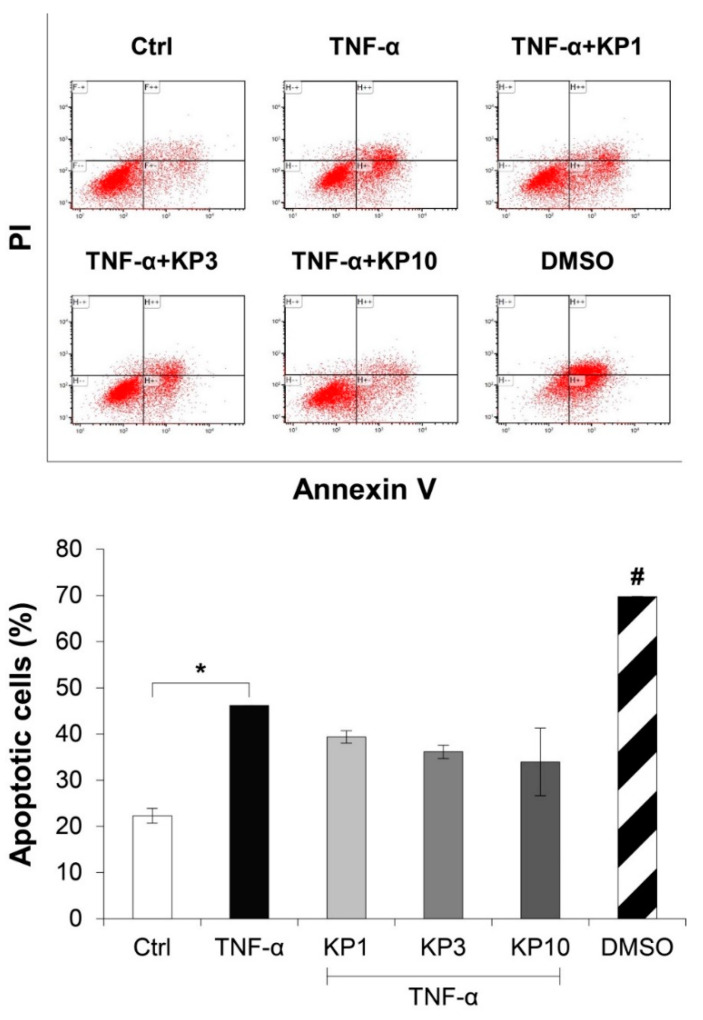
Kaempferia parviflora Wall. ex Baker (KP) has been reported to attenuate cartilage destruction in rat model of osteoarthritis. Previously, we demonstrated that KP rhizome extract and its active components effectively suppressed mechanisms associated with RA in SW982 cells. Here, we further evaluated the anti-arthritis potential of KP extract by using multi-level models, including a complete Freund's adjuvant-induced arthritis and a cartilage explant culture model, and to investigate the effects of KP extract and its major components on related gene expressions and underlying mechanisms within cells. In arthritis rats, the KP extract reduced arthritis indexes, with no significant changes in biological parameters. In the cartilage explant model, the KP extract exerted chondroprotective potential by suppressing sulfated glycosaminoglycans release while preserving high accumulation of proteoglycans. In human chondrocyte cell line, a mixture of the major components equal to their amounts in KP extract showed strong suppression the expression of genes-associated inflammatory joint disease similar to that of the extract. Additionally, KP extract significantly suppressed NF-κB and MAPK signaling pathways. The suppressing expression of necroptosis genes and promoted anti-apoptosis were also found. Collectively, these results provided supportive evidence of the anti-arthritis properties of KP extract, which are associated with its three major components.

Keywords: Kaempferia parviflora; anti-arthritis; anti-inflammation; chondroprotection; inflammatory joint disease.


Conflict of interest statement

The authors declare no conflict of interest. The funders had no role in the design of the study; in the collection, analyses, or interpretation of data; in the writing of the manuscript, or in the decision to publish the results.

Figures

Figure 1
Figure 1
KP extract attenuates CFA-induced rat arthritis. (A) A graph shows an increase in the right paw volume of all groups on day 0, indicating the onset of an experimental RA induced by CFA injection. Then, the induced rats were fed with 3 mg/kg/day of indomethacin (Indo), 150 mg/kg/day of KP extract (KP150), or 300 mg/kg/day of KP extract (KP300) for another 28 days. The results show that Indo, KP150, and KP300 relatively decreased rat paw edema since day 7 and significantly on day 28, compared to the arthritis control group (Arthritis). (B) A significant reduction of arthritis severity scores, which were evaluated on day 28, was observed in the arthritis rats treated with Indo, KP150, and KP300, when compared with the arthritis control. (C) The H&E-Scheme 150. and KP300 groups. Results represent mean ± SEM of 3 rats (normal group) or 6 rats (treated groups). One-way ANOVA followed by the post hoc Fisher’s test was used to analyze the statistical difference (* p < 0.05).
Figure 2
Figure 2
KP extract protects cartilage explant degradation. Porcine cartilage explant degradation was stimulated by a combination of 2 ng/mL of IL-1β and 4 ng/mL of IL-17A (Cyts) with or without KP extract (25 μg/mL) or 50 μM of diacerein (Dia) for 28 days. The control (Ctrl) explants were left untreated. A significant increase in accumulation of released S-GAGs induced by the combined cytokines was significantly decreased when co-treated with KP extract or diacerein (A). The non-significant differences in lactate dehydrogenase (LDH) accumulation in the culture media among all treatment groups and the control indicate the non-cytotoxic effects of the reagents and KP extract on the cartilage cells (B). Histopathological examination of cartilage disc (day 28) illustrates restoring of the viable cell numbers (C) and safranin O intensity (D) in co-treatment of the combined cytokines with KP extract or diacerein. Results represent mean ± SD of three independent experiments. One-way ANOVA followed by the post hoc Tukey multiple comparison test was used to analyze the statistical difference (* p < 0.05).
Figure 3
Figure 3
KP extract and its major components suppress arthritis-related gene expressions in SW1353 cells. SW1353 was activated by a combination of 2 ng/mL of IL-1β and 4 ng/mL of IL-17A (Cyts) for 24 h with or without diacerein (Dia; 50 μM), KP extract (1, 3, and 10 μg/mL), 5,7-dimethoxyflavone (D; 3.3 μg/mL), 5,7,4′-trimethoxyflavone (T; 2.6 μg/mL), 3,5,7,3′,4′-pentamethoxyflavone (P; 2.2 μg/mL) or a mixture of the three major compounds (DTP), which is equal to the estimated proportions in the KP extract at 10 μg/mL (D: T: P = 3.3: 2.6: 2.2 μg/mL). Ctrl is an untreated control. The dramatic increase in mRNA expressions of the arthropathy-associated inflammatory cytokines genes, IL1B (A), IL6 (B), and TNF (C), cartilage-degrading enzyme, MMP13 gene (D), and zinc transporter 8 (ZIP8) gene (E), was found when the cells were treated with the combined cytokines. These increased expressions were significantly suppressed by KP extract at a concentration of 10 μg/mL, which was similar to the effect of the mixture of the three major compounds that equal to their amounts in 10 μg/mL KP extract. Results represent mean ± SD of three independent experiments. One-way ANOVA followed by the post hoc Tukey multiple comparison test was used to analyze the statistical difference (* p < 0.05).
Figure 4
Figure 4
KP extract suppresses the NF-κB and MAPK pathways in SW1353 cells. SW1353 cells were pretreated for 2 h with KP extract (10 μg/mL), 5,7-dimethoxyflavone (D; 3.3 μg/mL), 5,7,4′-trimethoxyflavone (T; 2.6 μg/mL), 3,5,7,3′,4′-pentamethoxyflavone (P; 2.2 μg/mL) and a mixture of the three major compounds (DTP), which equal to the estimated proportions in the KP extract at 10 μg/mL (D: T: P = 3.3: 2.6: 2.2 μg/mL), and signaling inhibitors, including BAY11-7082 (BAY; 20 µM), SP600125 (SP; 10 µM), U0126 (U; 5 µM), and SB203580 (SB; 1 µM). The cells were then co-treated with IL-1β 2 ng/mL and IL-17A 4 ng/mL (Cyts) for 25–30 min. The untreated group was left as a control (Ctrl). The representative immunoblots against NF-κB and MAPK signaling molecules are shown in (A) and (B), respectively. Bar graphs are mean ± SD of two independent experiments in the ratios between the band intensities of phosphorylated form and total form of each condition. One-way ANOVA followed by the post hoc Fisher’s test was used to analyze the statistical difference (* p < 0.05). The band density for each sample was normalized to β-actin. The full-length blot images are provided in Supplementary Data (Figures S6 and S7).
Figure 5
Figure 5
KP extract suppresses the expressions of necroptosis-related genes but promotes the anti-apoptosis gene. SW1353 cells were pretreated with TNF-α 10 ng/mL for 2 h to trigger cell death-associated gene expression, followed by a co-treatment with KP extract (1, 3, and 10 μg/mL) for 24 h. The untreated group was left as a control (Ctrl). In KP extract co-treated groups, an increasing trend of anti-apoptosis process was observed from the upregulation of BCL2 gene expression (A) and the suppression of BAX gene expression (B), which contributed to the increasing ratio of BCL2:BAX gene expressions (C). The upregulation of necroptosis-related gene expression, RIPK1 (D) and RIPK3 (E), induced by TNF-α was significantly suppressed by the KP extract at 10 μg/mL. Results represent mean ± SD of three independent experiments. One-way ANOVA followed by the post hoc Tukey multiple comparison test was used to analyze the statistical difference (* p < 0.05).
Figure 6
Figure 6
KP extract reduces the number of apoptotic cells. SW1353 cells were pretreated with TNF-α 50 ng/mL for 2 h and followed by co-treatment with the KP extract (1, 3, and 10 μg/mL) for another 48 h. DMSO (2.5%) was used as a positive control. The untreated group was left as a control (Ctrl). The number of apoptotic cells was counted by flow cytometry. The significant increase in apoptotic cells induced by TNF-α is counteracted by the KP extract, lead to a decreasing trend of the apoptotic cell number. Results represent mean ± SD of two independent experiments. One-way ANOVA followed by the post hoc Tukey multiple comparison test was used to analyze the statistical difference (* p < 0.05 and # p < 0.001 compared to the untreated control).

Similar articles

Cited by

References

    1. Woetzel D., Huber R., Kupfer P., Pohlers D., Pfaff M., Driesch D., Häupl T., Koczan D., Stiehl P., Guthke R., et al. Identification of rheumatoid arthritis and osteoarthritis patients by transcriptome-based rule set generation. Arthritis Res. Ther. 2014;16:R84. doi: 10.1186/ar4526. - DOI - PMC - PubMed
    1. Laev S.S., Salakhutdinov N.F. Anti-arthritic agents: Progress and potential. Bioorganic Med. Chem. 2015;23:3059–3080. doi: 10.1016/j.bmc.2015.05.010. - DOI - PubMed
    1. Robinson W.H., Lepus C.M., Wang Q., Raghu H., Mao R., Lindstrom T.M., Sokolove J. Low-grade inflammation as a key mediator of the pathogenesis of osteoarthritis. Nat. Rev. Rheumatol. 2016;12:580–592. doi: 10.1038/nrrheum.2016.136. - DOI - PMC - PubMed
    1. Wojdasiewicz P., Poniatowski Ł.A., Szukiewicz D. The Role of Inflammatory and Anti-Inflammatory Cytokines in the Pathogenesis of Osteoarthritis. Mediat. Inflamm. 2014;2014:1–19. doi: 10.1155/2014/561459. - DOI - PMC - PubMed
    1. Alunno A., Carubbi F., Giacomelli R., Gerli R. Cytokines in the pathogenesis of rheumatoid arthritis: New players and therapeutic targets. BMC Rheumatol. 2017;1:1–13. doi: 10.1186/s41927-017-0001-8. - DOI - PMC - PubMed

MeSH terms

LinkOut - more resources