. 2021 Mar 11;22(6):2848.
doi: 10.3390/ijms22062848.

A Tryptophan 'Gate' in the CRISPR-Cas3 Nuclease Controls ssDNA Entry into the Nuclease Site, That When Removed Results in Nuclease Hyperactivity

Affiliations

A Tryptophan 'Gate' in the CRISPR-Cas3 Nuclease Controls ssDNA Entry into the Nuclease Site, That When Removed Results in Nuclease Hyperactivity

Liu He et al. Int J Mol Sci. .

Abstract

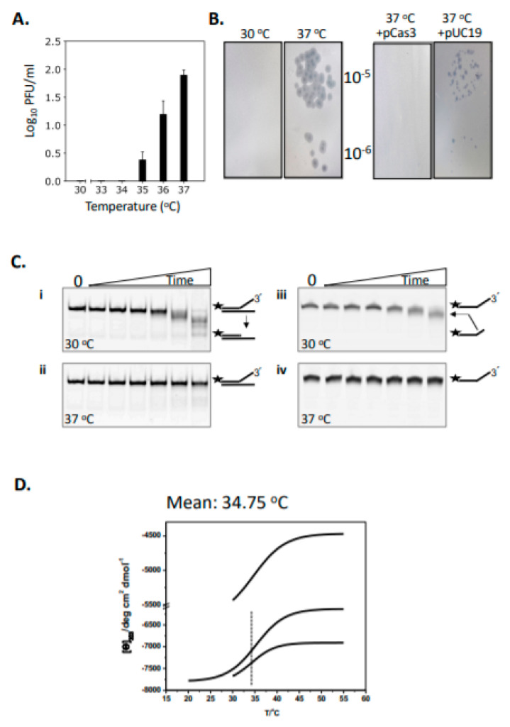
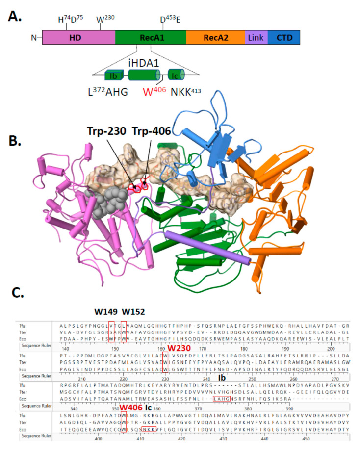
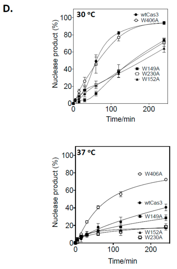
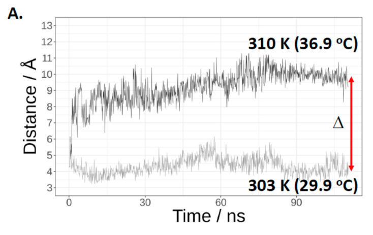
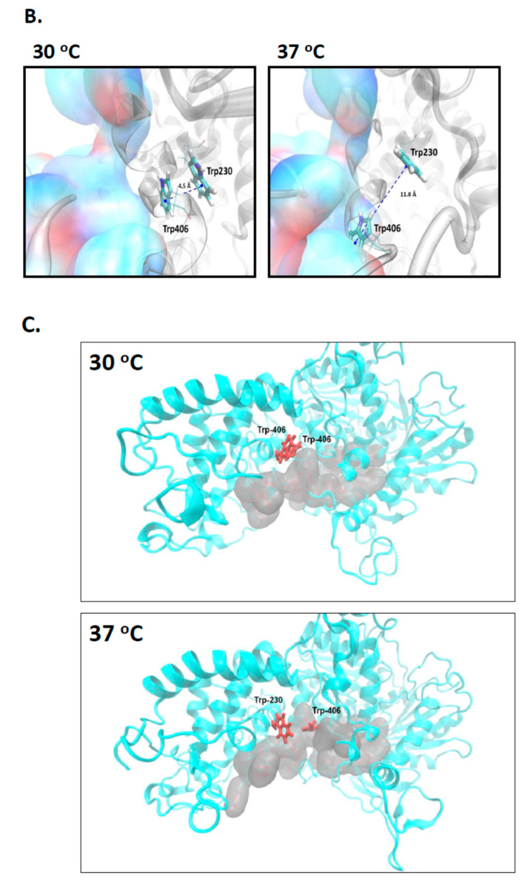
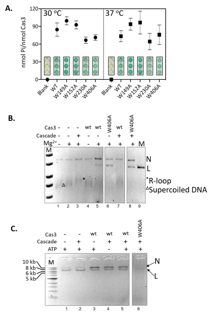
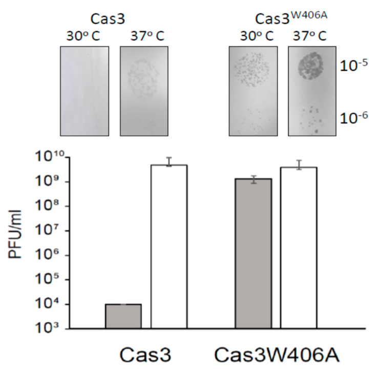
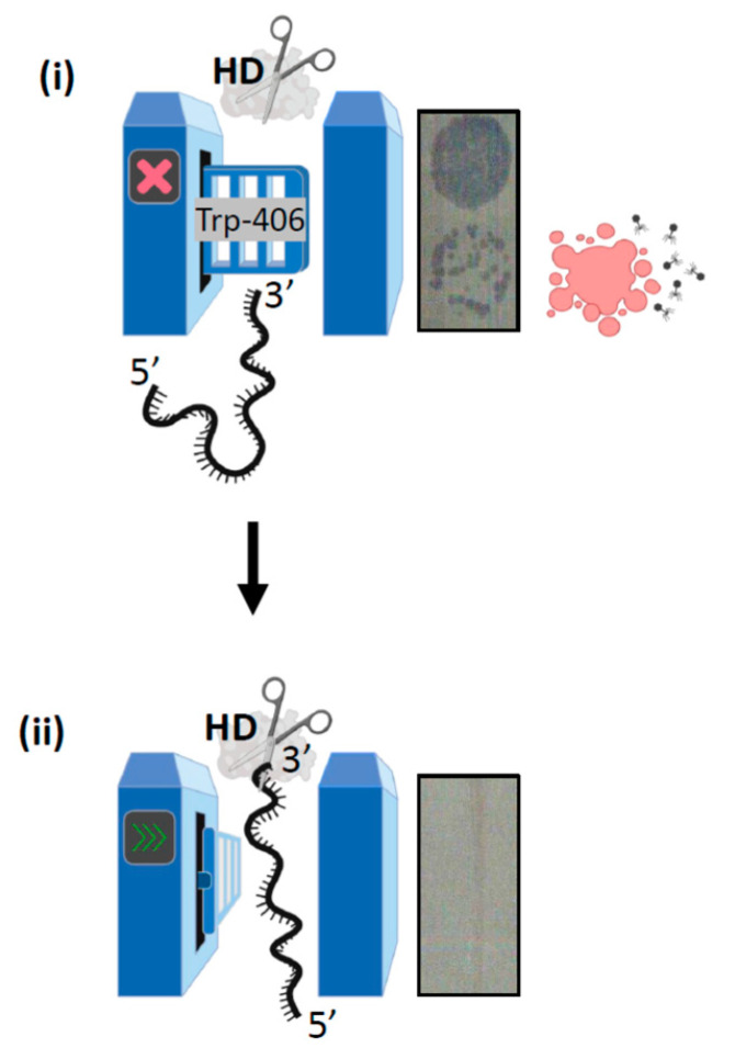
Cas3 is a ssDNA-targeting nuclease-helicase essential for class 1 prokaryotic CRISPR immunity systems, which has been utilized for genome editing in human cells. Cas3-DNA crystal structures show that ssDNA follows a pathway from helicase domains into a HD-nuclease active site, requiring protein conformational flexibility during DNA translocation. In genetic studies, we had noted that the efficacy of Cas3 in CRISPR immunity was drastically reduced when temperature was increased from 30 °C to 37 °C, caused by an unknown mechanism. Here, using E. coli Cas3 proteins, we show that reduced nuclease activity at higher temperature corresponds with measurable changes in protein structure. This effect of temperature on Cas3 was alleviated by changing a single highly conserved tryptophan residue (Trp-406) into an alanine. This Cas3W406A protein is a hyperactive nuclease that functions independently from temperature and from the interference effector module Cascade. Trp-406 is situated at the interface of Cas3 HD and RecA1 domains that is important for maneuvering DNA into the nuclease active site. Molecular dynamics simulations based on the experimental data showed temperature-induced changes in positioning of Trp-406 that either blocked or cleared the ssDNA pathway. We propose that Trp-406 forms a 'gate' for controlling Cas3 nuclease activity via access of ssDNA to the nuclease active site. The effect of temperature in these experiments may indicate allosteric control of Cas3 nuclease activity caused by changes in protein conformations. The hyperactive Cas3W406A protein may offer improved Cas3-based genetic editing in human cells.

Keywords: CRISPR; Cas3; genome editing; helicase.


Conflict of interest statement

The authors declare no conflict of interest.

Figures

Figure 1
Figure 1
Temperature-induced changes to Cas3 structure and function in E. coli cells and in vitro. (A) Δvir plaque formation was measured at 30-37 °C as a read-out for the efficacy of CRISPR interference. Plaques (PFU, plaque forming units) were readily observed at and above 35 °C (see also part B). Assays were in triplicate and standard errors from the mean are shown. (B) (i) Plaques formed as measured in part A at 37 °C were not observed if the cells also over-expressed Cas3 from a plasmid (ii, +pCas3), compared with the empty plasmid vector (pUC19) used as a control (iii). (C) Cas3 nuclease activity degrades Cy5 end-labelled (25 nM, label denoted by a star) DNA much more effectively at 30 °C compared to 37 °C, in reactions containing 56 nM of Cas3 with samples taken at 0, 5, 15, 30, 60, 120 and 240 min. Panels i and ii show native TBE polyacrylamide gels, and iii and iv are denaturing (urea) TBE gels. (D) Shows the Boltzmann curve traces from each of three independent experiments for E. coli Cas3, derived from circular dichroism spectroscopy measurements at 222 nm. The mean temperature for the inflection point is shown—34.75 °C—and see Supplementary Figure S2 for the full plotted data.
Figure 2
Figure 2
Structural positioning of two invariant tryptophan residues at the interface of Cas3 HD and RecA1 domains—a key role in Cas3 nuclease function for Trp-406. (A) Cartoon representation of Cas3 protein with amino acid residues indicated for the nuclease (HD) and Walker B ATPase active sites using numbering from the E. coli protein. Highlighted in the foreground is the region detailed in the results, located at the interface of HD and RecA1 domains (‘iHDA1’) and including the ssDNA binding helicase motifs Ib and Ic. (B) E. coli Cas3 structure deduced from T. fusca Cas3 (PDB: 4QQW) and T. terrenum (PDB: 4Q2C) (27,35) highlighting the tryptophan residue W406 (labelled) and the passage of ssDNA (tan cord). Cas3 regions are denoted as follows in the same way as in part A: HD (orchid), RecA1 (green), RecA2 (orange), an interdomain linker helix (purple) and the CTD (blue). Grey spheres indicate the HD nuclease active site residues. (C) Sequence alignment highlighting conservation of residues Trp-406 and Trp-230 in Escherichia coli K-12 (Eco) with the structurally determined Cas3 proteins from T. fusca (Tfu) and T. terrenum (Tter) proteins. Protein sequences were aligned using Clustal Omega and results were exported via Lasergene 17. (D) Nuclease activity of Cas3 and mutants (56 nM) measured on the DNA fork substrate (20 nM). Samples were collected at 0, 5, 15, 30, 60, 120 and 240 min. Reactions were carried out in triplicate at 30 °C or 37 °C as indicated, and data points show standard errors from the mean.
Figure 2
Figure 2
Structural positioning of two invariant tryptophan residues at the interface of Cas3 HD and RecA1 domains—a key role in Cas3 nuclease function for Trp-406. (A) Cartoon representation of Cas3 protein with amino acid residues indicated for the nuclease (HD) and Walker B ATPase active sites using numbering from the E. coli protein. Highlighted in the foreground is the region detailed in the results, located at the interface of HD and RecA1 domains (‘iHDA1’) and including the ssDNA binding helicase motifs Ib and Ic. (B) E. coli Cas3 structure deduced from T. fusca Cas3 (PDB: 4QQW) and T. terrenum (PDB: 4Q2C) (27,35) highlighting the tryptophan residue W406 (labelled) and the passage of ssDNA (tan cord). Cas3 regions are denoted as follows in the same way as in part A: HD (orchid), RecA1 (green), RecA2 (orange), an interdomain linker helix (purple) and the CTD (blue). Grey spheres indicate the HD nuclease active site residues. (C) Sequence alignment highlighting conservation of residues Trp-406 and Trp-230 in Escherichia coli K-12 (Eco) with the structurally determined Cas3 proteins from T. fusca (Tfu) and T. terrenum (Tter) proteins. Protein sequences were aligned using Clustal Omega and results were exported via Lasergene 17. (D) Nuclease activity of Cas3 and mutants (56 nM) measured on the DNA fork substrate (20 nM). Samples were collected at 0, 5, 15, 30, 60, 120 and 240 min. Reactions were carried out in triplicate at 30 °C or 37 °C as indicated, and data points show standard errors from the mean.
Figure 3
Figure 3
Molecular dynamics simulations indicate significant temperature-induced changes in positioning of Trp-230 and Trp-406. (A) Shows the distances between the centre of phenyl rings of Trp-230 and Trp-406 monitored during the MD simulations at 30 °C and 37 °C, as indicated. (B) Structures obtained during the final phase of MD simulations. The panel left side shows the Trp-230 and Trp-406 π-π interaction at 30 °C that is disrupted at 37 °C (panel right) because Trp-406 is moved into the pocket where it is stabilized by hydrophobic interactions within the ssDNA tunnel, shown as surfaces representing Thr-392, Gln-402, Gln-405, Leu-407, Gln-409, Lys-412, Lys-435, His-436, Asp-780 and Asp-778. (C) Temperature-induced changes in the positioning of Trp-230 and Trp-406 result in Trp-406 movement and blockage of the ssDNA binding channel (shown as grey surfaces representing the residues that form it), which we propose explains the lack of nuclease activity shown by E. coli Cas3 at the higher temperature, and its alleviation by replacing tryptophan with alanine. Cas3 protein is coloured in Cyan and Trp-230 and Trp-406 are coloured in red. At 37 °C Trp-406 moves to a location within a pocket in the ssDNA tunnel.
Figure 3
Figure 3
Molecular dynamics simulations indicate significant temperature-induced changes in positioning of Trp-230 and Trp-406. (A) Shows the distances between the centre of phenyl rings of Trp-230 and Trp-406 monitored during the MD simulations at 30 °C and 37 °C, as indicated. (B) Structures obtained during the final phase of MD simulations. The panel left side shows the Trp-230 and Trp-406 π-π interaction at 30 °C that is disrupted at 37 °C (panel right) because Trp-406 is moved into the pocket where it is stabilized by hydrophobic interactions within the ssDNA tunnel, shown as surfaces representing Thr-392, Gln-402, Gln-405, Leu-407, Gln-409, Lys-412, Lys-435, His-436, Asp-780 and Asp-778. (C) Temperature-induced changes in the positioning of Trp-230 and Trp-406 result in Trp-406 movement and blockage of the ssDNA binding channel (shown as grey surfaces representing the residues that form it), which we propose explains the lack of nuclease activity shown by E. coli Cas3 at the higher temperature, and its alleviation by replacing tryptophan with alanine. Cas3 protein is coloured in Cyan and Trp-230 and Trp-406 are coloured in red. At 37 °C Trp-406 moves to a location within a pocket in the ssDNA tunnel.
Figure 4
Figure 4
Nuclease hyperactivity of Cas3W406A is independent of ATPase activity and interaction with Cascade. (A) End-point measurements of ATP hydrolysis by Cas3 proteins (790 nM) was compared at 30 and 37 °C as indicated. Reactions were in triplicate, standard errors from the mean are shown, to give measurements of phosphate (Pi) released from per nM of Cas3 protein. These values were obtained from spectroscopic measurements of malachite green dye intensity, shown in the graph, each measurement was obtained after blanking the spectrophotometer with reactions lacking Cas3. (B) Shows Mg2+-dependent DNA nicking activity of Cas3 and Cas3W406A (850 nM) on M13 DNA in reactions that contain preformed Cascade (100 nM) R-loop substrates, indicated by an *. Reaction components are as indicated above the ethidium bromide stained agarose gel panel. In these reactions no deproteinising ‘stop’ solution was added, so that we were able to visualise that Cascade R-loops had formed (e.g., lane 3). (C) Shows Cas3 nicking and nuclease activity in the presence of Mg2+ and ATP and Cascade (100 nM), as indicated above the ethidium bromide stained agarose gel panel. These reactions were stopped by adding proteinase K ‘stop’ solution to dissipate R-loops so that nuclease products are more easily discerned. For panels (B,C) the topology of the DNA is highlighted to the right of the gel image by N (nicked) L (linear), * (R-Loop) and Δ (Supercoiled).
Figure 5
Figure 5
The allele cas3W406A does not provide resistance to λvir at 30°C. E. coli cell lawns of strain Δhnscas1) + λc + λT3 (IIB1309) and cas3W406A Δhnscas1) + λc + λT3 (IIB1342) were infected with phage dilutions (from 10−3 to 10−7) and incubated at 30°C and 37°C as indicated. Bars represent average and SD of the number of plaque forming units (PFUs) per ml from three independent experiments. Also shown are corresponding photographs of typical bacterial lawns and viral plaques giving the data.
Figure 6
Figure 6
Concluding summary of the Trp-406 nuclease ‘gate’ model. (i) When Trp-406 is positioned close to helicase motif Ic it blocks the ssDNA binding channel to the nuclease (HD) active site, resulting in a ‘closed’ gate. In this scenario CRISPR interference is inhibited because MGE DNA is not destroyed, resulting in cell lysis evident as plaques on a bacterial lawn. (ii) Conformational movement of Cas3 re-positions Trp-406 proximally to Trp-230, with which it interacts to form a lining to ssDNA channel in the gate being ‘open’. In this state Cas3 is able to degrade MGE DNA to fulfil its role in CRISPR adaptation reactions.

References

    1. Knott G.J., Doudna J.A. CRISPR-Cas guides the future of genetic engineering. Science. 2018;361:866–869. doi: 10.1126/science.aat5011. - DOI - PMC - PubMed
    1. Mojica F.J., Rodriguez-Valera F. The discovery of CRISPR in archaea and bacteria. FEBS J. 2016;283:3162–3169. doi: 10.1111/febs.13766. - DOI - PubMed
    1. Westra E.R., Levin B.R. It is unclear how important CRISPR-Cas systems are for protecting natural populations of bacteria against infections by mobile genetic elements. Proc. Natl. Acad. Sci. USA. 2020;117:27777–27785. doi: 10.1073/pnas.1915966117. - DOI - PMC - PubMed
    1. Makarova K.S., Wolf Y.I., Iranzo J., Shmakov S.A., Alkhnbashi O.S., Brouns S.J.J., Charpentier E., Cheng D., Haft D.H., Horvath P., et al. Evolutionary classification of CRISPR-Cas systems: A burst of class 2 and derived variants. Nat. Rev. MicroBiol. 2020;18:67–83. doi: 10.1038/s41579-019-0299-x. - DOI - PMC - PubMed
    1. Brouns S.J., Jore M.M., Lundgren M., Westra E.R., Slijkhuis R.J., Snijders A.P., Dickman M.J., Makarova K.S., Koonin E.V., van der Oost J. Small CRISPR RNAs guide antiviral defense in prokaryotes. Science. 2008;321:960–964. doi: 10.1126/science.1159689. - DOI - PMC - PubMed

MeSH terms

LinkOut - more resources