. 2021 Mar 11;26(6):1532.
doi: 10.3390/molecules26061532.

Chrysin Derivative CM1 and Exhibited Anti-Inflammatory Action by Upregulating Toll-Interacting Protein Expression in Lipopolysaccharide-Stimulated RAW264.7 Macrophage Cells

Affiliations

Chrysin Derivative CM1 and Exhibited Anti-Inflammatory Action by Upregulating Toll-Interacting Protein Expression in Lipopolysaccharide-Stimulated RAW264.7 Macrophage Cells

Eui-Baek Byun et al. Molecules. .

Abstract

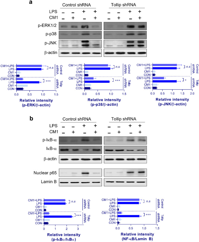
Although our previous study revealed that gamma-irradiated chrysin enhanced anti-inflammatory activity compared to intact chrysin, it remains unclear whether the chrysin derivative, CM1, produced by gamma irradiation, negatively regulates toll-like receptor (TLR) signaling. In this study, we investigated the molecular basis for the downregulation of TLR4 signal transduction by CM1 in macrophages. We initially determined the appropriate concentration of CM1 and found no cellular toxicity below 2 μg/mL. Upon stimulation with lipopolysaccharide (LPS), CM1 modulated LPS-stimulated inflammatory action by suppressing the release of proinflammatory mediators (cytokines TNF-α and IL-6) and nitric oxide (NO) and downregulated the mitogen-activated protein kinase (MAPK) and nuclear factor-κB (NF-κB) signaling pathways. Furthermore, CM1 markedly elevated the expression of the TLR negative regulator toll-interacting protein (Tollip) in dose- and time-dependent manners. LPS-induced expression of cell surface molecules (CD80, CD86, and MHC class I/II), proinflammatory cytokines (TNF-α and IL-6), COX-2, and iNOS-mediated NO were inhibited by CM1; these effects were prevented by the knockdown of Tollip expression. Additionally, CM1 did not affect the downregulation of LPS-induced expression of MAPKs and NF-κB signaling in Tollip-downregulated cells. These findings provide insight into effective therapeutic intervention of inflammatory disease by increasing the understanding of the negative regulation of TLR signaling induced by CM1.

Keywords: anti-inflammatory activity; chrysin derivative; macrophage; toll-interacting protein; toll-like receptor negative regulator.


Conflict of interest statement

The authors declare no conflict of interests.

Figures

Figure 1
Figure 1
Cell viability and anti-inflammatory effect of CM1 on RAW264.7 cells. HPLC chromatogram and chemical structure of CM1 are shown (a). RAW264.7 cells were treated with CM1 (0.5, 1, 1.5, and 2 μg/mL) for 24 h. Cell viability was assessed using the MTT assay (b). RAW264.7 cells were treated with CM1 (0.5, 1, 1.5, and 2 μg/mL) and 50 nM staurosporine for 24 h, and cytotoxicity was assessed by annexin V/PI staining and flow cytometry (c). Staurosporine was used as a positive control for measuring cell death. RAW264.7 cells were treated with LPS (200 ng/mL) or LPS and CM1 (1, 1.5, and 2 μg/mL) for 24 h, and TNF-α, IL-6, and NO levels in the culture medium were analyzed by ELISA and Griess assay (d). All bar graphs show the mean ± SD of three samples from one representative plot among three independent experiments. Statistical analysis was performed by one-way ANOVA in conjunction with Tukey’s multiple test; ** p < 0.01 and *** p < 0.001. CON: untreated RAW264.7. Abbreviations: MTT, 3-(4,5-dimethylthiazol-2-yl)-2,5-diphenyl-2H-tetrazolium bromide; PI, propidium iodide; LPS, lipopolysaccharide; TNF, tumor necrosis factor; IL, interleukin; NO, nitric oxide.
Figure 2
Figure 2
CM1-induced Tollip expression and inhibition of MAPK and NF-κB activation in RAW264.7 cells. RAW264.7 cells were pretreated with CM1 (1, 1.5, and 2 μg/mL) for 1 h and then stimulated with LPS (200 ng/mL) for 45 min (ac). The cytosolic and nuclear fractions were subjected to SDS-PAGE, and immunoblotting was performed using specific Abs against p-ERK1/2, p-p38, p-JNK, p-IκB-α, IκB-α, and p65 NF-κB. β-Actin and lamin B were used as the loading controls for the cytosolic and nuclear fractions, respectively. RAW264.7 cells were treated with CM1 (2 μg/mL) for 1 h (d). Total cellular proteins were resolved by SDS-PAGE, and immunoblotting was performed using specific SOCS1, IRAK-M, and Tollip Abs. RAW264. 7 cells were treated with CM1 for the indicated time periods (e) or at indicated concentrations (f), and Tollip levels were measured by immunoblotting. RAW264.7 cells were transfected with the control and Tollip shRNA vectors (g). Cells were treated with CM1, and Tollip and TLR4 levels were measured by immunoblotting. One representative blot from three independent experiments is shown. Statistical analysis was performed by one-way ANOVA in conjunction with Tukey’s multiple test (ac,e) or unpaired Student’s t test (d,g); n.s: no significance, * p < 0.05, ** p < 0.01, and *** p < 0.001. Abbreviations: MAPK, mitogen-activated protein kinase; NF, nuclear factor; Tollip, Toll-interacting protein; LPS, lipopolysaccharide; Abs, antibodies; p-ERK1/2, phosphor-extracellular signal-regulated kinase-1/2; p-p38, phosphor-p38; p-JNK; phosphor-c-Jun N-terminal kinase; IκB, inhibitor of κB; p-IκB; phosphor-IκB; SOCS1, anti-suppressor of cytokine signaling 1; IRAK-M, anti-interleukin-1 receptor-associated kinase; shRNA, short hairpin RNA; TLR4, Toll-like receptor-4.
Figure 3
Figure 3
CM1-mediated suppression of macrophage surface marker expression through upregulation of Tollip expression. Control and Tollip shRNA-transfected RAW264.7 cells were pretreated with CM1 (2 μg/mL) for 1 h and then exposed to LPS (200 ng/mL) for 24 h. Cells were stained with anti-CD80, anti-CD86, anti-MHC class I, and anti-MHC class II. The percentage of positive cells is shown in each panel. Data are presented as the mean ± SD for triplicate determinations of one representative plot among three independent experiments. Statistical analysis was performed by one-way ANOVA in conjunction with Tukey’s multiple test; n.s: no significance and *** p < 0.001, versus LPS treatment alone. Abbreviations: Tollip, Toll-interacting protein; shRNA, short hairpin RNA; LPS, lipopolysaccharide.
Figure 4
Figure 4
Tollip is indispensable for the inhibitory action of CM1 on proinflammatory mediators in LPS-treated RAW264.7 cells. Control and Tollip shRNA-transfected RAW264.7 cells were incubated with CM1 (2 μg/mL) for 1 h and then treated with LPS (200 ng/mL) for 24 h. TNF-α and IL-6 levels in the culture medium were measured by ELISA (a). RAW264.7 cells were pretreated with CM1 (2 μg/mL) for 1 h and then stimulated with LPS (200 ng/mL) for 45 min. NO level in the culture medium was measured using the Griess assay (b); *** p < 0.001. Data are presented as the mean ± SD for triplicate determinations of one representative plot among three independent experiments. Cytosolic fractions were analyzed to determine the COX-2 and iNOS protein levels using specific Abs (c). One representative blot from among three independent experiments is shown. Statistical analysis was performed by one-way ANOVA in conjunction with Tukey’s multiple test; n.s: no significance and *** p < 0.001. Abbreviations: Tollip, Toll-interacting protein; LPS, lipopolysaccharide; shRNA, short hairpin RNA; TNF, tumor necrosis factor; IL, interleukin; NO, nitric oxide; COX-2, cyclooxygenase-2; iNOS, inducible NO synthase; p-p38, phosphor-p38; p-JNK, phosphor-c-Jun N-terminal kinase; IκB, inhibitor of κB; p-IκB, phosphor-IκB; Abs, antibodies; NF, nuclear factor.
Figure 5
Figure 5
Tollip is indispensable for the inhibitory action of CM1 on MAPKs and NF-κB signaling in LPS-treated RAW264.7 cells. Control and Tollip shRNA-transfected RAW264.7 cells were incubated with CM1 (2 μg/mL) for 1 h and then treated with LPS (200 ng/mL) for 24 h. Control and Tollip shRNA-transfected RAW264.7 cells were pretreated with CM1 (2 μg/mL) for 1 h and then treated with LPS (200 ng/mL) for 45 min. Cytosolic fractions were analyzed to determine the p-ERK, p-p38, p-JNK, p-IκB-α, IκB-α, and β-actin protein levels using specific Abs (a). Nuclear fractions were analyzed to determine the p65 NF-κB and lamin B protein levels using specific Abs (b). One representative plot from among three independent experiments is shown. Statistical analysis was performed by one-way ANOVA in conjunction with Tukey’s multiple test; n.s: no significance, * p < 0.05, and *** p < 0.001. Abbreviations: Tollip, Toll-interacting protein; LPS, lipopolysaccharide; shRNA, short hairpin RNA; p-ERK1/2, phosphor-extracellular signal-regulated kinase-1/2, p-p38, phosphor-p38; p-JNK, phosphor-c-Jun N-terminal kinase; IκB, inhibitor of κB; p-IκB, phosphor-IκB; Abs, antibodies; NF, nuclear factor.

Similar articles

Cited by

References

    1. Cronkite D.A., Strutt T.M. The Regulation of Inflammation by Innate and Adaptive Lymphocytes. J. Immunol. Res. 2018;2018:1467538. doi: 10.1155/2018/1467538. - DOI - PMC - PubMed
    1. Takeuchi O., Akira S. Pattern recognition receptors and inflammation. Cell. 2010;140:805–820. doi: 10.1016/j.cell.2010.01.022. - DOI - PubMed
    1. Schaefer T.M., Desouza K., Fahey J.V., Beagley K.W., Wira C.R. Toll-like receptor (TLR) expression and TLR-mediated cytokine/chemokine production by human uterine epithelial cells. Immunology. 2004;112:428–436. doi: 10.1111/j.1365-2567.2004.01898.x. - DOI - PMC - PubMed
    1. Miron J., Picard C., Frappier J., Dea D., Theroux L., Poirier J. TLR4 Gene Expression and Pro-Inflammatory Cytokines in Alzheimer’s Disease and in Response to Hippocampal Deafferentation in Rodents. J. Alzheimers Dis. 2018;63:1547–1556. doi: 10.3233/JAD-171160. - DOI - PubMed
    1. Kuzmich N.N., Sivak K.V., Chubarev V.N., Porozov Y.B., Savateeva-Lyubimova T.N., Peri F. TLR4 Signaling Pathway Modulators as Potential Therapeutics in Inflammation and Sepsis. Vaccines. 2017;5:34. doi: 10.3390/vaccines5040034. - DOI - PMC - PubMed

MeSH terms

LinkOut - more resources