

Acute Pulmonary Embolism Severity Assessment Evaluated with Dual Energy CT Perfusion Compared to Conventional CT Angiographic Measurements
- PMID: 33799729
- PMCID: PMC8000326
- DOI: 10.3390/diagnostics11030495
Acute Pulmonary Embolism Severity Assessment Evaluated with Dual Energy CT Perfusion Compared to Conventional CT Angiographic Measurements
Abstract
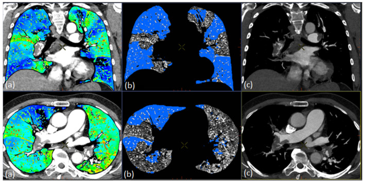
The purpose of the study was to investigate whether Dual Energy CT (DECT) can be used as a diagnostic tool to assess the severity of acute pulmonary embolism (PE) by correlating parenchymal perfusion defect volume, obstruction score and right ventricular-to-left ventricular (RV/LV) diameter ratio using CT angiography (CTA) and DECT perfusion imaging. A total of 43 patients who underwent CTA and DECT perfusion imaging with clinical suspicion of acute PE were retrospectively included in the study. In total, 25 of these patients had acute PE findings on CTA. DECT assessed perfusion defect volume (PDvol) were automatically and semiautomatically quantified. Overall, two CTA methods for risk assessment in patients with acute PE were assessed: the RV/LV diameter ratio and the Modified Miller obstruction score. Automatic PDvol had a weak correlation (r = 0.47, p = 0.02) and semiautomatic PDvol (r = 0.68, p < 0.001) had a moderate correlation to obstruction score in patients with confirmed acute PE, while only semiautomatic PDvol (r = 0.43, p = 0.03) had a weak correlation with the RV/LV diameter ratio. Our data indicate that PDvol assessed by DECT software technique may be a helpful tool to assess the severity of acute PE when compared to obstruction score and RV/LV diameter ratio.
Keywords: Dual Energy CT; Dual Source CT; lung perfusion; pulmonary embolism; pulmonary perfusion.
Conflict of interest statement
The authors declare no conflict of interest.
Figures


References
-
- Giri J., Sista A.K., Weinberg I., Kearon C., Kumbhani D.J., Desai N.D., Piazza G., Gladwin M.T., Chatterjee S., Kobayashi T., et al. Interventional Therapies for Acute Pulmonary Embolism: Current Status and Principles for the Development of Novel Evidence: A Scientific Statement From the American Heart Association. Circulation. 2019;140:e774–e801. doi: 10.1161/CIR.0000000000000707. - DOI - PubMed
-
- Meinel F.G., Graef A., Bamberg F., Thieme S.F., Schwarz F., Sommer W.H., Neurohr C., Kupatt C., Reiser M.F., Johnson T.R.C. Effectiveness of Automated Quantification of Pulmonary Perfused Blood Volume Using Dual-Energy CTPA for the Severity Assessment of Acute Pulmonary Embolism. Investig. Radiol. 2013;48:563–569. doi: 10.1097/RLI.0b013e3182879482. - DOI - PubMed
-
- Konstantinides S.V., Torbicki A., Agnelli G., Danchin N., Fitzmaurice D., Galiè N., Gibbs J.S.R., Huisman M.V., Humbert M., Kucher N., et al. 2014 ESC Guidelines on the Diagnosis and Management of Acute Pulmonary EmbolismThe Task Force for the Diagnosis and Management of Acute Pulmonary Embolism of the European Society of Cardiology (ESC)Endorsed by the European Respiratory Society (ERS) Eur. Heart J. 2014;35:3033–3080. doi: 10.1093/eurheartj/ehu283. - DOI - PubMed
LinkOut - more resources
Full Text Sources
Other Literature Sources
