. 2021 Mar 11;10(3):623.
doi: 10.3390/cells10030623.

PARP7 and Mono-ADP-Ribosylation Negatively Regulate Estrogen Receptor α Signaling in Human Breast Cancer Cells

Affiliations

PARP7 and Mono-ADP-Ribosylation Negatively Regulate Estrogen Receptor α Signaling in Human Breast Cancer Cells

Marit Rasmussen et al. Cells. .

Abstract

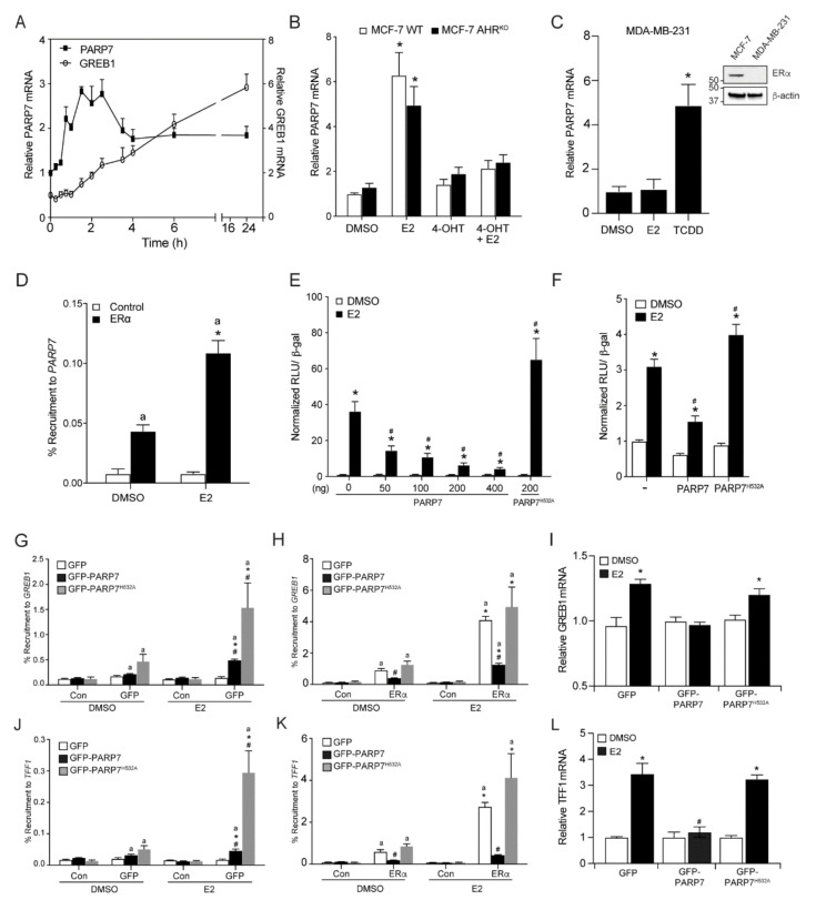
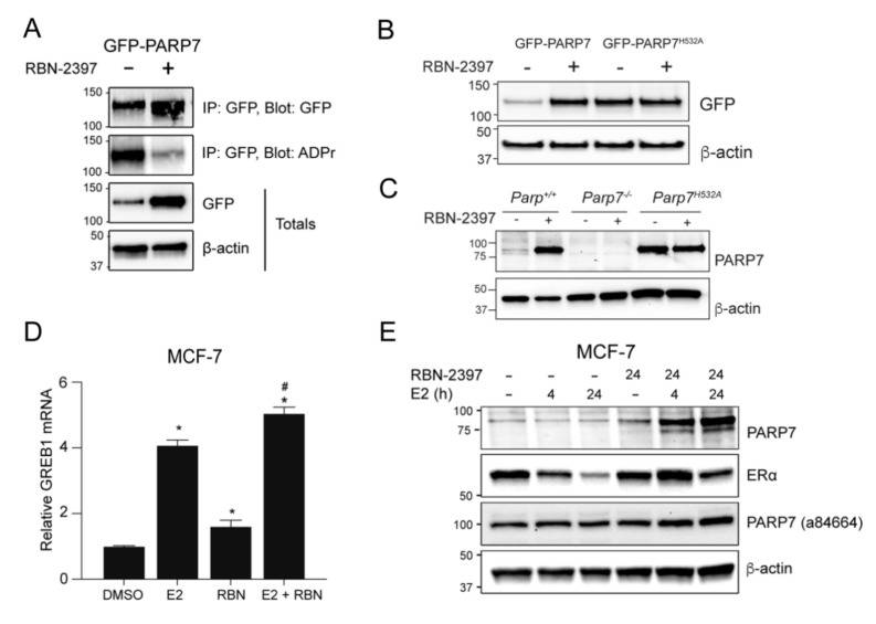
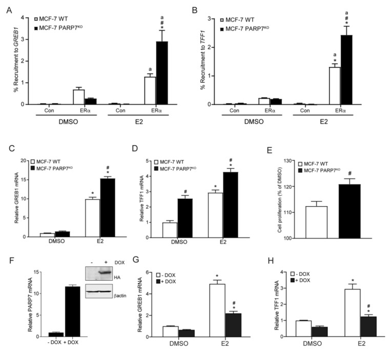
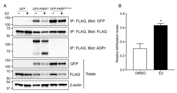
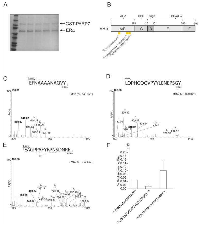
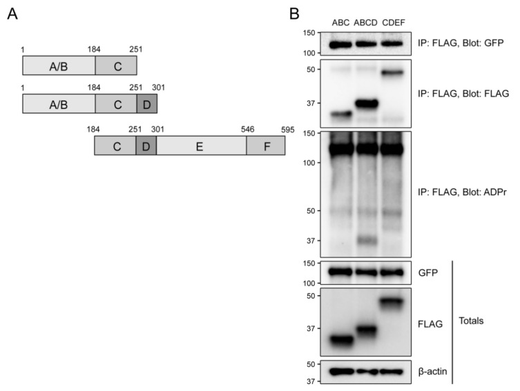
ADP-ribosylation is a post-translational protein modification catalyzed by a family of proteins known as poly-ADP-ribose polymerases. PARP7 (TIPARP; ARTD14) is a mono-ADP-ribosyltransferase involved in several cellular processes, including responses to hypoxia, innate immunity and regulation of nuclear receptors. Since previous studies suggested that PARP7 was regulated by 17β-estradiol, we investigated whether PARP7 regulates estrogen receptor α signaling. We confirmed the 17β-estradiol-dependent increases of PARP7 mRNA and protein levels in MCF-7 cells, and observed recruitment of estrogen receptor α to the promoter of PARP7. Overexpression of PARP7 decreased ligand-dependent estrogen receptor α signaling, while treatment of PARP7 knockout MCF-7 cells with 17β-estradiol resulted in increased expression of and recruitment to estrogen receptor α target genes, in addition to increased proliferation. Co-immunoprecipitation assays revealed that PARP7 mono-ADP-ribosylated estrogen receptor α, and mass spectrometry mapped the modified peptides to the receptor's ligand-independent transactivation domain. Co-immunoprecipitation with truncated estrogen receptor α variants identified that the hinge region of the receptor is required for PARP7-dependent mono-ADP-ribosylation. These results imply that PARP7-mediated mono-ADP-ribosylation may play an important role in estrogen receptor positive breast cancer.

Keywords: ARTD14; PARP7; TIPARP; breast cancer; estrogen receptor α; mono-ADP-ribosylation; poly ADP-ribose polymerase.


Conflict of interest statement

The authors declare no conflict of interest.

Figures

Figure 1
Figure 1
PARP7 is a target gene and repressor of ERα (A) PARP7 expression is induced by E2. MCF-7 cells were treated with E2, and RNA was isolated at various time points ranging from 15 min to 24 h. The relative mRNA levels of PARP7 (left axis) and GREB1 (right axis) were determined with RT-qPCR. (B) PARP7 is an ERα target gene and its expression is regulated by ERα, independent of AHR. MCF-7 wildtype and AHRKO cells were treated with 0.1% DMSO, 10 nM E2 or/and 100 nM 4-OHT for 2 h. The co-treated samples were treated with 4-OHT 2 h prior to E2 treatment. The relative mRNA levels were determined with RT-qPCR. The asterisk * denotes significant differences (p < 0.05) from DMSO. (C) PARP7 mRNA levels in MDA-MB-231 cells treated with 0.1% DMSO, 10 nM E2 or 10 nM TCDD for 2 h. Insert western blot of ERα levels in MCF-7 compared with MDA-MB-231 cells. The asterisk * denotes significant differences (p < 0.05) from DMSO. (D) ERα is recruited to the PARP7 promoter in an E2-dependent manner. Wildtype MCF-7 cells were treated with 0.1% DMSO or 10 nM E2 for one hour. The letter “a” denotes recruitment differences significantly greater than the control (p < 0.05), and significant differences (p < 0.05) from DMSO is denoted with the asterisk *. PARP7, but not the catalytically inactive H532A mutant, repressed ERα-regulated reporter gene activity. (E) HuH-7 or (F) MCF-7 cells were transfected with ERE-TK-Luc reporter, pSG5-ERα, β-galactosidase, PARP7, or the catalytically inactive mutant. Six hours after transfection, cells were treated with 0.1% DMSO or 10 nM E2 for 18 h. Changes in reporter gene activity are shown as normalized relative light units (RLU). The asterisk * denotes significant differences (p < 0.05) from DMSO. Hash mark # denotes significant differences (p < 0.05) when compared to the response to E2 treatment in the absence of PARP7. (G) Overexpressed GFP-PARP7, and GFP-PARP7H532A are recruited to GREB1 in response to E2. GFP-PARP7 but not GFP-PARP7H532A decreased (H) ERα binding to GREB1 and (I) reduced GREB1 mRNA levels in transfected HuH-7 cells. Overexpressed (J) GFP-PARP7 and GFP-PARP7H532A are recruited TFF1 in response to E2. GFP-PARP7 but not GFP-PARP7H532A decreased (K) ERα binding to TFF1 and reduced (L) TFF1 mRNA levels in transfected HuH-7 cells. For G, H, J and K, recruitment differences significantly greater than the control (p < 0.05) are denoted with the letter “a”. Significant differences greater than antibody matched DMSO (p < 0.05) are denoted with the asterisk *. Significant differences greater than treatment matched GFP (p < 0.05) are denoted with the hash mark #. For I and L, the asterisk * denotes significant differences (p < 0.05) from transfection matched DMSO. Significant differences greater than transfection matched E2 (p < 0.05) are denoted with the hash mark #. Data are shown as means ± S.E.M. from three independent experiments.
Figure 2
Figure 2
Inhibition of PARP7 activity stabilizes PARP7 protein levels and increases ERα activity. (A) RBN-2397 stabilizes PARP7 protein levels and decreases catalytic activity. COS-1 cells were transfected with GFP-PARP7 and treated with 0.1% DMSO or 100 nM RBN-2397 for 24 h. Samples were immunoprecipitated with anti-GFP, and membranes were blotted with anti-GFP and anti-ADP-ribose antibodies. (B) COS-1 cells were transfected with GFP-PARP7 or GFP-PARP7H532A and treated with 0.1% DMSO or 100 nM RBN-2397 for 24 h. (C) Parp7+/+, Parp7−/− or Parp7H532A MEFs were treated with 0.1% DMSO or 100 nM RBN-2397 for 24 h. The membrane was probed with our lab generated anti-PARP7. (D) Treatment with RBN-2397 increases mRNA expression of ERα target gene GREB1. Wildtype MCF-7 cells were treated with 0.1% DMSO, 10 nM E2 or co-treated with E2 and 100 nM RBN-2397 for 24 h. The asterisk * denotes significant differences (p < 0.05) from DMSO, and the hash mark # denotes significant differences (p < 0.05) compared to E2 treatment alone. (E) E2 stimulation increases PARP7 protein expression. MCF-7 cells were treated with 10 nM E2 for 0, 4 and 24 h, together with control (no treatment) or 24 h treatment with 100 nM RBN-2397. The membrane was blotted with our lab generated anti-PARP7, anti-ERα, or anti-PARP7 (Abcam; ab84664) antibodies. PARP7 bands are visible in samples co-treated with E2 and RBN-2397. Anti-PARP7 (ab84664), did not detect endogenous PARP7, but rather detected a protein at approximately 100 kDa in all lanes.
Figure 3
Figure 3
Confirmation of MCF-7 PARP7 knockout cells. (A) Schematic representation of the gRNA binding site, showing insertions/deletions resulting in frameshift mutations. The deleted bases are represented as dashes. The data are from 45 independent sequences. (B) PARP7 is not detected in the PARP7KO cells when blotting the membrane with our lab generated anti-PARP7 antibody. MCF-7 wildtype and PARP7KO cells were treated with DMSO, E2 or/and RBN-2397 for 24 h. Membranes were blotted with lab generated anti-PARP7, anti ERα and anti-PARP7 (Abcam; ab84664). Blotting with anti-PARP (ab84664) resulted in bands across all lanes at approximately 100 kDa. (C) Loss of PARP7 increases expression of AHR target gene CYP1A1. MCF-7 wildtype and PARP7KO cells were treated with 10 nM TCDD for 24 h. Relative mRNA levels of CYP1A1 was determined with RT-qPCR. Significant differences (p < 0.05) compared to DMSO are denoted with *, and differences due to PARP7 are denoted with the hash mark #. Data are shown as means ± S.E.M. for three independent experiments.
Figure 4
Figure 4
MCF-7 PARP7KO cells display increased ERα activity. Recruitment of ERα to the regulatory regions of (A) GREB1 and (B) TFF1 was increased in MCF-7 PARP7KO cells compared with MCF-7 wildtype cells. Cells were treated with 0.1% DMSO or 10 nM E2 for one hour. Chromatin immunoprecipitation was carried out with no antibody (control) or anti-ERα. Recruitment differences significantly greater than the control (p < 0.05) are denoted with the letter “a”, differences due to PARP7 (p < 0.05) are denoted with the hash mark #. Significant differences greater than DMSO (p < 0.05) are denoted with the asterisk *. (C) GREB1 and (D) TFF1 mRNA levels were significantly higher in MCF-7 PARP7KO cells treated with 10 nM E2 for 24 h compared with MCF-7 wildtype cells. The asterisk * denotes statistical significance (p < 0.05) when compared to the DMSO-treated wildtype, and hash mark # denotes significant (p < 0.05) differences due to PARP7. (E) MCF-7 PARP7KO cells exhibited increased proliferation in response to E2 compared with wildtype cells. Cells were treated with 0.1% DMSO or 10 nM E2 every day for 4 days. Cell proliferation was normalized to baseline (Day 1) and to the DMSO-treated samples. The hash mark # denotes statistical significance (p < 0.05) between the cell lines. (F) MCF7 cells expressing a Tet-ON regulated HA-tagged PARP7 were cultured in the presence or absence of doxycycline (DOX) to induce (F) PARP7 mRNA and protein levels (inset F). Cells were then incubated with or without DOX and treated with DMSO or 10 nM of E2 for 20 h. The mRNA expression levels of (G) GREB1 and (H) TFF1 were reduced in the presence of DOX. The data are representative of three independent experiments. The asterisk * denotes significant difference (p < 0.05) greater than DMSO, and the hash mark # denotes differences due to DOX. Data are shown as means ± SEM for three independent experiments.
Figure 5
Figure 5
Overexpressed PARP7 mono-ADP-ribosylates overexpressed ERα. (A) PARP7 and its catalytically inactive mutant interacts with ERα in COS-1 cells when treated with DMSO and E2. COS-1 cells were transfected with FLAG-ERα, and either GFP, GFP-PARP7 or GFP-PARP7H532A, and treated with DMSO or E2 for 24 h. Co-immunoprecipitation was carried out with anti-FLAG. The membranes were incubated with anti-FLAG, anti-GFP, and anti-ADP-ribose. Both wildtype PARP7 and ERα are mono-ADP-ribosylated, but not by the catalytically inactive PARP7H532A mutant. (B) Relative MARylation levels of immunoprecipitated FLAG-ERα in the presence of GFP-PARP7 after treatment with DMSO or E2 for 24 h. Quantification of protein bands normalized to β-actin revealed that mono-ADP-ribosylation of ERα was significantly (p < 0.05) increased upon treatment with E2 as indicated by an asterisk *.
Figure 6
Figure 6
Identification of mono-ADP-ribosylated peptides in bacterial expressed and purified ERα. (A) Representative SDS-PAGE of GST-PARP7 and ERα prior to LC/MS analysis. (B) A schematic representation of the domain structure of ERα. Location of ADP-ribosylated peptides are denoted by yellow rectangles. Peptide sequences are numbered from the unmodified full-length protein. (C) The MS2 spectrum of the trypsin generated ion at m/z 940.855. (D) The MS2 spectrum of the trypsin generated ion at m/z 920.705. (E) The MS2 spectrum of the trypsin generated ion at m/z 796.657. (F) Relative levels of modification (in percentage) of ADP-ribosylated peptides identified by M/S in ERα.
Figure 7
Figure 7
The hinge region of ERα is required for mono-ADP-ribosylation by PARP7. (A) A schematic representation of the truncated variants of ERα. (B) PARP7 co-immunoprecipitated with all three ERα variants. Only ERα ABCD was mono-ADP-ribosylated. COS-1 cells were transfected with GFP-PARP7 and either 3xFLAG-ERα ABC, 3xFLAG-ERα ABCD, 3xFLAG-ERα CDEF. Co-immunoprecipitation was carried out with anti-FLAG, and membranes were blotted with anti-FLAG, anti-GFP and anti-ADP-ribose.

Similar articles

Cited by

References

    1. Hassa P.O., Hottiger M.O. The diverse biological roles of mammalian PARPS, a small but powerful family of poly-ADP-ribose polymerases. Front. Biosci. 2008;13:3046–3082. doi: 10.2741/2909. - DOI - PubMed
    1. Hottiger M.O., Hassa P.O., Luscher B., Schuler H., Koch-Nolte F. Toward a unified nomenclature for mammalian ADP-ribosyltransferases. Trends Biochem. Sci. 2010;35:208–219. doi: 10.1016/j.tibs.2009.12.003. - DOI - PubMed
    1. Otto H., Reche P.A., Bazan F., Dittmar K., Haag F., Koch-Nolte F. In silico characterization of the family of PARP-like poly(ADP-ribosyl)transferases (pARTs) BMC Genom. 2005;6:139. doi: 10.1186/1471-2164-6-139. - DOI - PMC - PubMed
    1. Kleine H., Poreba E., Lesniewicz K., Hassa P.O., Hottiger M.O., Litchfield D.W., Shilton B.H., Luscher B. Substrate-assisted catalysis by PARP10 limits its activity to mono-ADP-ribosylation. Mol. Cell. 2008;32:57–69. doi: 10.1016/j.molcel.2008.08.009. - DOI - PubMed
    1. Corda D., Di Girolamo M. Functional aspects of protein mono-ADP-ribosylation. EMBO J. 2003;22:1953–1958. doi: 10.1093/emboj/cdg209. - DOI - PMC - PubMed

Publication types

MeSH terms