Review
. 2021 Mar 8;22(5):2719.
doi: 10.3390/ijms22052719.

The Role of Tumor Necrosis Factor Alpha (TNF-α) in Autoimmune Disease and Current TNF-α Inhibitors in Therapeutics

Affiliations
Review

The Role of Tumor Necrosis Factor Alpha (TNF-α) in Autoimmune Disease and Current TNF-α Inhibitors in Therapeutics

Dan-In Jang et al. Int J Mol Sci. .

Abstract

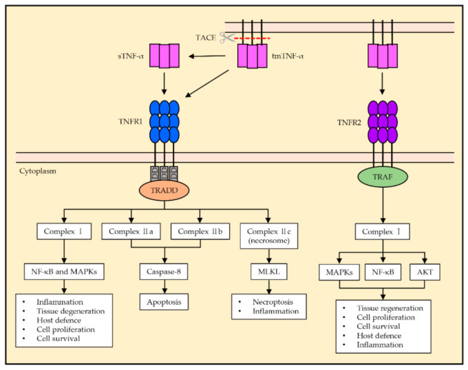
Tumor necrosis factor alpha (TNF-α) was initially recognized as a factor that causes the necrosis of tumors, but it has been recently identified to have additional important functions as a pathological component of autoimmune diseases. TNF-α binds to two different receptors, which initiate signal transduction pathways. These pathways lead to various cellular responses, including cell survival, differentiation, and proliferation. However, the inappropriate or excessive activation of TNF-α signaling is associated with chronic inflammation and can eventually lead to the development of pathological complications such as autoimmune diseases. Understanding of the TNF-α signaling mechanism has been expanded and applied for the treatment of immune diseases, which has resulted in the development of effective therapeutic tools, including TNF-α inhibitors. Currently, clinically approved TNF-α inhibitors have shown noticeable potency in a variety of autoimmune diseases, and novel TNF-α signaling inhibitors are being clinically evaluated. In this review, we briefly introduce the impact of TNF-α signaling on autoimmune diseases and its inhibitors, which are used as therapeutic agents against autoimmune diseases.

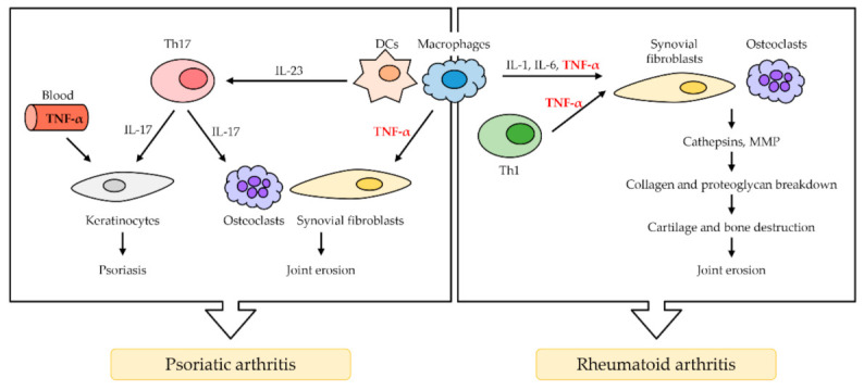
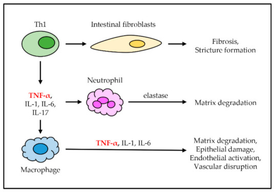
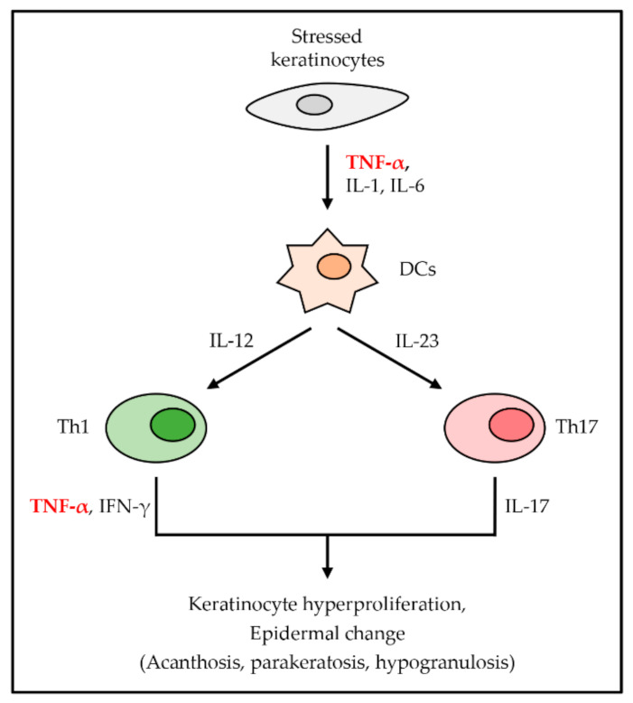
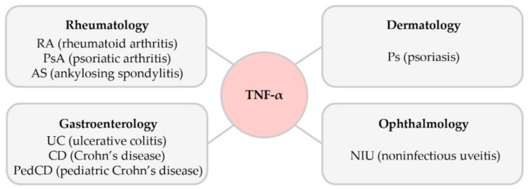
Keywords: TNF-α; TNF-α inhibitors; autoimmune diseases; inflammatory bowel disease; psoriatic arthritis; rheumatoid arthritis.


Conflict of interest statement

The authors declare no conflict of interest.

Figures

Figure 1
Figure 1
General tumor necrosis factor alpha (TNF-α) signaling pathway of TNFR1 and TNFR2. The TNFR1 signaling pathway is activated by the ligation of sTNF-α and tmTNF-α, and the death domain of TNFR1 recruits TRADD. Complex I activates NF-κB and MAPKs, which results in inflammation, tissue degeneration, host defense, cell proliferation, and cell survival. Complexes IIa and IIb activate caspase-8 and induce apoptosis. Complex IIc is known to induce necroptosis and inflammation via the activation of MLKL. The TNFR2 signaling pathway is mainly activated by tmTNF-α. TNFR2 does not possess a death domain and recruits TRAF via its TRAF domain, which activates the formation of complex I, resulting in NF-κB and MAPKs and AKT activation. TNFR2 activation is associated with homeostatic bioactivities such as tissue regeneration, cell proliferation, and cell survival, as well as host defense and inflammation.
Figure 2
Figure 2
The role of TNF-α in rheumatoid arthritis (RA) and psoriatic arthritis (PsA). TNF-α is mainly secreted by macrophage and Th1 cells. In RA, TNF-α activates synovial fibroblasts, which causes the overproduction of cathepsins and MMP. The breakdown of collagen and proteoglycan follows, resulting in cartilage and bone destruction, as well as joint erosion. Osteoclasts in RA induce synovial hyperplasia and angiogenesis. In PsA, activated dendritic cells (DCs) and macrophages secrete TNF-α and IL-23 excessively. IL-23 induces T cells to differentiate in Th17 cells, which secretes IL-17. IL-17 and TNF-α in the blood activate keratinocytes, resulting in psoriasis (PS). TNF-α also activates osteoclasts, leading to synovial fibroblast, which results in joint erosion.
Figure 3
Figure 3
The role of TNF-α in inflammatory bowel disease (IBD). TNF-α is secreted from Th1 cells along with other cytokines. These cytokines cause the accumulation of immune cells, including intestinal fibroblasts, neutrophils, and macrophages in the gut. Intestinal fibroblasts cause fibrosis and stricture formation. Neutrophils secrete elastase, which causes intestinal matrix degradation. Macrophages produce more inflammatory cytokines, which causes intestinal matrix degradation, epithelial damage, endothelial activation, and disruption.
Figure 4
Figure 4
The role of TNF-α in PS. TNF-α is first secreted from stressed keratinocytes with other proinflammatory cytokines. Secreted cytokines activate DCs, which induce the differentiation of T cells into Th1 cells and Th17 cells. They secrete TNF-α, IFN-γ, and IL-17, respectively, which cause keratinocyte hyperproliferation and several epidermal changes.
Figure 5
Figure 5
The role of TNF-α in uveitis. TNF-α is secreted from macrophages with other cytokines and activates DCs. Activated DCs secrete IL-12, IL-6, and TGF-β to induce differentiation of Th1 cells and Th17 cells from naïve T cells, respectively. Th1 cells and Th17 cells arrive at the uvea through migration and infiltration. Several leukocytes from blood are recruited by Th1 cells and Th17 cells. NIU occurs due to inflammation.
Figure 6
Figure 6
Timeline of the approval dates of TNF-α inhibitors in autoimmune diseases.
Figure 7
Figure 7
The association of TNF-α in various autoimmune diseases.

Similar articles

Cited by

References

    1. Bradley J.R. TNF-Mediated inflammatory disease. J. Pathol. 2008;214:149–160. doi: 10.1002/path.2287. - DOI - PubMed
    1. Horiuchi T., Mitoma H., Harashima S., Tsukamoto H., Shimoda T. Transmembrane TNF-alpha: Structure, function and interaction with anti-TNF agents. Rheumatology. 2010;49:1215–1228. doi: 10.1093/rheumatology/keq031. - DOI - PMC - PubMed
    1. Jiang Y., Yu M., Hu X., Han L., Yang K., Ba H., Zhang Z., Yin B., Yang X.P., Li Z., et al. STAT1 mediates transmembrane TNF-alpha-induced formation of death-inducing signaling complex and apoptotic signaling via TNFR1. Cell Death Differ. 2017;24:660–671. doi: 10.1038/cdd.2016.162. - DOI - PMC - PubMed
    1. Vandenabeele P., Declercq W., Beyaert R., Fiers W. Two tumour necrosis factor receptors: Structure and function. Trends Cell Biol. 1995;5:392–399. doi: 10.1016/S0962-8924(00)89088-1. - DOI - PubMed
    1. Bazzoni F., Beutler B. The tumor necrosis factor ligand and receptor families. N. Engl. J. Med. 1996;334:1717–1725. doi: 10.1056/NEJM199606273342607. - DOI - PubMed

MeSH terms

Substances