Review
. 2021 Mar 8;22(5):2728.
doi: 10.3390/ijms22052728.

Adaptive Changes in the Central Control of Energy Homeostasis Occur in Response to Variations in Energy Status

Affiliations
Review

Adaptive Changes in the Central Control of Energy Homeostasis Occur in Response to Variations in Energy Status

Cassandra Gastelum et al. Int J Mol Sci. .

Abstract

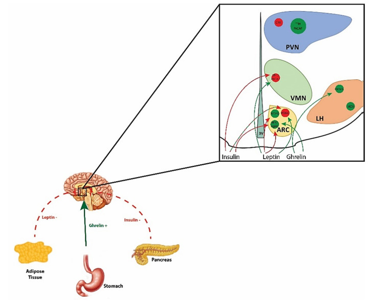
Energy homeostasis is regulated in coordinate fashion by the brain-gut axis, the homeostatic energy balance circuitry in the hypothalamus and the hedonic energy balance circuitry comprising the mesolimbcortical A10 dopamine pathway. Collectively, these systems convey and integrate information regarding nutrient status and the rewarding properties of ingested food, and formulate it into a behavioral response that attempts to balance fluctuations in consumption and food-seeking behavior. In this review we start with a functional overview of the homeostatic and hedonic energy balance circuitries; identifying the salient neural, hormonal and humoral components involved. We then delve into how the function of these circuits differs in males and females. Finally, we turn our attention to the ever-emerging roles of nociceptin/orphanin FQ (N/OFQ) and pituitary adenylate cyclase-activating polypeptide (PACAP)-two neuropeptides that have garnered increased recognition for their regulatory impact in energy homeostasis-to further probe how the imposed regulation of energy balance circuitry by these peptides is affected by sex and altered under positive (e.g., obesity) and negative (e.g., fasting) energy balance states. It is hoped that this work will impart a newfound appreciation for the intricate regulatory processes that govern energy homeostasis, as well as how recent insights into the N/OFQ and PACAP systems can be leveraged in the treatment of conditions ranging from obesity to anorexia.

Keywords: estradiol; fasting; nociceptin/orphanin FQ; obesity; pituitary adenylate cyclase-activating polypeptide; sex difference.


Conflict of interest statement

The authors declare no conflict of interest.

Figures

Figure 1
Figure 1
Schematic diagram illustrating the interplay between peripheral hormones and the homeostatic energy balance circuitry. Hormones like leptin and insulin released from adipose tissue and the endocrine pancreas, respectively, exert anorexigenic effects, whereas ghrelin released from the gastric mucosa exerts orexigenic effects. Leptin’s appetite-suppressing actions are mediated via excitatory effects on anorexigenic POMC and SF-1/PACAP neurons, as well as inhibitory effects on orexigenic NPY/AgRP and N/OFQ neurons. Insulin also inhibits NPY/AgRP neurons and, paradoxically, SF-1/PACAP neurons as well. Insulin’s effects on POMC neurons are dependent upon prevailing levels of tyrosine protein phosphatases. On the other hand, ghrelin’s appetite-promoting effects are due to its excitatory effects on NPY/AgRP and orexin neurons.
Figure 2
Figure 2
Fasting reverses the polarity of postsynaptic PACAP response in POMC neurons by switching the coupling of PAC1 receptors from TRPC5 to KATP channels. (AD), Representative current traces from voltage clamp recordings of POMC neurons that depict the response elicited by 100 nM PACAP under ad libitum and fasting conditions, the latter of which is abrogated by blockade of KATP channels with tolbutamide (100 μM) and PAC1 receptors with PACAP6–38 (200 nM). The inset I/V plots illustrate that the fasting-induced change in polarity is due to a switch from a mixed cation conductance to a PAC1 receptor-mediated K+ conductance via KATP channels. (E,F), Pie charts that show the proportion of POMC neurons that are excited by, inhibited by, or unresponsive to PACAP under ad libitum (to the left) and fasting (to the right) conditions. (G,H) highlight the PACAP-induced changes in membrane current (ΔI) and conductance (ΔG); alone (n = 12) and in conjunction with tolbutamide (n = 12) and PACAP6–38 (n = 8). Bars represent means and lines 1 SEM. #, p < 0.05 relative to PACAP under ad libitum conditions. *, p < 0.05 relative to PACAP under fasting conditions, one-way ANOVA/LSD. Figure adapted from [12].
Figure 3
Figure 3
E2 attenuates the PACAP-induced outward current in POMC neurons observed under fasting conditions. (A,B) are representative membrane current traces during voltage clamp recordings in EtOH vehicle- (0.01% (v:v); n = 11) and E2-treated (100 nM; n = 8) slices from OVX females that illustrate the estrogenic diminution of the robust and reversible PACAP-induced outward current and change in K+ conductance (as seen from the inset I/V plots). (C), Pie chart that indicates the percentage of POMC neurons from OVX females that are excited by, inhibited by, or unresponsive to PACAP under fasting conditions. (D,E) show the composite data underscoring the ability of E2 to negatively modulate the PACAP response. Bars represent means and lines 1 SEM of the PACAP-induced ΔI and ΔG in POMC neurons from OVX females under fasting conditions. *, p < 0.05 relative to EtOH vehicle, Student’s t-test.
Figure 4
Figure 4
The PACAP-induced outward current observed during fasting conditions is associated with a hyperpolarization and a decrease in firing. (A), Composite bar graph that demonstrates the more hyperpolarized RMP of POMC neurons under fasting conditions. (B,C), Representative current clamp traces from POMC neurons showing the reversible PACAP-induced depolarization and increase in firing under ad libitum-fed conditions (n = 10) and the reversible hyperpolarization and suppression of firing seen under fasting conditions (n = 10). Comparable effects are seen during recordings in vehicle pre-treated slices from OVX females. (D,E), Composite data illustrating the PACAP-induced changes in membrane potential (ΔV) and firing rate under ad libitum-fed and fasting conditions. Bars represent means and lines 1 SEM. * p < 0.05, relative to ad libitum-fed conditions, Student’s t-test (D); relative to baseline, Kruskal–Wallis/median-notched box-and-whisker analysis (E). Figure adapted from [12].
Figure 5
Figure 5
Optogenetic stimulation of VMN PACAP neurons depolarizes POMC neurons and increases their firing under ad libitum-fed conditions, effects which are flipped under fasting conditions. (A), Low power (4×) image of ARC POMC neurons taken from a PACAP-Cre/eGFP POMC mouse. (B), Photomicrograph (4×) showing the channel rhodopsin-2 expression in VMN PACAP neurons two weeks after AAV injection as visualized by eYFP. (C), Differential interference contrast image (40×) of a recorded POMC neuron and the corresponding eGFP fluorescence signal from the same neuron (D). (E), 40X image showing the eYFP-labeled fibers in the immediate vicinity of the recorded neuron. Photostimulation (10-ms pulses delivered at 20 Hz for 10 s) of male VMN PACAP neurons produces a reversible inward current linked to membrane depolarization and increase in the firing of ARC POMC under ad libitum-fed conditions neurons (F,H,J,LN); n = 7–11), and the exact opposite is seen under fasting conditions (G,I,KN); n = 11–13). Bars represent means, and lines 1 SEM of the light-induced change in ΔI (L), ΔV (M) and firing rate (N). * p < 0.05, relative to ad libitum-fed conditions, Student’s t-test (L,M); relative to baseline, Kruskal–Wallis/median-notched box-and-whisker analysis (N). Figure adapted from [12].
Figure 6
Figure 6
The fasting-induced switch in the polarity of the PACAP response in POMC neurons involves the activation of protein tyrosine phosphatases. Representative traces show that the PACAP-induced outward current (A; n = 12) and hyperpolarization (C; n = 10) seen under fasting conditions reverts back to an inward current (B; n = 9) and depolarization (D; n = 7) in the presence of the PTP1B/TCPTP inhibitor CX08005 (20 μM; B). This is further substantiated by the composite bar graphs in (EG) as well as the pie charts in (H,I). Bars represent means, and lines 1 SEM of the PACAP-induced change ΔI, ΔV or normalized firing rate under fasted conditions, alone and in combination with CX08005, Compound C or metformin. * p < 0.05, relative to PACAP alone, Student’s t-test (E,F); relative to baseline, Kruskal–Wallis/median-notched box-and-whisker analysis (G).
Figure 7
Figure 7
The fasting-induced reversal of the PACAP response in POMC neurons is also dependent upon activation of AMPK. The outward current caused by PACAP under fasting conditions in POMC neurons (A; n = 12) is transformed into an inward current in the presence of the AMPK inhibitor Compound C (30 μM; B; n = 8) The PACAP-induced outward current in (A) was reproduced under ad libitum-fed conditions in the presence of the AMPK activator metformin (500 μM; C; n = 10). The data from these representative traces is summarized in composite form by the bar graph in (D) and the pie charts in (EG). Bars represent means and lines 1 SEM. *, p < 0.05 relative to PACAP alone, one-way ANOVA/LSD.
Figure 8
Figure 8
Fasting augments the activity and expression of PTP1B and AMPK in the ARC. The four 20× images depict the PTP1B (A,B; 1:100) and pAMPK (D,E; 1:100) immunoreactivity under ad libitum-fed (A,D) and fasting (B,E) as visualized with AF546 (1:600). The composite data in the bar graphs summarize the fasting-induced increase in the number of PTP1B- (C) and pAMPK-immunoreactive (F) cells per capita in the ARC. Bars represent means and lines 1 SEM. * p < 0.05, relative to ad libitum-fed conditions, Student’s t-test.
Figure 9
Figure 9
Fasting completely reverses the effect of a direct injection of PACAP into the ARC on energy intake, which is attenuated by EB in OVX females. Under ad libitum-fed conditions, PACAP (30pmo; n = 6l) significantly decreases cumulative energy intake in wildtype males compared to saline-treated controls (0.2 μL; A; n = 6). This PACAP-induced decrease in energy intake is no longer apparent in fasted males, and PACAP actually causes an increase in cumulative consumption which is evident at six hours post-administration (B; n = 7–9). PACAP also decreases energy intake in ad libitum-fed OVX wildtype females, and this effect is potentiated by EB (20 μg/kg; s.c.; C; n = 6). Again, the effect of PACAP on consummatory behavior in fasted OVX females is exactly the opposite of that seen under ad libitum conditions, as is the modulatory effect of EB (D; n = 6). Bars represent means and lines 1 S.E.M. of the cumulative energy intake seen in ad libitum-fed or fasted mice injected with either PACAP or its saline vehicle. #, p < 0.05 relative to cumulative energy intake seen at three hours after PACAP injection, repeated-measures, multi-factorial ANOVA/LSD; *, p < 0.05 relative to saline vehicle, repeated measures, multi-factorial ANOVA/LSD; ^, p < 0.05 relative to sesame oil vehicle, repeated measures, multi-factorial ANOVA/LSD. Figure adapted from [12].
Figure 10
Figure 10
Schematic diagrams illustrating how PAC1 and NOP receptor signaling in POMC neurons is altered under various energy balance states. (A), In POMC neurons from ad libitum-fed animals, N/OFQ activates the NOP receptor that initiates Gi/o-mediated signaling and subsequent activation of GIRK channels through positive allosteric modification by the βγ complex. This in turn promotes K+ efflux and inhibition of POMC neurons, effects which are dampened by E2 acting through ERα and Gq-mER to stimulate PI3K and nNOS as well as PLC, PKC and PKA signaling pathways, respectively. (B), In POMC neurons from obese animals, NOP receptor/effector coupling is enhanced; leading to a greater inhibitory effect of N/OFQ on POMC neurons. This N/OFQ-induced inhibition of POMC neurons is once again abrogated by E2 in POMC neurons from obese females. (C), Under ad libitum conditions, PACAP activates its cognate PAC1 receptor to elicit Gq-mediated signaling; working through PI3K as well as PLC, IP3, DAG and PKC to promote Ca2+ mobilization from intracellular stores and the coupling of PAC1 receptors to TRCP5 channels. This leads to cation flux through the channel pore that depolarizes and thereby excites POMC neurons. In females, E2 can act via ERα and Gq-mER to potentiate PAC1 receptor/TRPC5 channel coupling and PACAP-induced excitation of POMC neurons. (D), Under conditions of diet-induced obesity, the PAC1 receptor-mediated activation of TRPC5 channels in male POMC neurons is attenuated. However, in obese females the PACAP-induced excitation of POMC neurons is maintained due to the potentiating effect of E2. (E), Under conditions of fasting, the expression and activity of AMPK and protein tyrosine phosphatases like PTP1B is elevated in POMC neurons. This triggers a switch in the coupling of PAC1 receptors, such that they now are no longer linked with TRPC5 channels and instead inhibit rather than excite POMC neurons via activation of KATP channels. This inhibitory effect of PACAP in POMC neurons from fasted animals is diminished by E2 in females.

Similar articles

Cited by

References

    1. Berthoud H.-R. The neurobiology of food intake in an obesogenic environment. Proc. Nutr. Soc. 2012;71:478–487. doi: 10.1017/S0029665112000602. - DOI - PMC - PubMed
    1. Majdic G., Young M., Gomez-Sanchez E., Anderson P., Szczepaniak L.S., Dobbins R.L., McGarry J.D., Parker K.L. Knockout mice lacking steroidogenic factor 1 are a novel genetic model of hypothalamic obesity. Endocrinology. 2002;143:607–614. doi: 10.1210/endo.143.2.8652. - DOI - PubMed
    1. Fordahl S.C., Jones S.R. High-fat-diet induced deficits in dopamine terminal function are reversed by restoring insulin signaling. ACS Chem. Neurosci. 2017;8:290–299. doi: 10.1021/acschemneuro.6b00308. - DOI - PMC - PubMed
    1. Volkow N.D., Wise R.A., Baler R. The dopamine motive system: Implications for drug and food addiction. Nat. Rev. Neurosci. 2017;18:741–752. doi: 10.1038/nrn.2017.130. - DOI - PubMed
    1. Hernandez J., Fabelo C., Perez L., Moore C., Chang R., Wagner E.J. Nociceptin/orphanin FQ modulates energy homeostasis through inhibition of neurotransmission at VMN SF-1/ARC POMC synapses in a sex- and diet-dependent manner. Biol. Sex Diff. 2019;10 doi: 10.1186/s13293-019-0220-3. - DOI - PMC - PubMed

Substances

LinkOut - more resources