Review
. 2021 Mar 28;22(7):3487.
doi: 10.3390/ijms22073487.

PGC-1s in the Spotlight with Parkinson's Disease

Affiliations
Review

PGC-1s in the Spotlight with Parkinson's Disease

Elena Piccinin et al. Int J Mol Sci. .

Abstract

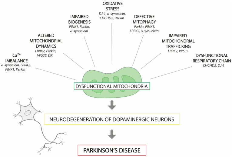
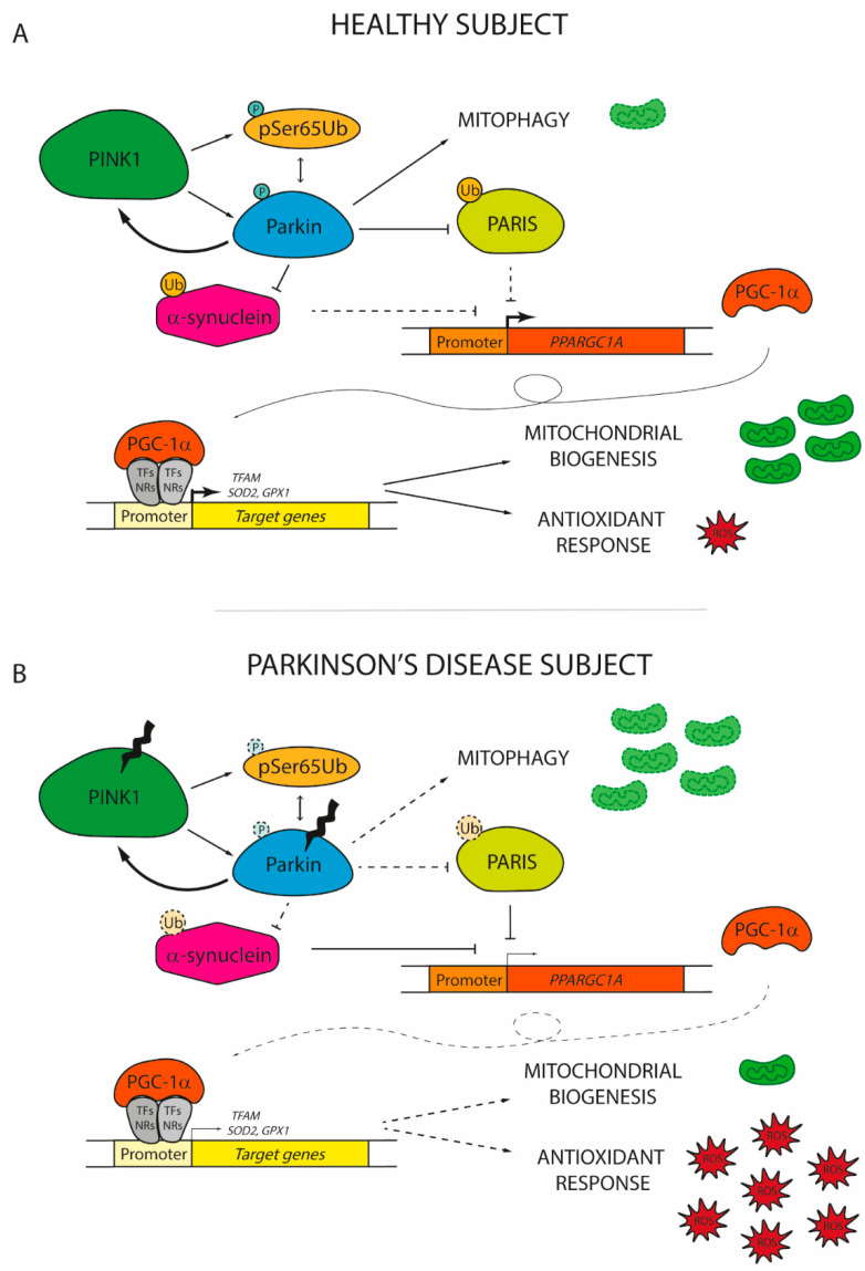
Parkinson's disease is one of the most common neurodegenerative disorders worldwide, characterized by a progressive loss of dopaminergic neurons mainly localized in the substantia nigra pars compacta. In recent years, the detailed analyses of both genetic and idiopathic forms of the disease have led to a better understanding of the molecular and cellular pathways involved in PD, pointing to the centrality of mitochondrial dysfunctions in the pathogenic process. Failure of mitochondrial quality control is now considered a hallmark of the disease. The peroxisome proliferator-activated receptor gamma coactivator 1 (PGC-1) family acts as a master regulator of mitochondrial biogenesis. Therefore, keeping PGC-1 level in a proper range is fundamental to guarantee functional neurons. Here we review the major findings that tightly bond PD and PGC-1s, raising important points that might lead to future investigations.

Keywords: PGC-1; Parkinson’s disease; coactivators; mitochondria; neurodegenerative disease.


Conflict of interest statement

The authors declare no conflict of interest.

Figures

Figure 1
Figure 1
Mitochondrial dysfunctions associated with Parkinson’s disease pathogenesis. The disruption of a gene (in italic) involved in mitochondrial homeostasis may result in the generation of dysfunctional mitochondria in the substantia nigra pars compacta, leading to the loss of pigmented dopaminergic neurons. The consequent dopamine deficit causes the motor symptoms associated with the disease. Abbreviations: PTEN-induced putative kinase 1 (PINK1), α-synuclein (SNCA), leucine-rich repeat kinase 2 (LRRK2), vacuolar protein sorting-associated protein 35 (VPS35), coiled-coil-helix-coiled-coil-helix domain containing 2 (CHCHD2).
Figure 2
Figure 2
The role of peroxisome proliferator-activated receptor gamma coactivator 1 (PGC-1α) in the onset of Parkinson’s disease. (A) In healthy subjects, damaged mitochondria are promptly replaced with new functional organelles. Mitochondrial depolarization induces PTEN-induced putative kinase 1 (PINK1) to phosphorylate serine 65 residue of ubiquitin (pSer65Ub) and of Parkin, which in turn interact together to increase the PINK1 phosphorylation rate of Parkin. The activation of the PINK1/Parkin pathway promotes the degradation of dysfunctional mitochondria via mitophagy and concomitantly induces the expression of PGC-1α by the ubiquitination of PARIS and α-synuclein. By coactivating nuclear receptors (NRs) or transcription factors (TFs), PGC-1α promotes the expression of genes involved in mitochondrial biogenesis (mitochondrial transcription factor A, TFAM) and antioxidant response (superoxide dismutase 2, SOD2; glutathione peroxidase 1, GPX1). (B) In Parkinson’s disease individuals, PINK1 and Parkin usually display loss-of-function mutations. Therefore, the PINK1/Parkin axis cannot prevent the accumulation of dysfunctional mitochondria due to its inability to sustain mitophagy. At the same time, altered Parkin fails to promote the degradation of both PARIS and α-synuclein, which start to accumulate in the nucleus inhibiting PGC-1α transcription. The low levels of PGC-1α observed in PD patients are not sufficient to induce the expression of genes involved in the renewal of mitochondria and in the antioxidant response. Thereby, reactive oxygen species (ROS) start to accumulate, finally leading to the damage and the death of dopaminergic neurons. Dashed lines and soft colours represent inhibited actions/pathways.

Similar articles

Cited by

References

    1. Diseases G.B.D., Injuries C. Global burden of 369 diseases and injuries in 204 countries and territories, 1990–2019: A systematic analysis for the Global Burden of Disease Study 2019. Lancet. 2020;396:1204–1222. doi: 10.1016/S0140-6736(20)30925-9. - DOI - PMC - PubMed
    1. Pezzoli G., Cereda E. Exposure to pesticides or solvents and risk of Parkinson disease. Neurology. 2013;80:2035–2041. doi: 10.1212/WNL.0b013e318294b3c8. - DOI - PubMed
    1. Langston J.W., Ballard P.A., Jr. Parkinson’s disease in a chemist working with 1-methyl-4-phenyl-1,2,5,6-tetrahydropyridine. N. Engl. J. Med. 1983;309:310. doi: 10.1056/nejm198308043090511. - DOI - PubMed
    1. Grunewald A., Kumar K.R., Sue C.M. New insights into the complex role of mitochondria in Parkinson’s disease. Prog. Neurobiol. 2019;177:73–93. doi: 10.1016/j.pneurobio.2018.09.003. - DOI - PubMed
    1. Green D.R., Van Houten B. SnapShot: Mitochondrial quality control. Cell. 2011;147:950–950 e951. doi: 10.1016/j.cell.2011.10.036. - DOI - PMC - PubMed

MeSH terms