. 2021 Mar 28;13(7):1551.
doi: 10.3390/cancers13071551.

Combination Assessment of Diffusion-Weighted Imaging and T2-Weighted Imaging Is Acceptable for the Differential Diagnosis of Lung Cancer from Benign Pulmonary Nodules and Masses

Affiliations

Combination Assessment of Diffusion-Weighted Imaging and T2-Weighted Imaging Is Acceptable for the Differential Diagnosis of Lung Cancer from Benign Pulmonary Nodules and Masses

Katsuo Usuda et al. Cancers (Basel). .

Abstract

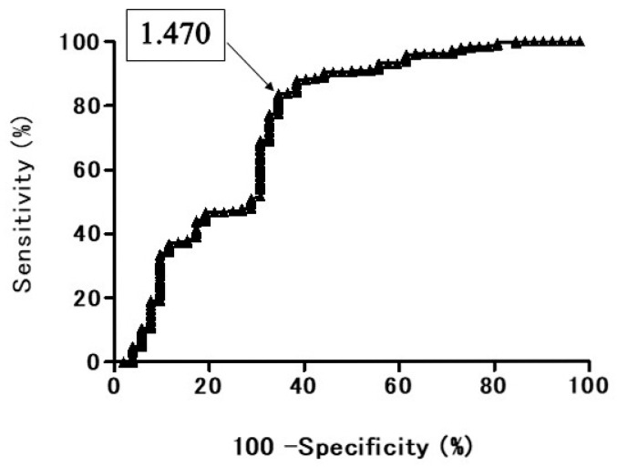
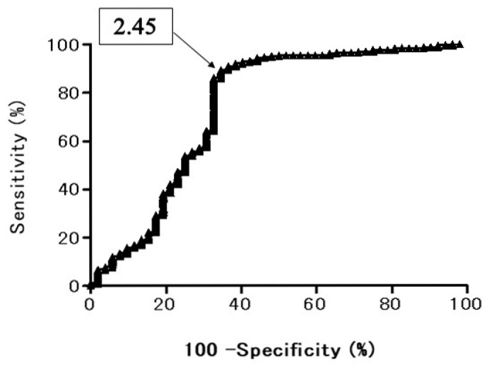
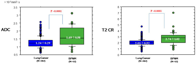
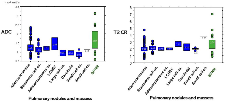
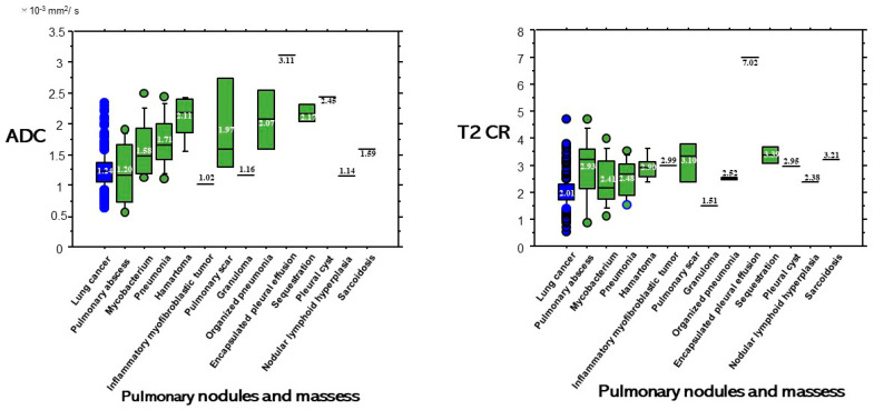
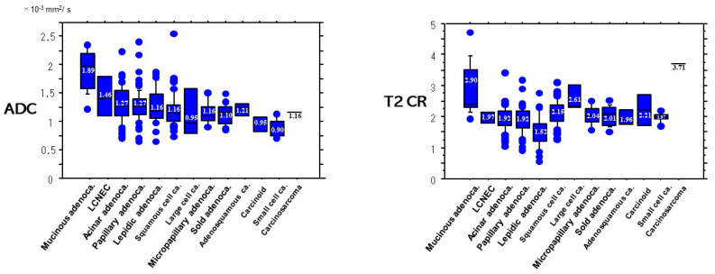
The purpose of this study is to determine whether the combination assessment of DWI and T2-weighted imaging (T2WI) improves the diagnostic ability for differential diagnosis of lung cancer from benign pulmonary nodules and masses (BPNMs). The optimal cut-off value (OCV) for differential diagnosis was set at 1.470 × 10⁻³ mm2/s for apparent diffusion coefficient (ADC), and at 2.45 for T2 contrast ratio (T2 CR). The ADC (1.24 ± 0.29 × 10⁻³ mm2/s) of lung cancer was significantly lower than that (1.69 ± 0.58 × 10⁻³ mm2/s) of BPNM. The T2 CR (2.01 ± 0.52) of lung cancer was significantly lower than that (2.74 ± 1.02) of BPNM. As using the OCV for ADC, the sensitivity was 83.9% (220/262), the specificity 63.4% (33/52), and the accuracy 80.6% (253/314). As using the OCV for T2 CR, the sensitivity was 89.7% (235/262), the specificity 61.5% (32/52), and the accuracy 85.0% (267/314). In 212 PNMs which were judged to be malignant by both DWI and T2WI, 203 PNMs (95.8%) were lung cancers. In 33 PNMs which were judged to be benign by both DWI and T2WI, 23 PNMs (69.7%) were BPNMs. The combined assessment of DWI and T2WI could judge PNMs more precisely and would be acceptable for differential diagnosis of PNMs.

Keywords: T2-weighted imaging (T2WI); apparent diffusion coefficient (ADC); diffusion-weighted magnetic resonance imaging (DWI); lung cancer; magnetic resonance imaging (MRI); pulmonary abscess; pulmonary nodule and mass (PNM).


Conflict of interest statement

All authors have no conflict of interest to declare.

Figures

Figure 1
Figure 1
Receiver operating characteristic (ROC) curve shows the diagnostic performance of diffusion-weighted magnetic resonance imaging (DWI) for distinguishing benign pulmonary nodule and mass (BPNM) from lung cancer. Area under the ROC curve 74.8%. Apparent diffusion coefficient (ADC) = 1.470 × 10⁻³ mm2/s, sensitivity 84.2%, specificity 63.5%.
Figure 2
Figure 2
Receiver operating characteristic (ROC) curve shows the diagnostic performance of T2 contrast ratio (T2 CR) for distinguishing BPNM from lung cancer. Area under the ROC curve 74.3%. T2 CR = 2.45, sensitivity 89.5%, specificity 65.4%.
Figure 3
Figure 3
(a) CT, (b) DWI, (c) ADC map, (d) T2 WI. Case 1: Adenocarcinoma. ADC 1.39 × 10⁻³ mm2/s, T2 CR: 1.67. Case2: Squamous cell carcinoma. ADC 1.04 × 10⁻³ mm2/s, T2 CR: 1.25.
Figure 4
Figure 4
(a) CT, (b) DWI, (c) ADC map, (d) T2 WI. Case 3: Hamartoma, ADC 2.43 × 10⁻³ mm2/s, T2 CR: 3.61. Case 4: Pulmonary abscess. ADC 0.837 × 10⁻³ mm2/s, T2 CR: 3.64. Case 5: Pulmonary tuberculosis. ADC 1.85 × 10⁻³ mm2/s, T2 CR:1.87.
Figure 5
Figure 5
ADC and T2 Contrast ratio (CR) between lung cancers and BPNMs. The ADC (1.24 ± 0.29 × 10⁻³ mm2/s) of lung cancers was significantly lower than that (1.69 ± 0.58 × 10⁻³ mm2/s) of BPNMs (p < 0.0001). T2 CR was the ratio of T2 signal intensity of the pulmonary nodule divided by T2 signal intensity of a rhomboid muscle. The T2 CR (2.01 ± 1.02) of lung cancers was significantly lower than that (2.74 ± 1.02) of BPNMs (p < 0.0001).
Figure 6
Figure 6
ADC and T2 CR based on diagnosis of PNMs.
Figure 7
Figure 7
The ADC (1.20 ± 0.53 × 10⁻³ mm2/s) of pulmonary abscesses was not significantly lower than that (1.24 ± 0.29 × 10⁻³ mm2/s) of lung cancers (p = 0.695). The ADC (1.58 ± 0.47 × 10⁻³ mm2/s) of mycobacterial infections was significantly higher than that (1.24 ± 0.29 × 10⁻³ mm2/s) of lung cancers (p < 0.0001). The T2 CR (2.93 ± 1.26) of pulmonary abscesses was significantly higher than that (2.01 ± 0.52) of lung cancers (p = 0.010) and the T2 CR (2.41 ± 0.86) of mycobacterial infections was significantly higher than (2.01 ± 0.52) of lung cancers (p = 0.010).
Figure 8
Figure 8
ADC and T2 CR based on pathologic subtypes of lung cancer. Both the ADC and the T2CR of mucinous adenocarcinoma were significantly higher than those of other pathologic subtypes.

Similar articles

Cited by

References

    1. Could M.K., Maclean C.C., Kuschner W.G., Rydzak C.E., Owens D.K. Accuracy of positron emission tomography for diagnosis of pulmonary nodules and mass lesions: A meta-analysis. JAMA. 2001;285:914–924. - PubMed
    1. Cheran S.K., Nielsen N.D., Patz E.F. False-negative findings for primary lung tumors on FDG positron emission tomography. Staging and prognostic implications. Am. J. Roentgenol. 2004;182:1129–1132. doi: 10.2214/ajr.182.5.1821129. - DOI - PubMed
    1. Satoh Y., Ichikawa T., Motosugi U., Kimura K., Sou H., Sano K., Araki T. Diagnosis of peritoneal dissemination. Comparison of 18F-DDG PET/CT, diffusion-weighted MRI, and contrast-enhanced MDCT. Am. J. Roentgenol. 2011;196:447–453. doi: 10.2214/AJR.10.4687. - DOI - PubMed
    1. Goo J.M., Im J.-G., Do K.-H., Yeo J.S., Seo J.B., Kim H.Y., Chung J.-K. Pulmonary tuberculoma evaluated by means of FDG PET. Findings in 10 cases. Radiology. 2000;216:117–121. doi: 10.1148/radiology.216.1.r00jl19117. - DOI - PubMed
    1. Webb W.R., Gatsonis C., Zerhouni E.A., Heelan R.T., Glazer G.M., Francis I.R., McNeil B.J. CT and MR imaging in staging non-small cell bronchogenic carcinoma: Radiologic Diagnostic Oncology Group. Radiology. 1991;178:705–713. doi: 10.1148/radiology.178.3.1847239. - DOI - PubMed

LinkOut - more resources