. 2021 Mar 6;22(5):2665.
doi: 10.3390/ijms22052665.

A Genomic Blueprint of Flax Fungal Parasite Fusarium oxysporum f. sp. lini

Affiliations

A Genomic Blueprint of Flax Fungal Parasite Fusarium oxysporum f. sp. lini

Anastasia Samsonova et al. Int J Mol Sci. .

Abstract

Fusarium wilt of flax is an aggressive disease caused by the soil-borne fungal pathogen Fusarium oxysporum f. sp. lini. It is a challenging pathogen presenting a constant threat to flax production industry worldwide. Previously, we reported chromosome-level assemblies of 5 highly pathogenic F. oxysporum f. sp. lini strains. We sought to characterize the genomic architecture of the fungus and outline evolutionary mechanisms shaping the pathogen genome. Here, we reveal the complex multi-compartmentalized genome organization and uncover its diverse evolutionary dynamics, which boosts genetic diversity and facilitates host adaptation. In addition, our results suggest that host of functions implicated in the life cycle of mobile genetic elements are main contributors to dissimilarity between proteomes of different Fusaria. Finally, our experiments demonstrate that mobile genetics elements are expressed in planta upon infection, alluding to their role in pathogenicity. On the whole, these results pave the way for further in-depth studies of evolutionary forces shaping the host-pathogen interaction.

Keywords: Fusarium oxysporum f. sp. lini; comparative genomics; flax; genome architecture; genome evolution; transposable elements.


Conflict of interest statement

The authors declare no conflict of interest.

Figures
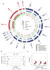
Figure 1
Figure 1
Genomic architecture and properties of Fusarium oxysporum f. sp. lini. (a) Genomic architecture of Fusarium oxysporum f. sp. lini MI39 isolate visualized with Circos software. The outermost ring depicts chromosome ideogram where chromosomes were assigned to either Core (blue) or Variable (red) compartments of the genome. The next two rings (violet and red ticks) show effector protein loci and MIMPs genomic positions, respectively. Protein-coding genes (p.-c genes), SNP, and repetitive element densities computed in 50 K bins along the chromosomes are presented as blue, red, and green tracks. Color intensity gradient reflects changes in density. The darkest color shade corresponds to maximum density values. The most inner track (grey with circle glyphs) depicts location of CAZymes in MI39 F. oxysporum f. sp. lini genome. CAZyme types are color-coded as follows: blue—carbohydrate-binding, red—auxillary activity, violet—glycoside hydrolase, green—carbohydrate esterase, orange—glycosyl transferase, brown—polysaccharide lyase. (b) Fraction of genomic features associated with either Core (blue) or Variable (red) parts of the genome. Values in a dumbbell chart correspond to percentage of features attributed to either of them. (c) Distribution of SNP frequency values computed in various genomic regions (as described in (a)) and parts of the genome. Core and variable part is shown in red and blue, correspondingly.
Figure 2
Figure 2
Compartmentalization of F. oxysporum f. sp. lini genome. (a) Hierarchical clustering of genomic regions in accordance with genomic characteristics associated with genome stability. Regions attributed to the Core part are marked with blue, whereas loci from the variable part are shown in red. (b) Visualization of genomic region clustering with uniform manifold approximation and projection (UMAP) reveals three groups of loci. (c). Homology heatmap for comparisons of the percentage identity between flax strains. The percentage identity is indicated by different box colors, whereas the numbers shown is the boxes correspond to percentage of overlap between chromosomes. Chromosome labels are highlighted according to genome compartmentalization.
Figure 3
Figure 3
Functional annotation of F. oxysporum genomes. Heatmap of PFAM domain frequencies observed in variable part of F. oxysporum genomes. (a) The heatmap shows 35 PFAM domains significantly overrepresented (adj. p-value < 10−6) in variable genome of MI39 F. oxysporum f. sp. lini isolate. Frequency values associated with each domain were mean-centered by rows. Each row of the heatmap represents log10-transformed frequency values of one PFAM domain across all F. oxysporum formae speciales (yellow, high frequency; brown, low frequency). Ivory color represents missing data points i.e., situations when a domain has not been detected in a pathogen’s genome. Domain enrichment significance is indicated in shades of blue on a separate panel on the left of the heatmap. The next two panels present domain name for each PFAM accession and information regarding domain function and its relevance to genome mobilization. The abbreviated F. oxysporum formae specialis names shown at the bottom of the heatmap stand for: MI39, FOCU—f. sp. cubense, FOME—f. sp. meloni, FOLY—f. sp. lycopersici, FOPI—f. sp. pisi, FOVA—f. sp. vasinfectum, FOCT—f. sp. cotton, FORP—f. sp. raphani, FO47—FO47 strain, FORA—f. sp. radicis, FOCO—f. sp. conglutinans, FOVE—Fusarium verticilloides. (b) Significantly overrepresented GO terms encompassing processes associated with proteins in the variable compartment of the MI39 genome. Spheres represent GO term enrichment with size and color as indicated in the inset.
Figure 4
Figure 4
Bayesian phylogeny of Fusarium oxysporum isolates from flax and other hosts. The phylogeny is inferred from the maximum likelihood analysis of concatenated alignment of RPB2-TUB2-EF1α and is rooted on the outgroup comprised of F. solani and F. graminearum branch). Tree tips and label colors reflect the pathogen’s host. The color-coded nucleotide sequence alignments are shown to the right of the tree.
Figure 5
Figure 5
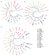
Consensus trees from the Bayesian phylogenetic analysis of SIX genes from the Fusarium clade. Tree tips and label colors reflect the pathogen’s host organism. List of hosts is shown separately. (a) SIX1 consensus tree consensus tree, (b) SIX7 consensus tree, and (c) SIX13 consensus tree.
Figure 6
Figure 6
PCR analyses showing the presence of F.oxysporum f. sp. lini gene products. (a) PCR amplification of E1Fα (reference), SIX1, SIX7, and SIX13; (b) PCR amplification of genes involved in a life cycle of transposable elements. Control cDNA, extracted from non-infected plant is labeled with Cntrl, whereas Fus marks cDNA obtained from infected flax roots as described in the Materials and Methods section.

References

    1. Zhang Y., Ma L.-J. Deciphering Pathogenicity of Fusarium oxysporum From a Phylogenomics Perspective. Adv. Genet. 2017;100:179–209. - PubMed
    1. Ma L.-J., van der Does H.C., Borkovich K.A., Coleman J.J., Daboussi M.-J., Di Pietro A., Dufresne M., Freitag M., Grabherr M., Henrissat B., et al. Comparative genomics reveals mobile pathogenicity chromosomes in Fusarium. Nature. 2010;464:367–373. doi: 10.1038/nature08850. - DOI - PMC - PubMed
    1. Williams A.H., Sharma M., Thatcher L.F., Azam S., Hane J.K., Sperschneider J., Kidd B.N., Anderson J.P., Ghosh R., Garg G., et al. Comparative genomics and prediction of conditionally dispensable sequences in legume–infecting Fusarium oxysporum formae speciales facilitates identification of candidate effectors. BMC Genom. 2016;17:311–324. doi: 10.1186/s12864-016-2486-8. - DOI - PMC - PubMed
    1. Armitage A.D., Taylor A., Sobczyk M.K., Baxter L., Greenfield B.P.J., Bates H.J., Wilson F., Jackson A.C., Ott S., Harrison R.J., et al. Characterisation of pathogen-specific regions and novel effector candidates in Fusarium oxysporum f. sp. cepae. Sci. Rep. 2018;8:13530. doi: 10.1038/s41598-018-30335-7. - DOI - PMC - PubMed
    1. Dong S., Raffaele S., Kamoun S. The two-speed genomes of filamentous pathogens: Waltz with plants. Curr. Opin. Genet. Dev. 2015;35:57–65. doi: 10.1016/j.gde.2015.09.001. - DOI - PubMed

Supplementary concepts

LinkOut - more resources