Review
. 2021 Mar 6;10(5):1107.
doi: 10.3390/jcm10051107.

Peri-Implantitis: A Clinical Update on Prevalence and Surgical Treatment Outcomes

Affiliations
Review

Peri-Implantitis: A Clinical Update on Prevalence and Surgical Treatment Outcomes

Andrea Roccuzzo et al. J Clin Med. .

Abstract

Dental implants may be considered a reliable routine procedure in clinical practice for the replacement of missing teeth. Results from long-term studies indicate that implant-supported dental prostheses constitute a predictable treatment method for the management of fully and partially edentulous patients. Implants and their restorations, however, are not free from biological complications. In fact, peri-implantitis, defined as progressive bone loss associated to clinical inflammation, is not a rare finding nowadays. This constitutes a concern for clinicians and patients given the negative impact on the quality of life and the sequelae originated by peri-implantitis lesions. The purpose of this narrative review is to report on the prevalence of peri-implantitis and to overview the indications, contraindications, complexity, predictability and effectiveness of the different surgical therapeutic modalities to manage this disorder.

Keywords: biological complications; bone regeneration; dental implants; peri-implantitis.


Conflict of interest statement

The authors declare no conflict of interest.

Figures

Figure 1
Figure 1
(a) Mandibular left premolar implant showing an increase in probing pocket depth as compared with previous records, bleeding and pus exhibiting shortly after probing. Note, the shallow vestibulum at the buccal aspect of the infected implant; (b) Radiographic image revealing significant bone loss. Note, the remaining particles of an anorganic bovine bone previously used for grafting; (c) Intra-operative appearance of the peri-implant infra-osseous defect after debridement. Note, the remaining particles of an anorganic bovine bone previously used for grafting simultaneously at implant placement stage.
Figure 2
Figure 2
(a) Clinical appearance of implants placed in bone augmented with anorganic bovine bone and autogenous bone 5 years after placement in a smoker patient. Note, the poor plaque control and the inadequate prosthesis design that precluded adequate self-performed oral hygiene measures; (b) Intra-operative appearance of the peri-implant defects after debridement. Note, the predominant horizontal pattern of bone resorption.
Figure 3
Figure 3
(a) Clinical appearance at several implant-supported fixed prosthesis involved affected of advanced peri-implantitis; (b) clinical probing indicates advanced attachment loss; (c) implant removal is suggested in advanced forms of peri-implantitis.
Figure 4
Figure 4
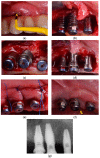
(a) Clinical presentation of peri-implantitis; (b) Access flap reveals moderate bone loss (<50%) (frontal view). Note the supra-crestal defect morphology; (c) Occlusal view of moderate bone loss; (d) Implantoplasty was performed as adjunct to the surgical resective therapy of peri-implantitis (frontal view); (e) Occlusal view of the implantoplasty and bone topography after osteoplasty to reach a flat bone architecture; (f) clinical resolution of peri-implantitis at 6-month follow-up; (g) bone stability is noted upon radiographic assessment.
Figure 5
Figure 5
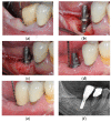
(a) Mandibular right premolar implant installed in pristine bone showing increased probing pocket depth as compared with previous records, bleeding and pus exhibiting shortly after probing; (b) Intra-operative appearance of the peri-implant infra-osseous defect after debridement; (c) Anorganic bovine bone mineral with 10% collagen is applied in the infra-osseous component; (d) Six-month follow-up after non-submerged healing, no signs of inflammation and peri-implant probing depth was noted to be consistent with health; (e) One-year follow-up after delivery of the final restoration. Note, peri-implant stability; (f) Radiographic image, at 1-year follow-up, reveals substantial bone fill.
Figure 6
Figure 6
(a) Clinical presentation of peri-implantitis; (b) Radiographic image compatible showing moderate (<50%) bone loss; (c) Inadequate prosthesis emergence profile; (d) Partial-thickness apical position flap; (e) Soft tissue conditioning by means of free epithelial graft; (f) Prosthesis contour modification to facilitate proximal access during self-performed oral hygiene; (g) Clinical resolution of peri-implantitis associated with a gain of keratinized mucosa.
Figure 6
Figure 6
(a) Clinical presentation of peri-implantitis; (b) Radiographic image compatible showing moderate (<50%) bone loss; (c) Inadequate prosthesis emergence profile; (d) Partial-thickness apical position flap; (e) Soft tissue conditioning by means of free epithelial graft; (f) Prosthesis contour modification to facilitate proximal access during self-performed oral hygiene; (g) Clinical resolution of peri-implantitis associated with a gain of keratinized mucosa.

Similar articles

Cited by

References

    1. Berglundh T., Armitage G., Araujo M.G., Gustavo A.-O., Juan B., Paulo M.C., Stephen C., David C., Jan D., Elena F., et al. Peri-implant diseases and conditions: Consensus report of workgroup 4 of the 2017 World Workshop on the Classification of Periodontal and Peri-Implant Diseases and Conditions. J. Periodontol. 2018;89:S313–S318. doi: 10.1002/JPER.17-0739. - DOI - PubMed
    1. Derks J., Schaller D., Håkansson J., Wennström J.L., Tomasi C., Berglundh T. Effectiveness of implant therapy analyzed in a Swedish population: Prevalence of peri-implantitis. J. Dent. Res. 2016;95:43–49. doi: 10.1177/0022034515608832. - DOI - PubMed
    1. Derks J., Tomasi C. Peri-implant health and disease. A systematic review of current epidemiology. J. Clin. Periodontol. 2015;42:S158–S171. doi: 10.1111/jcpe.12334. - DOI - PubMed
    1. Aguirre-Zorzano L.A., Estefania-Fresco R., Telletxea O., Bravo M. Prevalence of peri-implant inflammatory disease in patients with a history of periodontal disease who receive supportive periodontal therapy. Clin. Oral Implant Res. 2015;26:1338–1344. doi: 10.1111/clr.12462. - DOI - PubMed
    1. Daubert D.M., Weinstein B.F., Bordin S., Leroux B.G., Flemming T.F. Prevalence and predictive factors for peri-implant disease and implant failure: A cross-sectional analysis. J. Periodontol. 2015;86:337–347. doi: 10.1902/jop.2014.140438. - DOI - PubMed