. 2021 Mar 6;22(5):2673.
doi: 10.3390/ijms22052673.

Excess TGF-β1 Drives Cardiac Mesenchymal Stromal Cells to a Pro-Fibrotic Commitment in Arrhythmogenic Cardiomyopathy

Affiliations

Excess TGF-β1 Drives Cardiac Mesenchymal Stromal Cells to a Pro-Fibrotic Commitment in Arrhythmogenic Cardiomyopathy

Angela Serena Maione et al. Int J Mol Sci. .

Abstract

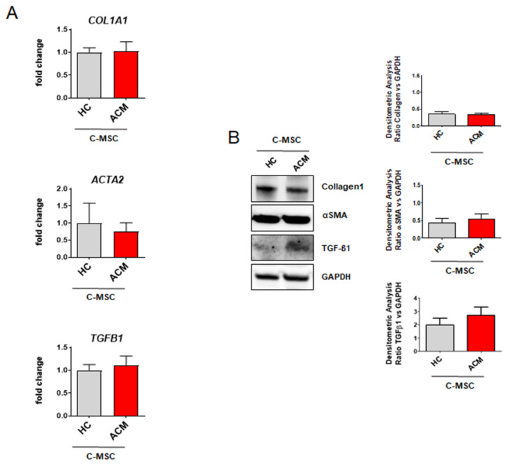
Arrhythmogenic Cardiomyopathy (ACM) is characterized by the replacement of the myocardium with fibrotic or fibro-fatty tissue and inflammatory infiltrates in the heart. To date, while ACM adipogenesis is a well-investigated differentiation program, ACM-related fibrosis remains a scientific gap of knowledge. In this study, we analyze the fibrotic process occurring during ACM pathogenesis focusing on the role of cardiac mesenchymal stromal cells (C-MSC) as a source of myofibroblasts. We performed the ex vivo studies on plasma and right ventricular endomyocardial bioptic samples collected from ACM patients and healthy control donors (HC). In vitro studies were performed on C-MSC isolated from endomyocardial biopsies of both groups. Our results revealed that circulating TGF-β1 levels are significantly higher in the ACM cohort than in HC. Accordingly, fibrotic markers are increased in ACM patient-derived cardiac biopsies compared to HC ones. This difference is not evident in isolated C-MSC. Nevertheless, ACM C-MSC are more responsive than HC ones to TGF-β1 treatment, in terms of pro-fibrotic differentiation and higher activation of the SMAD2/3 signaling pathway. These results provide the novel evidence that C-MSC are a source of myofibroblasts and participate in ACM fibrotic remodeling, being highly responsive to ACM-characteristic excess TGF-β1.

Keywords: TGF-β1; arrhythmogenic cardiomyopathy; cardiac remodeling; cardiac-mesenchymal stromal cells; fibrosis.


Conflict of interest statement

The authors declare no conflict of interest.

Figures

Figure 1
Figure 1
Fibrotic profile characterization of ACM-patient-derived tissues. (A) TGF-β1 plasma levels of HC donors and ACM patients. n = 52/group. Student’s t-test: ** p < 0.01. (B) Expression of fibrosis-associated genes (COL1A1, ACTA2, TGFB1) in total RNA extracts of RV endomyocardial bioptic samples from HC donors and ACM patients. GAPDH was used as a house-keeping gene and qRT-PCR data are presented as the fold change of target gene expression with respect to HC C-MSC. n = 4/group. Student’s t-test: * p < 0.05, ** p < 0.01. (C) Representative images of Western blot analysis of fibrosis-associated proteins (Collagen1, αSMA, TGF-β1) in total protein extracts of RV endomyocardial bioptic samples from HC donors and ACM patients. Quantification of the protein abundance relative to Ponceau Red is shown in the graphs. n = 4/group. Student’s t-test: * p < 0.05, ** p < 0.01. (D) Representative images of immunostaining of COL1A1 and CD44 sections from biopsies from HC donors and ACM patients. Nuclei are stained with Hoechst 33342. Magnification is 63X and the scale bar indicates 50 μm. n = 3/group.
Figure 2
Figure 2
Analysis of fibrotic markers in ACM C-MSC. (A) COL1A1, ACTA2, TGFB1 gene expression in total RNA extracts of cardiac mesenchymal stromal cells isolated from HC donors and ACM patients in culture condition. GAPDH was used as a house-keeping gene and qRT-PCR data are presented as the fold change of target gene expression respect to HC C-MSC. n = 7/group. Student’s t-test: no significant difference. (B) Western blot analysis of Collagen1, αSMA, and TGF-β1 proteins in total extracts of cardiac mesenchymal stromal cells isolated from HC donors and ACM patients in culture condition. Quantification of the protein abundance relative to GAPDH is shown in the graphs. n = 4/group. Student’s t-test: no significant difference.
Figure 3
Figure 3
Effect of TGF-β1 stimulation on gene expression of pro-fibrotic markers. C-MSC isolated from HC donors and ACM patients were grown in low serum (2%) overnight and were stimulated or not with TGF-β1 for 24 h in the presence or absence of LY364947 treatment. Comparison of fibrosis-associated gene expression (TGFB1, CTGF, COL1A1, COL1A2, COL3A1, ACTA2) in total RNA extracts from ACM (A) and HC C-MSC (B). GAPDH was used as a house-keeping gene and qRT-PCR data are presented as the fold change of target gene expression with respect to the untreated. n = 3/group. One-way ANOVA and Bonferroni’s post-test: * p < 0.05, ** p < 0.01.
Figure 4
Figure 4
TGF-β1 induces the overexpression of fibrosis-associated proteins. Following overnight low serum (2%) growth, cardiac mesenchymal stromal cells isolated from HC donors and ACM patients were stimulated with TGF-β1 for 5 days in the presence or absence of LY364947 treatment. Representative images of Western blot analysis on fibrosis-associated proteins (Collagen1, αSMA) in total protein extracts of treated cells. Quantification of the protein abundance relative to GAPDH has been shown as fold change respect to untreated protein in the graphs and results are expressed as mean ± SEM, (n = 3/group). One-way ANOVA and Bonferroni’s post-test: * p < 0.05.
Figure 5
Figure 5
TGF-β1 leads to increased collagen production and α-SMA positive stress fibers in ACM C-MSC. C-MSC isolated from HC donors and ACM patients were treated as described in Figure 4. Representative images of immunostaining for COL1A1 (A) and αSMA (B). Nuclei are stained with Hoechst 33342. Magnification is 40× and the scale bar indicates 50 μm (n = 3/group). Quantification of the images has been shown as fold change respect to untreated in the graphs and results are expressed as mean ± SEM, (n = 3/group). One-way ANOVA and Bonferroni’s post-test: * p < 0.05, ** p < 0.01.
Figure 6
Figure 6
ACM C-MSC pro-fibrotic commitment depends on the TGF-β1 canonical signaling pathway. Cardiac mesenchymal stromal cells isolated from HC donors and ACM patients were grown in low serum (2%) overnight and were stimulated or not with TGF-β1 for 30 min in the presence or absence of LY364947 treatment. (A) Representative images of Western blot analysis on active phosphorylated form and total SMAD2/3 in total protein extracts of treated cells. Phospho-SMAD2/3 levels were corrected by total SMAD2/3 densitometry. Western blot data are presented as the fold change with respect to untreated target protein expression. The results are expressed as mean ± SEM (n = 3/group). One-way ANOVA and Bonferroni’s post-test: * p < 0.05. (B) Representative images of immunostaining for SMAD2 to visualize nuclear translocation. Nuclei are stained with Hoechst 33342. Magnification is 40× and the scale bar indicates 50 μm (n = 3/group).

References

    1. Corrado D., Link M.S., Calkins H. Arrhythmogenic Right Ventricular Cardiomyopathy. N. Engl. J. Med. 2017;376:61–72. doi: 10.1056/NEJMra1509267. - DOI - PubMed
    1. Stadiotti I., Pompilio G., Maione A.S., Pilato C.A., D’Alessandra Y., Sommariva E. Arrhythmogenic cardiomyopathy: What blood can reveal? Heart Rhythm. 2019;16:470–477. doi: 10.1016/j.hrthm.2018.09.023. - DOI - PubMed
    1. Casella M., Gasperetti A., Sicuso R., Conte E., Catto V., Sommariva E., Bergonti M., Vettor G., Rizzo S., Pompilio G., et al. Characteristics of Patients With Arrhythmogenic Left Ventricular Cardiomyopathy: Combining Genetic and Histopathologic Findings. Circ. Arrhythm. Electrophysiol. 2020;13:e009005. doi: 10.1161/CIRCEP.120.009005. - DOI - PubMed
    1. Hoorntje E.T., Te Rijdt W.P., James C.A., Pilichou K., Basso C., Judge D.P., Bezzina C.R., van Tintelen J.P. Arrhythmogenic cardiomyopathy: Pathology, genetics, and concepts in pathogenesis. Cardiovasc. Res. 2017;113:1521–1531. doi: 10.1093/cvr/cvx150. - DOI - PubMed
    1. Lin C.Y., Lin Y.J., Li C.H., Chung F.P., Lo M.T., Lin C., Chang H.C., Chang S.L., Lo L.W., Hu Y.F., et al. Heterogeneous distribution of substrates between the endocardium and epicardium promotes ventricular fibrillation in arrhythmogenic right ventricular dysplasia/cardiomyopathy. Eurospace. 2018;20:501–511. doi: 10.1093/europace/euw393. - DOI - PubMed

MeSH terms