Review
. 2021 Mar 21;11(3):466.
doi: 10.3390/biom11030466.

Neuropathology of Animal Prion Diseases

Affiliations
Review

Neuropathology of Animal Prion Diseases

Leonor Orge et al. Biomolecules. .

Abstract

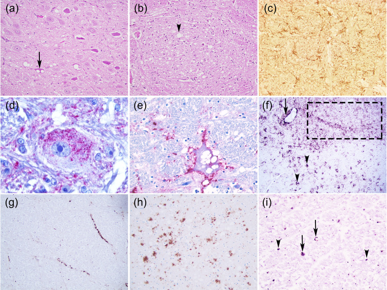
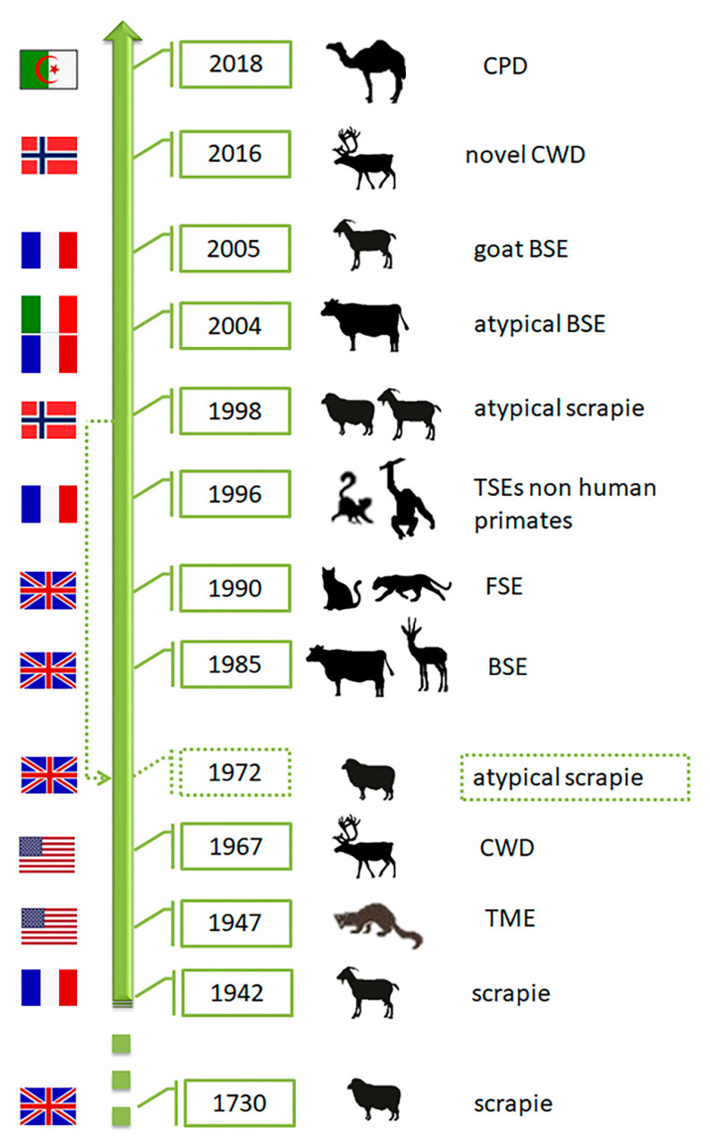
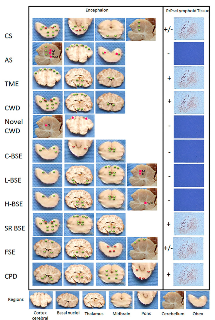
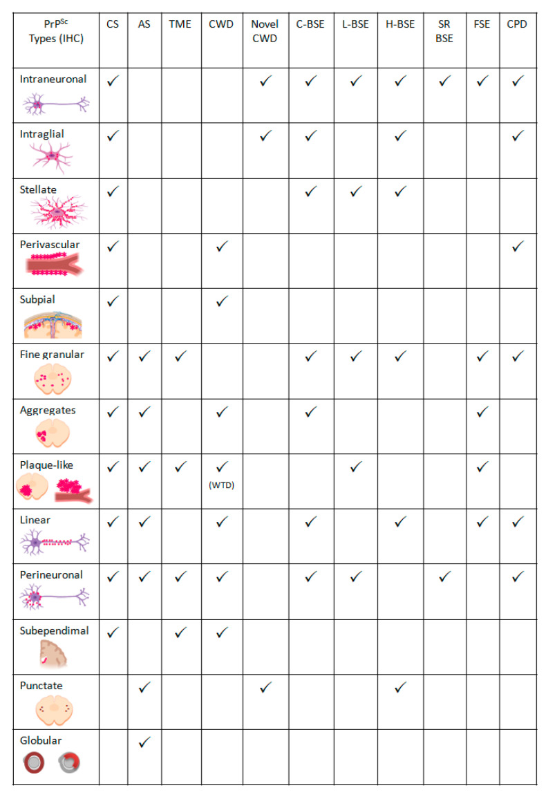
Transmissible Spongiform Encephalopathies (TSEs) or prion diseases are a fatal group of infectious, inherited and spontaneous neurodegenerative diseases affecting human and animals. They are caused by the conversion of cellular prion protein (PrPC) into a misfolded pathological isoform (PrPSc or prion- proteinaceous infectious particle) that self-propagates by conformational conversion of PrPC. Yet by an unknown mechanism, PrPC can fold into different PrPSc conformers that may result in different prion strains that display specific disease phenotype (incubation time, clinical signs and lesion profile). Although the pathways for neurodegeneration as well as the involvement of brain inflammation in these diseases are not well understood, the spongiform changes, neuronal loss, gliosis and accumulation of PrPSc are the characteristic neuropathological lesions. Scrapie affecting small ruminants was the first identified TSE and has been considered the archetype of prion diseases, though atypical and new animal prion diseases continue to emerge highlighting the importance to investigate the lesion profile in naturally affected animals. In this report, we review the neuropathology and the neuroinflammation of animal prion diseases in natural hosts from scrapie, going through the zoonotic bovine spongiform encephalopathy (BSE), the chronic wasting disease (CWD) to the newly identified camel prion disease (CPD).

Keywords: animal TSE; gene PRNP; neuroinflammation; neuropathology; prion; spongiform degeneration.


Conflict of interest statement

The authors declare no conflict of interest.

Figures

Figure 1
Figure 1
Neurohistopathological features of animal prion diseases. (a) Neuronal vacuolation (arrow) (Classical scrapie, sheep, medulla oblongata, dorsal vagal nucleus); (b) Neuropil vacuolation (arrow head) (Classical BSE, bovine, medulla oblongata, nucleus of the solitary tract); (c) Astrogliosis (Atypical scrapie, sheep, medulla oblongata, GFAP Polyclonal antibody, DAKO, 1:1000 dilution, x200); (di) Some PrPSc deposition types: (d) Intraneuronal (Feline spongiform encephalopathy, cat, medulla oblongata, reticular formation, 3F4 PrP Residues 109–111 Monoclonal antibody, DAKO, 1:300 dilution, x1000); (e) Perineuronal (BSE, bovine, medulla oblongata, reticular formation, 6H4 PrP Residues 144–152 Monoclonal antibody, Prionics AG, dilution 1:1000 x400); (f) Fine granular (dot line), stellate (arrow head), perivascular (arrow) (Classical scrapie, sheep, medulla oblongata, 2G11 PrP Residues 146-R154-R171-182 Monoclonal antibody, Pourquier Institute, 1:200 dilution, x100); (g) Linear (Classical BSE, bovine, medulla oblongata, reticular formation, 12F10 PrP Residues 142–160 Monoclonal antibody, SPBIO, 1:200 dilution, x200); (h) Plaque-like (cerebral cortex section from the CWD Proficiency testing 2008 organized by the European Reference Laboratory for TSEs-APHA, Weybridge, 2G11 PrP Monoclonal antibody, 1:200 dilution, x100; (i) Punctuate (arrow head) and globular (arrow) (Atypical scrapie, sheep, medula oblongata, pyramidal tract, 2G11 PrP Monoclonal antibody, 1:200 dilution). (ab) Haematoxylin and eosin (H & E) stain, x200; (de) StreptABC-AP, Nova fucsina, Mayer’s Haematoxylin; (c, fi) Vectastain Elite –HRP, DAB, Mayer’s Haematoxylin.
Figure 2
Figure 2
Multiple alignment of PrP protein from Ovis aries (ENSOARP00020014736), Capra hircus (CAA63050.1), Bos Taurus (ENSBTAP00000069134), Canis familiaris (ENSCAFP00000009107), Felis catus (ENSFCAP00000030786), Rangifer tarandus (ABS87897.1), Cervus elaphus (AAU93885.1), Alces alces (AZB50215.1) and Camelus dromedarius (CAA70901.1). The alignment was performed using T-Coffee program [9] and edited using Genedoc software [10].
Figure 3
Figure 3
Missense mutations of prion protein (PrP) in Ovis aries. The coloured bar represents structural features of the protein according to UniProt (P23907), including α-helix (orange), β-strand (green) and turn (red). Variants are classified above and below the bar as ”Risk factor” and ”Resistance” according to in vivo and in vitro analyses. The other variants were analysed using an in-silico approach through PROVEAN algorithm, and classified as ”Deleterious” and ”Neutral”.
Figure 4
Figure 4
Chronology of identification of animal prion diseases. TME-transmissible mink encephalopathy; CWD—chronic wasting disease; BSE-bovine spongiform encephalopathy; FSE—feline spongiform encephalopathy; CPD—camel prion disease. The flags represent the country of the first report of the disease. The dashed arrow indicates retrospective identification of atypical scrapie in 1972 (Silhouettes from Freepik.com and img.inkfrog.com).
Figure 5
Figure 5
Major distinctive distribution of lesions and PrPSc in animal prion diseases. CS—classical scrapie; AS—atypical scrapie; TME-transmissible mink encephalopathy; CWD—chronic wasting disease; C-BSE—classical bovine spongiform encephalopathy; L-BSE-atypical Low BSE; H-BSE—atypical High BSE; SR BSE—small ruminant BSE; FSE—feline spongiform encephalopathy; CPD—camel prion disease. M Molecular layer; G Granular layer; WM white matter; +, presence of PrPSc by immunohistochemistry; +/−, presence of PrPSc depending on PRNP genotype or species; −, absence of detectable PrPSc by immunohistochemistry; red dots, neuronal vacuolation; green dots, neuropil vacuolation; red asterisk, PrPSc deposition.
Figure 6
Figure 6
Summary of the most frequent PrPSc immunolabelling types in animal prion diseases. CS—classical scrapie; AS—atypical scrapie; TME-transmissible mink encephalopathy; CWD—chronic wasting disease; C-BSE—classical bovine spongiform encephalopathy; L-BSE-atypical Low BSE; H-BSE—atypical High BSE; SR BSE—small ruminant BSE; FSE—feline spongiform encephalopathy; CPD—camel prion disease. WTD—white-tailed deer. The figures of PrPSc types were created in BioRender.com.

References

    1. Budka H. Neuropathology of Prion Diseases. Br. Med. Bull. 2003;66:121–130. doi: 10.1093/bmb/66.1.121. - DOI - PubMed
    1. VanDeVelde M., Higgins R.J., Oevermann A. Veterinary Neuropathology: Essentials of Theory and Practice. Wiley-Blackwell; Oxford, UK: 2012.
    1. Spiropoulos J., Simmons M.M. Pathology of Animal Transmissible Spongiform Encephalopathies (TSEs) Food Saf. 2017;5:1–9. doi: 10.14252/foodsafetyfscj.2016027. - DOI - PMC - PubMed
    1. Soto C., Satani N. The Intricate Mechanisms of Neurodegeneration in Prion Diseases. Trends Mol. Med. 2011;17:14–24. doi: 10.1016/j.molmed.2010.09.001. - DOI - PMC - PubMed
    1. Carroll J., Chesebro B. Neuroinflammation, Microglia, and Cell-Association during Prion Disease. Viruses. 2019;11:65. doi: 10.3390/v11010065. - DOI - PMC - PubMed

Publication types

LinkOut - more resources