Expanding the Junction: New Insights into Non-Occluding Roles for Septate Junction Proteins during Development
- PMID: 33801162
- PMCID: PMC8006247
- DOI: 10.3390/jdb9010011
Expanding the Junction: New Insights into Non-Occluding Roles for Septate Junction Proteins during Development
Abstract
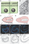
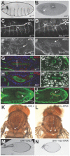
The septate junction (SJ) provides an occluding function for epithelial tissues in invertebrate organisms. This ability to seal the paracellular route between cells allows internal tissues to create unique compartments for organ function and endows the epidermis with a barrier function to restrict the passage of pathogens. Over the past twenty-five years, numerous investigators have identified more than 30 proteins that are required for the formation or maintenance of the SJs in Drosophila melanogaster, and have determined many of the steps involved in the biogenesis of the junction. Along the way, it has become clear that SJ proteins are also required for a number of developmental events that occur throughout the life of the organism. Many of these developmental events occur prior to the formation of the occluding junction, suggesting that SJ proteins possess non-occluding functions. In this review, we will describe the composition of SJs, taking note of which proteins are core components of the junction versus resident or accessory proteins, and the steps involved in the biogenesis of the junction. We will then elaborate on the functions that core SJ proteins likely play outside of their role in forming the occluding junction and describe studies that provide some cell biological perspectives that are beginning to provide mechanistic understanding of how these proteins function in developmental contexts.
Keywords: Drosophila; apical/basal polarity; dorsal closure; epithelia; morphogenesis; organogenesis; planar polarity; secretion; septate junction; wound healing.
Conflict of interest statement
The authors declare no conflict of interest.
Figures


Similar articles
-
Septate junction proteins are required for cell shape changes, actomyosin reorganization and cell adhesion during dorsal closure in Drosophila.Front Cell Dev Biol. 2022 Sep 27;10:947444. doi: 10.3389/fcell.2022.947444. eCollection 2022. Front Cell Dev Biol. 2022. PMID: 36238688 Free PMC article.
-
Septate Junction Proteins Play Essential Roles in Morphogenesis Throughout Embryonic Development in Drosophila.G3 (Bethesda). 2016 Aug 9;6(8):2375-84. doi: 10.1534/g3.116.031427. G3 (Bethesda). 2016. PMID: 27261004 Free PMC article.
-
The transmembrane protein Macroglobulin complement-related is essential for septate junction formation and epithelial barrier function in Drosophila.Development. 2014 Feb;141(4):899-908. doi: 10.1242/dev.102160. Development. 2014. PMID: 24496626
-
Molecular organization and function of invertebrate occluding junctions.Semin Cell Dev Biol. 2014 Dec;36:186-93. doi: 10.1016/j.semcdb.2014.09.009. Epub 2014 Sep 17. Semin Cell Dev Biol. 2014. PMID: 25239398 Review.
-
Occluding junctions of invertebrate epithelia.J Comp Physiol B. 2016 Jan;186(1):17-43. doi: 10.1007/s00360-015-0937-1. Epub 2015 Oct 28. J Comp Physiol B. 2016. PMID: 26510419 Review.
Cited by
-
Formation and remodelling of septate junctions in the epidermis of isopod Porcellioscaber during development.Zookeys. 2022 May 18;1101:159-181. doi: 10.3897/zookeys.1101.78711. eCollection 2022. Zookeys. 2022. PMID: 36760974 Free PMC article.
-
Septate junction proteins are required for cell shape changes, actomyosin reorganization and cell adhesion during dorsal closure in Drosophila.Front Cell Dev Biol. 2022 Sep 27;10:947444. doi: 10.3389/fcell.2022.947444. eCollection 2022. Front Cell Dev Biol. 2022. PMID: 36238688 Free PMC article.
-
A role for Myo-II zipper and spaghetti squash in Gliotactin-dependent Drosophila melanogaster wing hair planar cell polarity.PLoS One. 2025 Jul 23;20(7):e0328970. doi: 10.1371/journal.pone.0328970. eCollection 2025. PLoS One. 2025. PMID: 40700419 Free PMC article.
-
ESCRT-III-dependent adhesive and mechanical changes are triggered by a mechanism detecting alteration of septate junction integrity in Drosophila epithelial cells.Elife. 2024 Feb 2;13:e91246. doi: 10.7554/eLife.91246. Elife. 2024. PMID: 38305711 Free PMC article.
-
Mechanosensitive recruitment of Vinculin maintains junction integrity and barrier function at epithelial tricellular junctions.Curr Biol. 2024 Oct 21;34(20):4677-4691.e5. doi: 10.1016/j.cub.2024.08.060. Epub 2024 Sep 27. Curr Biol. 2024. PMID: 39341202
References
Publication types
Grants and funding
LinkOut - more resources
Full Text Sources
Other Literature Sources
Molecular Biology Databases
Miscellaneous

