. 2021 Mar 2;13(5):1041.
doi: 10.3390/cancers13051041.

Insulin-Like Growth Factor Binding Protein-3 Exerts Its Anti-Metastatic Effect in Aerodigestive Tract Cancers by Disrupting the Protein Stability of Vimentin

Affiliations

Insulin-Like Growth Factor Binding Protein-3 Exerts Its Anti-Metastatic Effect in Aerodigestive Tract Cancers by Disrupting the Protein Stability of Vimentin

Huong Thuy Le et al. Cancers (Basel). .

Abstract

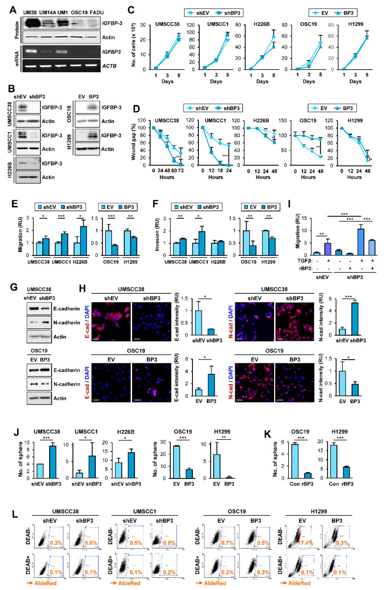
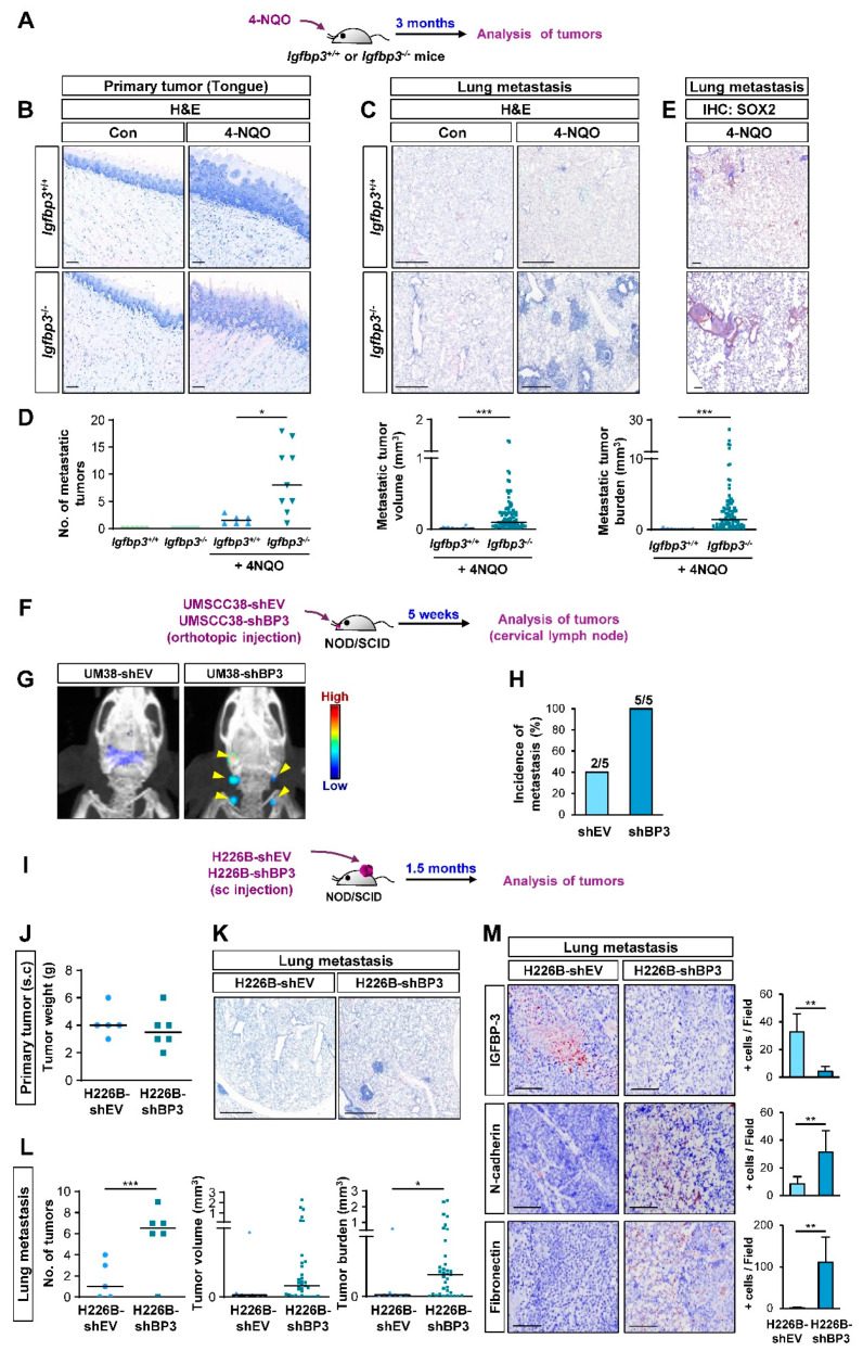
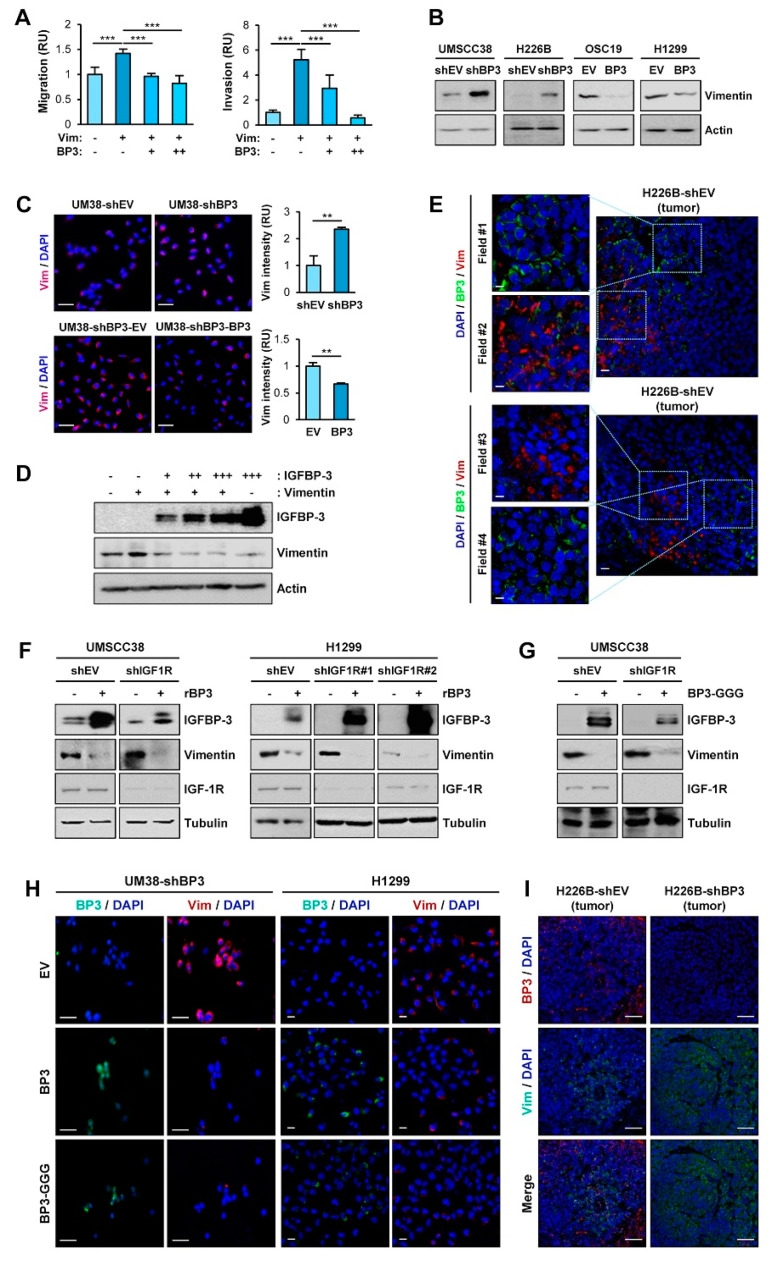
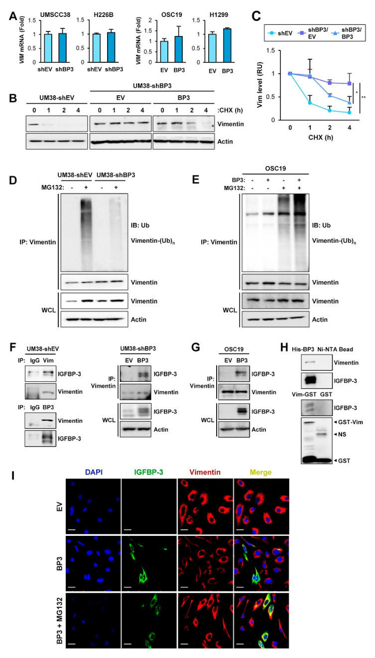
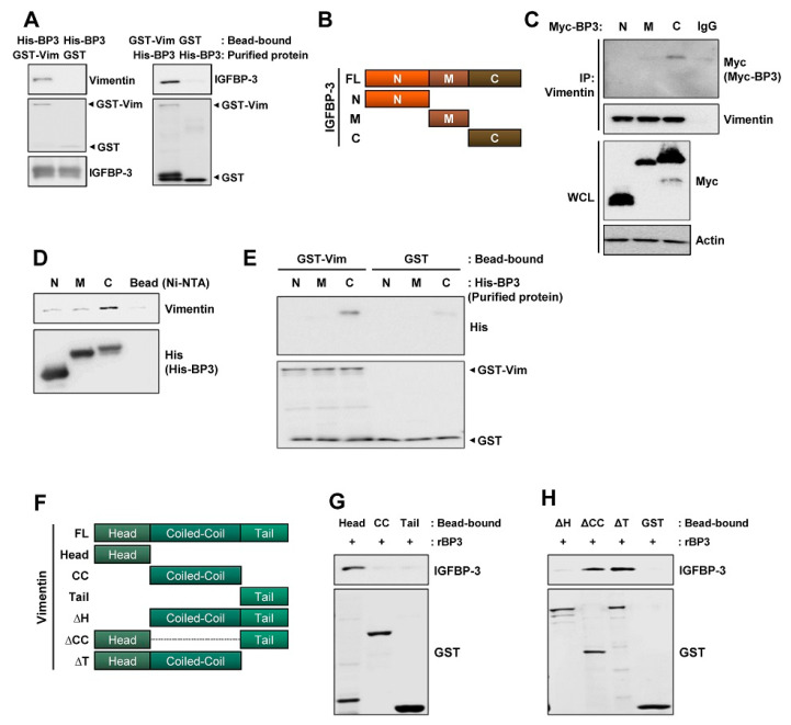
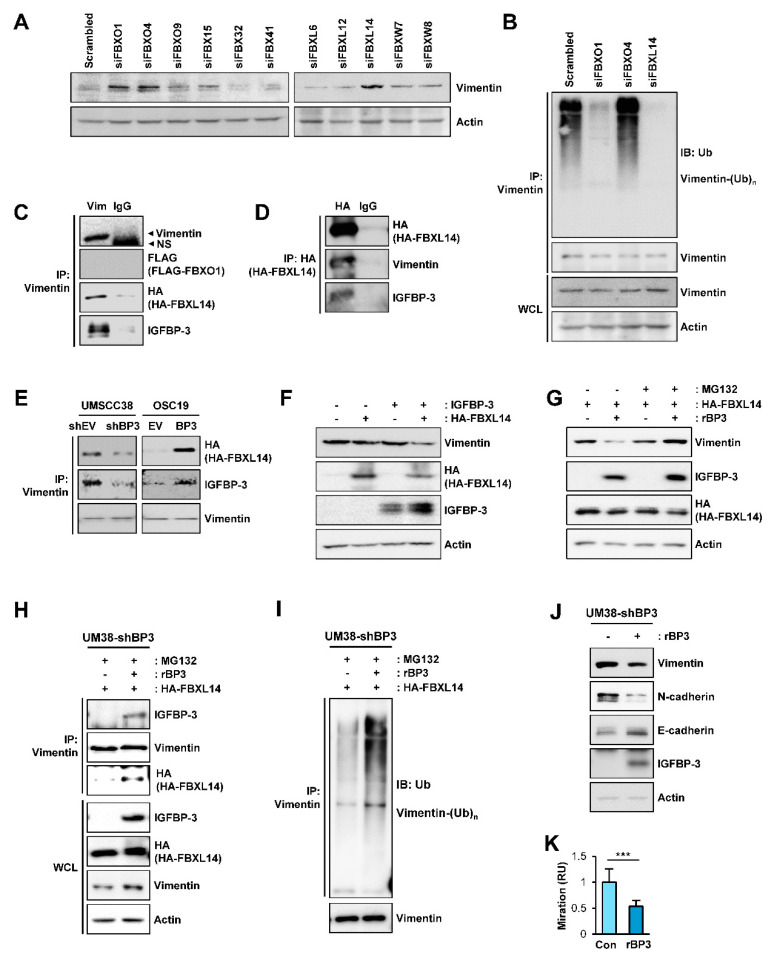
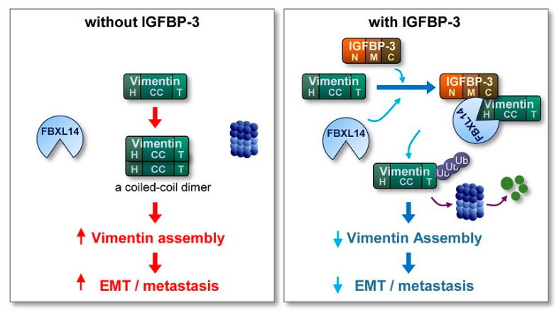
The proapoptotic, antiangiogenic, and antimetastatic activities of insulin-like growth factor binding protein-3 (IGFBP-3) through IGF-dependent or -independent mechanisms have been suggested in various types of human cancers. However, a mechanistic explanation of and downstream targets involved in the antimetastatic effect of IGFBP-3 is still lacking. In this study, by applying various in vitro and in vivo models, we show that IGFBP-3 suppresses migration and invasion of human head and neck squamous carcinoma (HNSCC) and non-small cell lung cancer (NSCLC) cells. Silencing IGFBP-3 expression elevated the migration and invasion of NSCLC and HNSCC cells in vitro and their local invasion and metastasis in vivo, whereas overexpression of IGFBP-3 decreased such prometastatic changes. Local invasion of 4-nitroquinoline-1-oxide (4-NQO)-induced HNSCC tumors was consistently significantly potentiated in Igfbp3 knockout mice compared with that in wild-type mice. Mechanistically, IGFBP-3 disrupted the protein stability of vimentin via direct binding and promoting its association with the E3 ligase FBXL14, causing proteasomal degradation. The C-terminal domain of IGFBP-3 and the head domain of vimentin are essential for their interaction. These results provide a molecular framework for IGFBP-3's IGF-independent antimetastatic and antitumor activities.

Keywords: insulin-like growth factor binding protein-3; metastasis; vimentin.


Conflict of interest statement

The authors declare no conflict of interest.

Figures

Figure 1
Figure 1
IGFBP-3 inhibits the acquisition of EMT and CSC-like phenotypes in HNSCC and NSCLC cells. (A,B) Western blot (WB) (A,B) and RT-PCR (A) analyses of IGFBP-3 expression in the indicated HNSCC cell lines (A) and HNSCC and NSCLC cell lines wherein IGFBP-3 expression was either silenced of enforced by stable transfection with shRNA or expression vector (B). (C) Effect of IGFBP-3 on cell proliferation was accessed by cell counting assay. (DF) Effects of IGFBP-3 expression on the migration and invasion of the indicated cancer cells evaluated by a scratch assay (D) and by the Transwell migration (E) and invasion (F) assays. (G,H) WB (G) and IF (H) analyses for the protein expression of E-cadherin and N-cadherin in the indicated cancer cells with manipulation of IGFBP-3 expression. Quantification of the fluorescence intensity was analyzed by ImageJ software (H). Scale bars: 50 μm (H). (I) UMSCC38-shEV and UMSCC38-shBP3 cells unstimulated or stimulated with TGF-β (10 ng/mL) in the absence or presence of recombinant IGFBP-3 (10 μg/mL) for 48 h were subjected to the Transwell migration assay (JL) Regulation of sphere formation (J,K) and ALDH activity (L) by manipulation of IGFBP-3 expression (J,L) or treatment with recombinant IGFBP-3 protein (K). The bar represents mean ± SD. * p < 0.05, ** p < 0.01, and *** p < 0.001, as determined by the two-tailed Student’s t-test compared with the corresponding control (CF,H,J,K) and one-way ANOVA followed by Dunnett’s post-hoc test (I). UM38: UMSCC38; UM14A: UMSCC14A; UM1: UMSCC1; OSC19: OSC19-Luc. EV: empty vector; BP3: IGFBP-3.
Figure 2
Figure 2
IGFBP-3 suppresses metastasis in vivo. (A) Schematic diagram illustrating the protocol to assess the impact of IGFBP-3 expression in metastasis of mouse HNSCC using the 4-NQO-induced HNSCC tumorigenesis model (Igfbp3+/+ Con: n = 6; Igfbp3+/+ 4-NQO: n = 7; Igfbp3−/− Con: n = 6; Igfbp3−/− 4-NQO: n = 9). (B,C) Primary (B) and metastatic (C) tumor formation in 4-NQO-administered mice was determined by H&E staining of the tongue (B) and lung (C) tissues. Scale bars: 25 μm (B), 1 mm (C). (D) Microscopic evaluation of the metastatic tumors in the lungs. (E) IHC analysis to determine the level of SOX2 expression in lung tissues. Scale bars: 25 μm. (F) Schematic diagram illustrating the protocol to assess the impact of IGFBP-3 expression in metastasis of human HNSCC using orthotopic tongue xenograft tumor model (n = 5 per group). (G) Representative IVIS images showing metastatic tumor formation in mice bearing UMSCC38-shBP3 tumor xenografts. (H) Microscopic evaluation showing increased metastasis in cervical lymph nodes in mice bearing UMSCC38-shBP3 cells compared with those in UMSCC38-shEV cells. Three cervical lymph nodes per mouse were evaluated. (I) Schematic diagram illustrating the protocol to assess the impact of IGFBP-3 expression in metastasis of human NSCLC using NSCLC xenograft tumor model (shEV: n = 5; shBP3: n = 6). (J) Changes in the weight of primary tumors between H226B-shEV and H226B-shBP3 tumor xenografts. (K,L) Upregulated metastatic tumor formation in mice bearing H226B-shBP3 tumor xenografts compared with those bearing 226B-shEV tumor xenografts, as determined by H&E staining of lung tissue sections (K) and microscopic evaluation of H&E-stained tissues (L). (M) The level of tumoral expression of N-cadherin and fibronectin in H226B-shEV and H226B-shBP3 xenograft tumors was determined by IHC analysis. Scale bars: 1 mm (K), 0.5 mm (M). The bar represents mean ± SD. * p < 0.05, ** p < 0.01, and *** p < 0.001, as determined by Mann-Whitney test (D,L (middle and right), M) and the two-tailed Student’s t-test (L, left) compared with the corresponding control. EV: empty vector; BP3: IGFBP-3.
Figure 3
Figure 3
IGFBP-3 negatively regulates vimentin protein expression in an IGF-independent manner. (A) Transwell migration and invasion analyses of OSC19-Luc cells in which pEGFP-vimentin (Vim) was transiently transfected, either alone or together with increasing concentrations of expression vector carrying IGFBP-3 (BP3). (B,C) Western blot (WB) (B) and IF (C) analyses of vimentin expression in the indicated cancer cells with enforced overexpression or knockdown of IGFBP-3. Scale bars: 50 μm (C). (D) Dose-dependent inhibition of vimentin protein expression by overexpression of IGFBP-3. OSC19-Luc cells were transiently transfected with empty vectors (-, pEGFP and pCMV6), pEGFP-vimentin and increasing amounts of pCMV6-IGFBP-3 for 48 h. The expression levels of IGFBP-3 and vimentin were determined by WB analysis. (E) Inverse correlation between IGFBP-3 and vimentin expression in H226B-shEV tumor xenografts was determined by IF analysis. Scale bars: 10 μm (left), 20 μm (right). (F–H) WB (F,G) and IF(H) analyses of vimentin expression in the indicated cancer cells following treatment with recombinant IGFBP-3 protein (F) or transient transfection with empty vector [- (G) or EV (H)], pCMV6-IGFBP3 (BP3), or pCMV6-IGFBP3-GGG (BP3-GGG) (G,H). Scale bars: 50 μm (H, left), 20 μm (H, right). (I) IF analysis showing upregulation of vimentin expression in H226B-shBP3 tumors compared with H226B-EV tumors. Scale bars: 50 μm. The bar represents mean ± SD. ** p < 0.01 and *** p < 0.001, as determined by one-way ANOVA followed by Dunnett’s post-hoc test (A) and the two-tailed Student’s t-test compared with the corresponding control (C). Vim: vimentin; BP3: IGFBP-3; OSC19: OSC19-Luc; EV: empty vector.
Figure 4
Figure 4
IGFBP-3 destabilizes vimentin via induction of ubiquitin-mediated proteasomal degradation of the vimentin protein. (A) Real-time PCR analysis of VIM mRNA levels in the indicated cancer cells with manipulations of IGFBP-3 expression. (B,C) UMSCC38-shEV and UMSCC38-shBP3 cells transfected with empty (EV) or pCMV6-IGFBP3 (BP3) vectors were treated with cycloheximide (CHX; 100 μg/mL) for the indicated time intervals and subjected to Western blot (WB) analysis on vimentin expression. (C) The relative expression level of vimentin was determined by densitometric analysis of vimentin expression at each time point normalized by the level of actin expression using the ImageJ software. (D,E) Paired UMSCC38-shEV and UMSCC38-shBP3 cells (D) and OSC19-EV and OSC19-BP3 cells (E) were treated with MG132 (10 µM) for 6 h. Cell lysates were immunoprecipitated with the anti-vimentin antibody, followed by WB analysis with the anti-ubiquitin antibody. Whole cell lysates (WCL) were also included for WB analysis. (F) UMSCC38-shEV (left) and UMSCC-shBP3 transfected with control (EV) or IGFBP-3 expression vector (right) were immunoprecipitated with preimmune serum (IgG) or anti-vimentin or anti-IGFBP-3 antibodies. The interaction between IGFBP-3 and vimentin was determined by WB analysis. (G) OSC19-Luc cells were transiently transfected with empty (EV) or pCMV6-IGFBP3 (BP3) vectors. Cell lysates were immunoprecipitated with the anti-vimentin antibody. Vimentin immunoprecipitates or whole-cell lysates (WCL) were subjected to WB analysis to determine the interaction between IGFBP-3 and vimentin. (H) Pull-down assays to determine the interaction between IGFBP-3 and vimentin. Ni-NTA agarose-bound recombinant His-IGFBP-3 (top) or glutathione (GSH)-agarose-bound recombinant GST-vimentin (bottom) proteins were incubated with UMSCC38 cell lysates. Ni-NTA agarose (top) or GSH-agarose-bound GST (bottom) were used to ensure specific interaction. (I) UMSCC38-shBP3 cells were transiently transfected with pCMV6 (EV) or pCMV6-IGFBP3 (BP3) and treated with MG132 (10 μM) for 6 h. The co-localization between IGFBP-3 and vimentin was determined by IF analysis. Scale bars: 20 µm. The bar represents mean ± SD. * p < 0.05 and ** p < 0.01, as determined by one-way ANOVA followed by Dunnett’s post-hoc test (C). Vim: vimentin; BP3: IGFBP-3; OSC19: OSC19-Luc; Ub: ubiquitin; NS: nonspecific band.
Figure 5
Figure 5
C-terminal domain of IGFBP-3 and the head domain of vimentin are critical for the binding of IGFBP-3 to vimentin. (A) Pull-down assays to determine the interaction between IGFBP-3 and vimentin. Purified and bead-bound proteins in each pull-down set were indicated. (B) Schematic diagram illustrating full-length and domain constructs (N: N-terminal domain; M: middle domain; C: C-terminal domain) of the IGFBP-3 protein. (C) HEK293T cells were transfected with Myc-tagged IGFBP-3 domain constructs for 48 h. Interaction between vimentin and C domain of IGFBP-3 was determined by immunoprecipitation with the anti-vimentin antibody and subsequent Western blot (WB) analysis. Bottom. Expression of each IGFBP-3 domain was determined by WB analysis. (D) Pull-down assay for the interaction between N-, M-, C-terminal domain of IGFBP-3 and endogenous vimentin protein. The Ni-NTA agarose-bound His-tagged IGFBP-3 domains (N, M, and C) were incubated with UMSCC38 cell lysates. The interaction between each IGFBP-3 domain and vimentin was determined by WB analysis. (E) Direct interaction between glutathione-agarose-bound GST-vimentin (GST-Vim) and C-terminal domain of IGFBP-3 was determined by a pull-down assay. (F) Schematic diagram illustrating full-length (FL) and domain constructs [Head: head domain; CC: coiled-coil domain; Tail: tail domain; ΔH: head domain deletion mutant; ΔCC: coiled-coil domain deletion mutant; ΔT: tail domain deletion mutant] of the vimentin protein. (G) Direct interaction between recombinant IGFBP-3 (rBP3) and the head domain of vimentin was determined by a pull-down assay. Bottom. Expressions of each vimentin domain were determined by WB analysis using the anti-GST antibody. (H) Disruption of the direct interaction with IGFBP-3 by deletion of the head domain (ΔH domain) of vimentin was determined by a pull-down assay. Bottom. Expressions of each deletion mutants were determined by WB analysis using the anti-GST antibody. Vim: vimentin; BP3: IGFBP-3.
Figure 6
Figure 6
Association with FBXL14 is involved in the IGFBP-3-mediated proteasomal degradation of the vimentin protein. (A) UMSCC38 cells were transiently transfected with various siRNAs targeting F-box proteins. Expression of vimentin was assessed by western blot (WB) analysis. (B) UMSCC38 cells were transiently transfected with the indicated siRNA for 48 h, treated with MG132 for 6 h, and then followed by immunoprecipitation with the anti-vimentin antibody. Polyubiquitinated vimentin was detected by WB analysis. (C,D) HEK293T cells were transiently transfected with Flag-tagged FBXO1(C) or HA-tagged FBXL14 (C,D) expression vectors and then treated with MG132 (10 µM) for 6 h. The interaction among vimentin, IGFBP-3, and FBXL14 was determined by immunoprecipitation (IP) of HEK293T lysates with anti-vimentin (C) or anti-HA (D) antibodies, followed by WB analyses on the indicated proteins. (C) No overt interaction among vimentin, IGFBP-3, and FBXO1 was determined by immunoprecipitation with the anti-vimentin antibody, followed by WB analysis. (E) Cells were transfected with a HA-FBXL14 expression vector and then treated with MG132 (10 µM) for 6 h. Vimentin association with FBXL14, IGFBP-3 was determined by IP of indicated cell lysates with the anti-vimentin antibody, followed by WB analysis on the indicated proteins. (F) WB analysis on the indicated proteins in HEK293T cells in which empty vectors (-), pCMV6-IGFBP3, a HA-FBXL14 expression vector, or their combination were transiently transfected. (GI) UM38-shBP3 cells were transiently transfected with HA-FBXL14 expression vector, treated with vehicle or rBP3 (5 µg/mL) for 48 h, and then untreated or treated with with MG132 (10 μM) for 6 h. WB analyses for the indicated protein expressions (G) and IP analyses for the interaction among vimentin, rBP3 and FBXL14 (H) and induction of the polyubiquitination of vimentin (I) are shown. (J) WB analysis showing regulation of EMT-associated markers by treatment with rBP3. (K) The Transwell migration assay for the regulation of UM38-shBP3 cell migration by treatment with rBP3. The bar represents mean ± SD. *** p < 0.001, as determined by two-tailed Student’s t-test. Vim: vimentin; BP3: IGFBP-3; Ub: ubiquitin; NS: nonspecific band.
Figure 7
Figure 7
Schematic model of the mechanism underlying the antimetastatic effect of IGFBP-3. In the absence of IGFBP-3, vimentin protein, which is composed of a central α-helical coiled-coil (CC) domain capped on each side by amino (head; H) and carboxyl (tail; T) domains, forms a coiled-coil dimer, the basic subunit of vimentin assembly, eventually stimulating the EMT program and metastasis of cancer cells. In the presence of IGFBP-3, the vimentin head domain makes a direct interaction with the IGFBP-3 C-terminal domain, resulting in the recruitment of the ubiquitin ligase FBXL14 and proteasomal degradation of vimentin. Consequently, assembled vimentin proteins required for the formation of intermediate filament is reduced, eventually suppressing the EMT program and metastasis.

References

    1. Bray F., Ferlay J., Soerjomataram I., Siegel R.L., Torre L.A., Jemal A. Global cancer statistics 2018: GLOBOCAN estimates of incidence and mortality worldwide for 36 cancers in 185 countries. CA Cancer J. Clin. 2018;68:394–424. doi: 10.3322/caac.21492. - DOI - PubMed
    1. Vigneswaran N., Williams M.D. Epidemiologic trends in head and neck cancer and aids in diagnosis. Oral Maxillofac Surg. Clin. North Am. 2014;26:123–141. doi: 10.1016/j.coms.2014.01.001. - DOI - PMC - PubMed
    1. Chen T., Luo J., Wang R., Gu H., Gu Y., Huang Q., Wang Y., Zheng J., Gu C., Pan X., et al. Visceral pleural invasion predict a poor survival among lung adenocarcinoma patients with tumor size </= 3cm. Oncotarget. 2017;8:66576–66583. doi: 10.18632/oncotarget.16476. - DOI - PMC - PubMed
    1. Xu Z., Yang Q., Chen X., Zheng L., Zhang L., Yu Y., Chen M., You Q., Sun J. Clinical associations and prognostic value of site-specific metastases in non-small cell lung cancer: A population-based study. Oncol. Lett. 2019;17:5590–5600. doi: 10.3892/ol.2019.10225. - DOI - PMC - PubMed
    1. Zhu J., Zhou R., Wang Y., Yu M. Perineural invasion as a prognostic factor in head and neck squamous cell carcinoma: A systematic review and meta-analysis. Acta Oto-Laryngol. 2019;139:1038–1043. doi: 10.1080/00016489.2019.1655167. - DOI - PubMed

LinkOut - more resources