. 2021 Mar 2;11(3):371.
doi: 10.3390/biom11030371.

The Effect of Herbal Medicinal Products on Psoriasis-Like Keratinocytes

Affiliations

The Effect of Herbal Medicinal Products on Psoriasis-Like Keratinocytes

Fabian Gendrisch et al. Biomolecules. .

Abstract

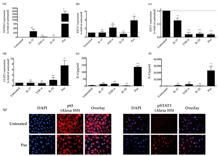
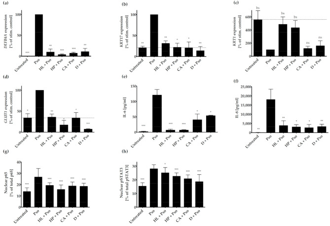
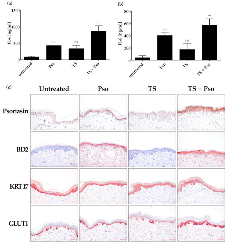
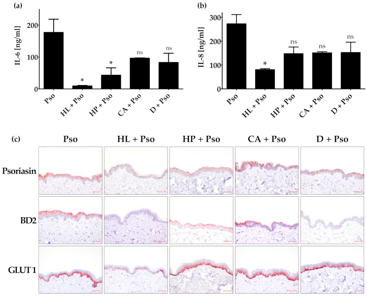
Psoriasis is a chronic inflammatory skin disease characterized by hyperproliferation of keratinocytes and expression of pro-inflammatory cytokines in the epidermis. New biological drugs were developed for the systemic treatment of moderate to severe psoriasis. However, products for the topical treatment of mild psoriasis are still required. Here, we examined the effect of natural compounds on psoriasis-like keratinocytes in vitro and ex vivo. Psoriasis-like keratinocytes were generated by treating human primary keratinocytes with the psoriasis-associated cytokines IL-17A, TNF-α and IL-22. Initially, 10 botanical extracts from Ayurvedic Medicine, Traditional Chinese Medicine, Northern American traditional medicine and Occidental Monastic Medicine were investigated using BrdU assays and IL-6 and IL-8 ELISAs. Curcuma amada, Humulus lupulus and Hypericum perforatum turned out to be the most effective plant extracts. In vitro, the plant extracts inhibited the expression of anti-microbial peptides (β-defensin 2), the hyperproliferation marker keratin 17, the glucose transporter 1 and downregulated the nuclear translocation of NF-κB and pSTAT3. In an ex vivo psoriasis model, Humulus lupulus displayed the most prominent anti-proliferative and anti-inflammatory effect. In conclusion, among the plant extracts investigated, Humulus lupulus showed the most promising anti-psoriatic effect. It is an interesting candidate for topical psoriasis treatment that should be further studied in clinical trials.

Keywords: Curcuma amada; Humulus lupulus; Hypericum perforatum; inflammation; interleukin 17A; interleukin 22; psoriasis; β-defensin 2.


Conflict of interest statement

The authors declare no conflict of interest. The founding sponsors had no role in the design of the study, in the collection, analyses, or interpretation of data, and in the writing the manuscript.

Figures

Figure 1
Figure 1
IL-17A-, TNF-α- and IL-22-induced synergy by expression of psoriasis-related genes or proteins. HPK were stimulated with IL-17A (20 ng/mL), TNF-α (20 ng/mL), IL-22 (20 ng/mL) or a combination of these cytokines (Pso) for 24 h. (a) DEFB4A, (b) KRT17, (c) KRT1 and (d) GLUT1 mRNA expression was analyzed by real time qRT-PCR (n = 3). ACTB was used as reference gene for normalization. Protein level of secreted IL-6 (e) and IL-8 (f) was measured in the cell culture supernatant by ELISA (n = 3). The dotted line indicates the value of the untreated sample. Data shown as mean ± SD of three independent experiments. (g) HPK were stimulated with the cytokine combination (20 ng/mL of IL-17A, IL-22 and TNF-α, Pso) for 24 h and immunofluorescence stainings of p65 NFκB and pSTAT3 were performed. The nucleus is stained with DAPI. Representative pictures of p65 NFκB and pSTAT3 staining in untreated and cytokine-treated cells. The % of total protein in the nucleus was measured using the Intensity Ratio Nuclei Cytoplasm Tool (RRID:SCR_018573) in ImageJ. Data shown as mean ± SD of five independent experiments with two pictures per experiment. Statistics were always one-way ANOVA with Newman-Keuls post-test. * p ≤ 0.05, ** p ≤ 0.01, *** p ≤ 0.001, ns-not significant (p > 0.1).
Figure 2
Figure 2
Effect of plant extracts on psoriasis-related genes or proteins. HPK were incubated with or without plant extracts (1 µg/mL HL (Humulus lupulus), 1 µg/mL HP (Hypericum perforatum), 2 µg/mL CA (Curcuma amara) or 0.3 µg/mL D (dithranol) for 2 h and then stimulated with psoriasis cytokines (IL-17A, TNF-α and IL-22; 20 ng/mL each) for 22 h (Pso). (a) DEFB4A, (b) KRT17, (c) KRT1 and (d) GLUT1 mRNA expression was analyzed by real time qRT-PCR (n = 3). ACTB was used as reference gene for normalization. Protein level of secreted IL-6 (e) and IL-8 (f) was measured in the cell culture supernatant by ELISA (n = 3). The dotted line indicates the value of the untreated sample. Data shown as mean ± SD of three independent experiments. (g) Effect of plant extracts on p65 subunit of NF-κB and (h) pSTAT3 nuclear translocation. HPK were pre-treated with either 1 µg/mL HL (Humulus lupulus), 1 µg/mL HP (Hypericum perforatum), 2 µg/mL CA (Curcuma amara) or 0.3 µg/mL D (dithranol) for 2 h prior to stimulation with 20 ng/mL of IL-17A, IL-22 and TNF-α. Cells were fixed, permeabilized and the p65 subunit of NF-κB and pSTAT3 were stained using the antibodies noted in the Materials and Methods Section, while DAPI was used for nuclear staining. The % of total protein in the nucleus was measured using the Intensity Ratio Nuclei Cytoplasm Tool (RRID:SCR_018573) in ImageJ. Data shown as mean ± SD of five independent experiments with two pictures per experiment. Statistics were always one-way ANOVA with Newman-Keuls post-test. * p ≤ 0.05, ** p ≤ 0.01, *** p ≤ 0.001, bs (borderline significant): 0.1 > p > 0.05, ns-not significant (p > 0.1).
Figure 3
Figure 3
Generation of an ex vivo psoriasis model. Skin models have been cultured for 4 days at the air-liquid interface using appropriate differentiation medium. The skin models were left untreated, stimulated with psoriasis cytokines (IL-17A, TNF-α, IL-22, 20 ng/mL each; Pso), tape stripped (TS) or both actions were performed (TS + Pso). The protein level of secreted IL-6 (a) and IL-8 (b) was measured in the cell culture supernatant by ELISA (n = 4). (c) Skin models were fixed, embedded in paraffin and 3 µm sections were stained with antibodies against psoriasin (S100A7), β-defensin 2 (BD2), keratin 17 (KRT17) or glucose transporter 1 (GLUT 1) (n = 4). Scale bar = 100 µm. ** p ≤ 0.01, *** p ≤ 0.001, ns-not significant (p > 0.1).
Figure 4
Figure 4
Effect of plant extracts in the ex vivo psoriasis model. The skin models were incubated with or without filter paper soaked with 40 µL plant extract solution on top of the skin explants (1.5 µg/mL HL (Humulus lupulus), 1.5 µg/mL HP (Hypericum perforatum), 3 µg/mL CA (Curcuma amara) or 0.4 µg/mL D (dithranol) for 24 h and then stimulated with psoriasis cytokines (IL-17A, TNF-α and IL-22; 20 ng/mL each (Pso)) for 72 h. The protein level of secreted IL-6 (a) and IL-8 (b) was measured in the cell culture supernatant by ELISA (n = 2). (c) Skin models were fixed, embedded in paraffin and 3 µm sections were stained with antibodies against psoriasin (S100A7), β-defensin 2 (BD2), keratin 17 (KRT17) or glucose transporter 1 (GLUT1). Scale bar = 100 µm. * p ≤ 0.05, ns-not significant (p > 0.1).

References

    1. Melnikova I. Psoriasis market. Nat. Rev. Drug Discov. 2009;8:767–768. doi: 10.1038/nrd2996. - DOI - PubMed
    1. Benezeder T., Wolf P. Resolution of plaque-type psoriasis: What is left behind (and reinitiates the disease) Semin. Immunopathol. 2019;41:633–644. doi: 10.1007/s00281-019-00766-z. - DOI - PMC - PubMed
    1. Hoffjan S., Stemmler S. On the role of the epidermal differentiation complex in ichthyosis vulgaris, atopic dermatitis and psoriasis. Br. J. Dermatol. 2007;157:441–449. doi: 10.1111/j.1365-2133.2007.07999.x. - DOI - PubMed
    1. Yang L., Fan X., Cui T., Dang E., Wang G. Nrf2 Promotes Keratinocyte Proliferation in Psoriasis through Up-Regulation of Keratin 6, Keratin 16, and Keratin 17. J. Investig. Dermatol. 2017;137:2168–2176. doi: 10.1016/j.jid.2017.05.015. - DOI - PubMed
    1. Herster F., Bittner Z., Archer N.K., Dickhöfer S., Eisel D., Eigenbrod T., Knorpp T., Schneiderhan-Marra N., Löffler M.W., Kalbacher H., et al. Neutrophil extracellular trap-associated RNA and LL37 enable self-amplifying inflammation in psoriasis. Nat. Commun. 2020;11:1–13. doi: 10.1038/s41467-019-13756-4. - DOI - PMC - PubMed

Publication types

LinkOut - more resources