. 2021 Mar 2;9(3):247.
doi: 10.3390/biomedicines9030247.

Retinal Microvascular Impairment in COVID-19 Bilateral Pneumonia Assessed by Optical Coherence Tomography Angiography

Affiliations

Retinal Microvascular Impairment in COVID-19 Bilateral Pneumonia Assessed by Optical Coherence Tomography Angiography

Jorge González-Zamora et al. Biomedicines. .

Abstract

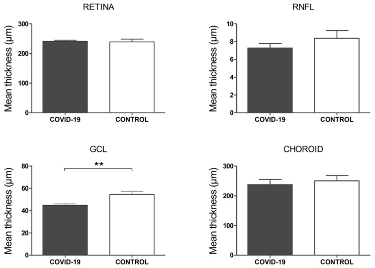
The purpose of this study was to evaluate the presence of retinal and microvascular alterations in COVID-19 patients with bilateral pneumonia due to SARS-COV-2 that required hospital admission and compare this with a cohort of age- and sex-matched controls. COVID-19 bilateral pneumonia patients underwent retinal imaging 14 days after hospital discharge with structural optical coherence tomography (OCT) and optical coherence tomography angiography (OCTA) measurements. Vessel density (VD) and foveal avascular zone (FAZ) area were evaluated in the superficial, deep capillary plexus (SCP, DCP), and choriocapillaris (CC). After exclusion criteria, only one eye per patient was selected, and 50 eyes (25 patients and 25 controls) were included in the analysis. COVID-19 patients presented significantly thinner ganglion cell layer (GCL) (p = 0.003) and thicker retinal nerve fiber layer (RNFL) compared to controls (p = 0.048), and this RNFL thickening was greater in COVID-19 cases with cotton wool spots (CWS), when compared with patients without CWS (p = 0.032). In both SCP and DCP, COVID-19 patients presented lower VD in the foveal region (p < 0.001) and a greater FAZ area than controls (p = 0.007). These findings suggest that thrombotic and inflammatory phenomena could be happening in the retina of COVID-19 patients. Further research is warranted to analyze the longitudinal evolution of these changes over time as well as their correlation with disease severity.

Keywords: COVID-19; OCT; OCTA; SARS-COV-2; coronavirus; cotton wool spot; microvascular; optical coherence tomography; optical coherence tomography angiography; retina.


Conflict of interest statement

The authors declare no conflict of interest.

Figures

Figure 1
Figure 1
Consolidated standard for reporting trials (CONSORT)-style flow chart describing included and excluded eyes in the study.
Figure 2
Figure 2
Regions of interest in the macula, defined by the early treatment diabetic retinopathy (ETDRS) grid subfields. The evaluated fields on each of the structural optical coherence tomography (OCT) and OCT angiography (OCTA) are presented in gray (A). Structural OCT, macula. Central ETDRS subfield measurements of foveal, retinal nerve fiber layer (RNFL), ganglion cell layer (GCL), and choroid thickness, respectively (B). Structural OCT, optic nerve head. Four quadrants (C). OCTA, central and inner ring ETDRS subfields. Vessel density measurements were obtained in the superficial capillary plexus (SCP), deep capillary plexus (DCP), and choriocapillaris (CC) layers. F: fovea. IS: inner superior. IN: inner nasal. II: inner inferior. IT: inner temporal. OS: outer superior. ON: outer nasal. OI: outer inferior. OT: outer temporal. S: superior. N: nasal. I: inferior. T: temporal.
Figure 3
Figure 3
Structural optical coherence tomography (OCT) analysis, macula. Mean foveal thickness (central subfield of the ETDRS grid) in microns of the retina, retinal nerve fiber layer (RNFL), ganglion cell layer (GCL), and choroid layers. p-values correspond to a comparison between COVID-19 patients and controls. Error bars correspond to the SEM. ** p < 0.01.
Figure 4
Figure 4
Structural optical coherence tomography (OCT) analysis, optic nerve head. Mean thickness in microns of the retinal nerve fiber layer (RNFL). Left p-values correspond to a comparison between COVID-19 patients and controls. Right p-values correspond to a comparison between COVID-19 patients with cotton wool spots (CWS) and without CWS. Error bars correspond to the SEM. * p < 0.05.
Figure 5
Figure 5
Optical coherence tomography angiography (OCTA) analysis. Top row: Superficial capillary plexus (SCP) analysis. Mean vessel density (%) of the foveal and parafoveal regions. Foveal avascular zone (FAZ) area. Middle row: Deep capillary plexus (DCP) analysis. Mean vessel density (%) of the foveal and parafoveal regions. Foveal avascular zone (FAZ) area. Bottom row: Choriocapillaris (CC) analysis. Mean vessel density (%) of the foveal and parafoveal regions. p-values correspond to a comparison between COVID-19 patients and controls. Error bars correspond to the SEM. ** p < 0.01, *** p < 0.001.
Figure 6
Figure 6
Composite images of case-control comparison. Top row: COVID-19 patient with bilateral pneumonia due to SARS-COV-2 case. Bottom row: Age-, sex- and laterality-matched control. Fundus retinography (left column), optical coherence tomography angiography (OCTA) vessel density maps (middle column), and OCTA image of the superficial capillary plexus (SCP) are depicted. In this case, a cotton wool spot is presented in the fundus retinography (green arrowhead). Reduced vessel density (15.38 vs. 30.08) and enlarged foveal avascular zone (FAZ) (225.8 vs. 189.2 µm2) are observed in the case compared to the matched control. 4.5 × 4.5 mm en face angiogram of the SCP are shown. Manually delineated FAZ are shown in green.

References

    1. Hoffmann M., Kleine-Weber H., Schroeder S., Krüger N., Herrler T., Erichsen S., Schiergens T.S., Herrler G., Wu N.H., Nitsche A., et al. SARS-CoV-2 Cell Entry Depends on ACE2 and TMPRSS2 and Is Blocked by a Clinically Proven Protease Inhibitor. Cell. 2020;181:271–280. doi: 10.1016/j.cell.2020.02.052. - DOI - PMC - PubMed
    1. Recalde-Zamacona B., García-Tobar L., Argueta A., Álvarez L., De Andrea C.E., Fernández Alonso M., Ezponda A., Carmona Torre F., Jordán Iborra C., Quiroga J.A., et al. Histopathological findings in fatal COVID-19 severe acute respiratory syndrome: Preliminary experience from a series of 10 Spanish patients. Thorax. 2020 doi: 10.1136/thoraxjnl-2020-215577. - DOI - PubMed
    1. Kasal D.A., De Lorenzo A., Tibiriçá E. COVID-19 and Microvascular Disease: Pathophysiology of SARS-CoV-2 Infection With Focus on the Renin-Angiotensin System. Hear. Lung Circ. 2020;29:1596–1602. doi: 10.1016/j.hlc.2020.08.010. - DOI - PMC - PubMed
    1. Varga Z., Flammer A.J., Steiger P., Haberecker M., Andermatt R., Zinkernagel A.S., Mehra M.R., Schuepbach R.A., Ruschitzka F., Moch H. Endothelial cell infection and endotheliitis in COVID-19. Lancet. 2020;395:1417–1418. doi: 10.1016/S0140-6736(20)30937-5. - DOI - PMC - PubMed
    1. Paniz-Mondolfi A., Bryce C., Grimes Z., Gordon R.E., Reidy J., Lednicky J., Sordillo E.M., Fowkes M. Central nervous system involvement by severe acute respiratory syndrome coronavirus-2 (SARS-CoV-2) J. Med. Virol. 2020;92:699–702. doi: 10.1002/jmv.25915. - DOI - PMC - PubMed

LinkOut - more resources