

Management of the Sequelae of a Sport-Related Traumatic Dental Injury Using Ultrasound Examination in the Diagnosis and Follow-Up
- PMID: 33801327
- PMCID: PMC8000130
- DOI: 10.3390/dj9030027
Management of the Sequelae of a Sport-Related Traumatic Dental Injury Using Ultrasound Examination in the Diagnosis and Follow-Up
Abstract
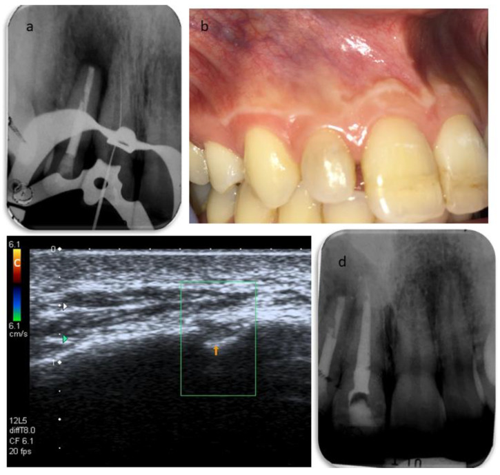
About a quarter of all oral pathologies involving the oral cavity and dental apparatus are traumatic injuries, and a substantial number of these cases are the result of sports injuries affecting adolescents and young adults. Here, we report the case of a 25-year-old healthy female referred to the department of Endodontics for the evaluation and management of teeth 1.2 and 1.1 because of a chronic apical abscess in an area involved in a sport-related dental trauma in the past. A multi-modular diagnostic assessment, comprising conventional periapical radiographs, CBCT imaging, ultrasound, and histopathologic examination, led to a final diagnosis of an apical granulomatous lesion connected to both teeth, and an associated sinus tract. During the follow-up period of three years, the patient was reviewed twice a year and showed progressive healing of the bone and absence of the sinus tract. The present report shows the challenges of diagnosing complications arising from past dental trauma. Furthermore, it is the first documented traumatic case where ultrasound examination was fruitfully used. Emphasis should be put on introducing diagnostic ultrasound for the management of both apical periodontitis and the related sinus tract.
Keywords: apical periodontitis; sport; traumatic dental injury; ultrasound examination.
Conflict of interest statement
The authors declare no conflict of interest.
Figures


Similar articles
-
Multimodular Assessment of a Traumatic Bone Cyst Overlapped with Apical Periodontitis.Case Rep Dent. 2020 Nov 25;2020:8829305. doi: 10.1155/2020/8829305. eCollection 2020. Case Rep Dent. 2020. PMID: 33294232 Free PMC article.
-
Association between the Presence of Apical Periodontitis and Clinical Symptoms in Endodontic Patients Using Cone-beam Computed Tomography and Periapical Radiographs.J Endod. 2015 Nov;41(11):1824-9. doi: 10.1016/j.joen.2015.06.004. Epub 2015 Sep 5. J Endod. 2015. PMID: 26349581
-
An evaluation of the periapical status of teeth with necrotic pulps using periapical radiography and cone-beam computed tomography.Int Endod J. 2014 Apr;47(4):387-96. doi: 10.1111/iej.12159. Epub 2013 Jul 26. Int Endod J. 2014. PMID: 23889592
-
Treatment of a large cystlike inflammatory periapical lesion associated with mature necrotic teeth using regenerative endodontic therapy.J Endod. 2014 Dec;40(12):2081-6. doi: 10.1016/j.joen.2014.07.027. Epub 2014 Oct 5. J Endod. 2014. PMID: 25292168
-
Endodontic management of immature teeth with spontaneous apical closure and periapical lesions: case series and review of the literature.Dent Traumatol. 2015 Aug;31(4):324-7. doi: 10.1111/edt.12164. Epub 2015 Mar 2. Dent Traumatol. 2015. PMID: 25731672 Review.
Cited by
-
Diagnostic Value of Specialist Systems in Sports Knee Injuries.Scanning. 2022 Aug 31;2022:1892877. doi: 10.1155/2022/1892877. eCollection 2022. Scanning. 2022. Retraction in: Scanning. 2023 Dec 13;2023:9876203. doi: 10.1155/2023/9876203. PMID: 36105552 Free PMC article. Retracted.
-
Management strategies for sport-related traumatic dental injuries: a systematic review based on case reports.BMC Sports Sci Med Rehabil. 2025 Jul 19;17(1):208. doi: 10.1186/s13102-025-01188-1. BMC Sports Sci Med Rehabil. 2025. PMID: 40684222 Free PMC article.
References
-
- Spinas E., Aresu M., Giannetti L. Use of mouth guard in basketball: Observational study of a group of teenagers with and without motivational reinforcement. Eur. J. Paediatr. Dent. 2014;15:392–396. - PubMed
-
- Spinas E., Giannetti L., Mameli A., Re D. Dental injuries in young athletes, a five-year follow-up study. Eur. J. Paediatr. Dent. 2018;19:187–193. - PubMed
Publication types
LinkOut - more resources
Full Text Sources
Other Literature Sources
Miscellaneous
