

CXCL10 Is an Agonist of the CC Family Chemokine Scavenger Receptor ACKR2/D6
- PMID: 33801414
- PMCID: PMC7958614
- DOI: 10.3390/cancers13051054
CXCL10 Is an Agonist of the CC Family Chemokine Scavenger Receptor ACKR2/D6
Abstract
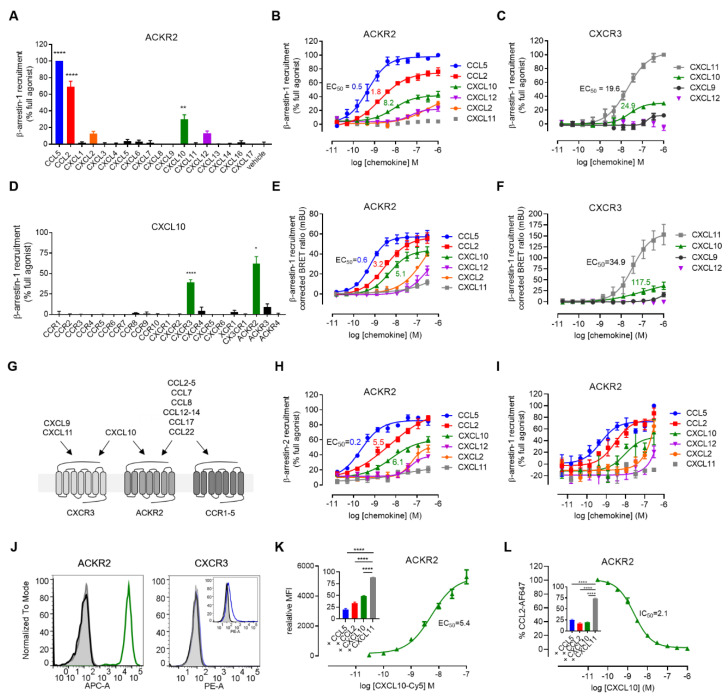
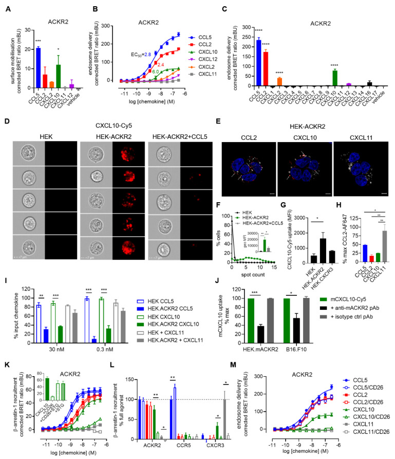
Atypical chemokine receptors (ACKRs) are important regulators of chemokine functions. Among them, the atypical chemokine receptor ACKR2 (also known as D6) has long been considered as a scavenger of inflammatory chemokines exclusively from the CC family. In this study, by using highly sensitive β-arrestin recruitment assays based on NanoBiT and NanoBRET technologies, we identified the inflammatory CXC chemokine CXCL10 as a new strong agonist ligand for ACKR2. CXCL10 is known to play an important role in the infiltration of immune cells into the tumour bed and was previously reported to bind to CXCR3 only. We demonstrated that ACKR2 is able to internalize and reduce the availability of CXCL10 in the extracellular space. Moreover, we found that, in contrast to CC chemokines, CXCL10 activity towards ACKR2 was drastically reduced by the dipeptidyl peptidase 4 (DPP4 or CD26) N-terminal processing, pointing to a different receptor binding pocket occupancy by CC and CXC chemokines. Overall, our study sheds new light on the complexity of the chemokine network and the potential role of CXCL10 regulation by ACKR2 in many physiological and pathological processes, including tumour immunology. Our data also testify that systematic reassessment of chemokine-receptor pairing is critically needed as important interactions may remain unexplored.
Keywords: ACKR2; ACKR3; CD26; CXCL10; CXCL12; CXCL2; CXCR3; D6; DPP4; IP-10; NanoBRET; NanoBiT; scavenger.
Conflict of interest statement
A patent application has been filed on “Specific ACKR2 modulators for use in therapy” (Applicant: Luxembourg Institute of Health).
Figures
Similar articles
-
Bystander Expression of Atypical Chemokine Receptor 2 Protects T Cells from Chemoattraction towards Cancer-Associated Fibroblasts.Eur J Immunol. 2025 Feb;55(2):e202451215. doi: 10.1002/eji.202451215. Eur J Immunol. 2025. PMID: 39931761 Free PMC article.
-
Extended repertoire of CXC chemokines acting as agonists and antagonists of the human and murine atypical chemokine receptor ACKR2.J Leukoc Biol. 2025 Apr 23;117(4):qiaf013. doi: 10.1093/jleuko/qiaf013. J Leukoc Biol. 2025. PMID: 39903605 Free PMC article.
-
Role of atypical chemokine receptor ACKR2 in experimental oral squamous cell carcinogenesis.Cytokine. 2019 Jun;118:160-167. doi: 10.1016/j.cyto.2018.03.001. Epub 2018 Mar 15. Cytokine. 2019. PMID: 29550065
-
New pairings and deorphanization among the atypical chemokine receptor family - physiological and clinical relevance.Front Immunol. 2023 Apr 20;14:1133394. doi: 10.3389/fimmu.2023.1133394. eCollection 2023. Front Immunol. 2023. PMID: 37153591 Free PMC article. Review.
-
The Role of Atypical Chemokine Receptor D6 (ACKR2) in Physiological and Pathological Conditions; Friend, Foe, or Both?Front Immunol. 2022 May 23;13:861931. doi: 10.3389/fimmu.2022.861931. eCollection 2022. Front Immunol. 2022. PMID: 35677043 Free PMC article. Review.
Cited by
-
β-arrestin1 and 2 exhibit distinct phosphorylation-dependent conformations when coupling to the same GPCR in living cells.Nat Commun. 2022 Sep 26;13(1):5638. doi: 10.1038/s41467-022-33307-8. Nat Commun. 2022. PMID: 36163356 Free PMC article.
-
The Role of Post-Translational Modifications of Chemokines by CD26 in Cancer.Cancers (Basel). 2021 Aug 24;13(17):4247. doi: 10.3390/cancers13174247. Cancers (Basel). 2021. PMID: 34503058 Free PMC article. Review.
-
Bystander Expression of Atypical Chemokine Receptor 2 Protects T Cells from Chemoattraction towards Cancer-Associated Fibroblasts.Eur J Immunol. 2025 Feb;55(2):e202451215. doi: 10.1002/eji.202451215. Eur J Immunol. 2025. PMID: 39931761 Free PMC article.
-
Transcriptional characterization of sepsis in a LPS porcine model.Mol Genet Genomics. 2025 Jun 5;300(1):57. doi: 10.1007/s00438-025-02261-7. Mol Genet Genomics. 2025. PMID: 40471388
-
Dual role of CXCL10 in cancer progression: implications for immunotherapy and targeted treatment.Cancer Biol Ther. 2025 Dec;26(1):2538962. doi: 10.1080/15384047.2025.2538962. Epub 2025 Aug 4. Cancer Biol Ther. 2025. PMID: 40760734 Free PMC article. Review.
References
-
- Vacchini A., Cancellieri C., Milanesi S., Badanai S., Savino B., Bifari F., Locati M., Bonecchi R., Borroni E.M. Control of Cytoskeletal Dynamics by beta-Arrestin1/Myosin Vb Signaling Regulates Endosomal Sorting and Scavenging Activity of the Atypical Chemokine Receptor ACKR2. Vaccines. 2020;8:542. doi: 10.3390/vaccines8030542. - DOI - PMC - PubMed
Grants and funding
- Pathfinder "Interceptor" 19/14260467, INTER/FWO "Nanokine" grant 15/10358798, INTER/FNRS grants 20/15084569, CORE "COMBATIC" 18/12670304 and PoC "Megakine" 19/14209621, grants AFR-3004509, PRIDE-11012546 "NextImmune" and PRIDE-10675146 "CANBIO"/Fonds National de la Recherche Luxembourg
- grants 7.4593.19, 7.4529.19 and 7.8504.20/Fonds De La Recherche Scientifique - FNRS
LinkOut - more resources
Full Text Sources
Other Literature Sources
Miscellaneous
