. 2021 Mar 2;11(3):618.
doi: 10.3390/nano11030618.

Nanomagnetic Actuation of Hybrid Stents for Hyperthermia Treatment of Hollow Organ Tumors

Affiliations

Nanomagnetic Actuation of Hybrid Stents for Hyperthermia Treatment of Hollow Organ Tumors

Benedikt Mues et al. Nanomaterials (Basel). .

Abstract

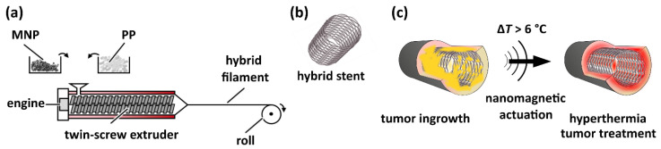
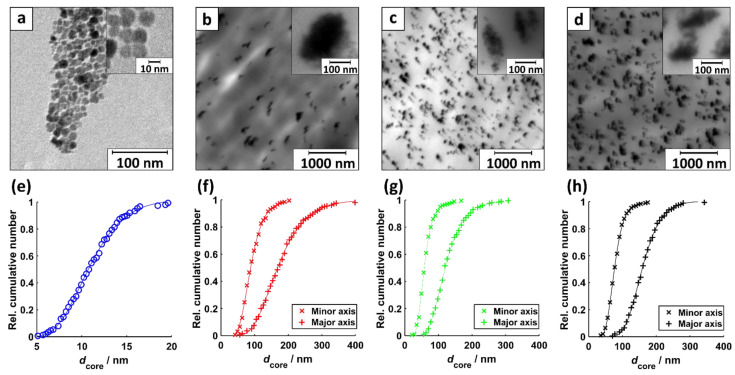
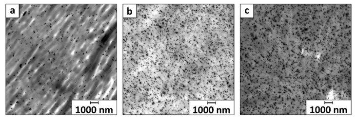
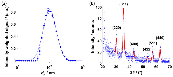
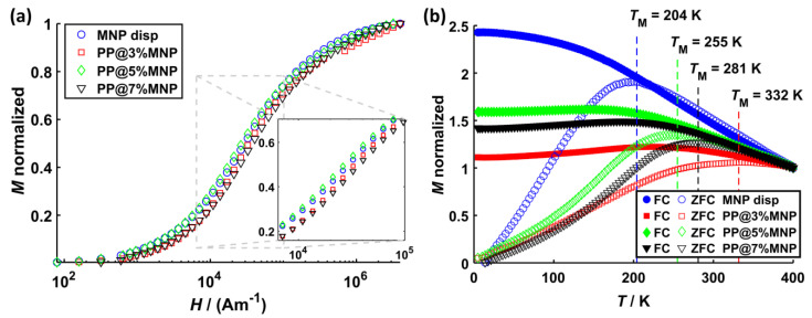
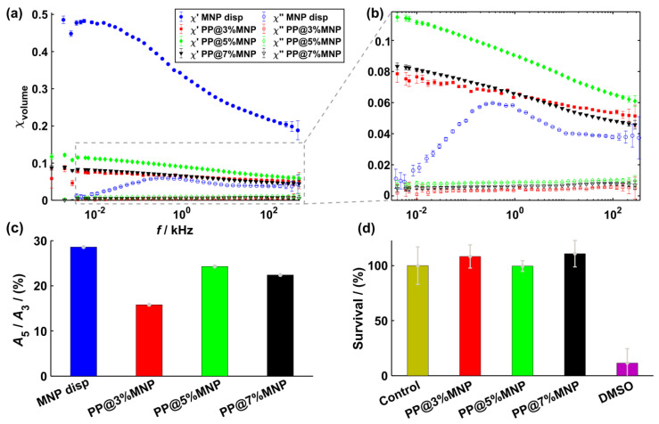
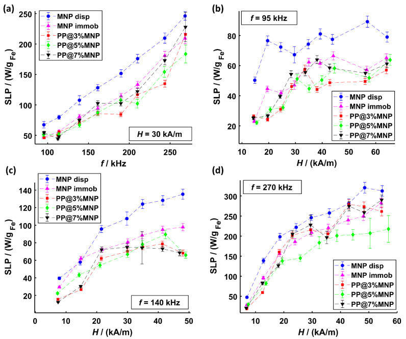
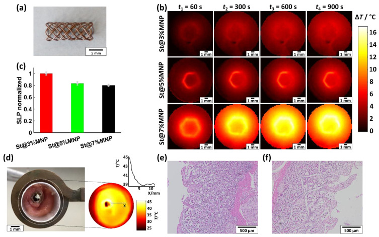
This paper describes a magnetic nanotechnology that locally enables hyperthermia treatment of hollow organ tumors by using polymer hybrid stents with incorporated magnetic nanoparticles (MNP). The hybrid stents are implanted and activated in an alternating magnetic field to generate therapeutically effective heat, thereby destroying the tumor. Here, we demonstrate the feasibility of nanomagnetic actuation of three prototype hybrid stents for hyperthermia treatment of hollow organ tumors. The results show that the heating efficiency of stent filaments increases with frequency from approximately 60 W/gFe (95 kHz) to approximately 250 W/gFe (270 kHz). The same trend is observed for the variation of magnetic field amplitude; however, heating efficiency saturates at approximately 30 kA/m. MNP immobilization strongly influences heating efficiency showing a relative difference in heating output of up to 60% compared to that of freely dispersed MNP. The stents showed uniformly distributed heat on their surface reaching therapeutically effective temperatures of 43 °C and were tested in an explanted pig bile duct for their biological safety. Nanomagnetic actuation of hybrid stents opens new possibilities in cancer treatment of hollow organ tumors.

Keywords: Brownian relaxation; Néel relaxation; hybrid implants; hyperthermia efficiency; magnetic nanoparticles; stents; tumor therapy.


Conflict of interest statement

The authors declare no conflict of interest.

Figures

Figure 1
Figure 1
(a) Illustration of the production process of hybrid filaments via melt-spinning of PP pellets and MNP with a twin-screw extruder. (b) Sketch of a hybrid stent. (c) Illustration of hyperthermia approach to treat hollow organ tumors using a hybrid stent: The MNP inside the stent are actuated in an alternating magnetic field, leading to a temperature increase by more than 6 °C at the stent surface. This local overheating leads to the destruction of the tumor (colored in yellow) near the stent surface.
Figure 2
Figure 2
TEM images of (a) MNP disp, (b) MNP inside PP@3%MNP, (c) PP@5%MNP, and (d) PP@7%MNP filaments. The insets show an exemplary magnification of one part of the same image. The corresponding core diameter distribution with a fit to a log-normal CDF of (e) spherical shaped dispersed MNP (R2 = 0.9987), (f) minor and major axis of ellipsoidal shaped MNP agglomerated inside PP@3%MNP (R2min = 0.9984, R2maj = 0.9992), (g) PP@5%MNP (R2min = 0.9991, R2maj = 0.9991), and (h) PP@7%MNP filaments (R2min = 0.9995, R2maj = 0.9992).
Figure 3
Figure 3
Overview TEM images of (a) MNP inside PP@3%MNP, (b) PP@5%MNP, and (c) PP@7%MNP filaments displayed with a magnification factor of 600. The displayed area is approximately 225 µm2.
Figure 4
Figure 4
(a) Intensity-weighted size distribution of the hydrodynamic diameters with a fit (R2 = 0.9973) to the PDF of a log-normal distribution for the MNP dispersed in water. (b) XRD intensity profile for MNP dispersed in water with a fit of the Pseudo-Voigt function to determine the corresponding Bragg angles (see Supplementary Materials S4 for R2 values).
Figure 5
Figure 5
(a) Virgin magnetization curves as well as (b) ZFC (open symbols) and FC (filled symbols) magnetization curves for dispersed MNP, PP@3%MNP, PP@5%MNP, and PP@7%MNP. The virgin curves are normalized to the saturation magnetization, while ZFC/FC curves are normalized to the initial magnetization value of the FC curve.
Figure 6
Figure 6
(a) Real part χ′ (filled symbols) and imaginary part χ″ (open symbols) of the complex dynamic susceptibility for dispersed MNP, MNP inside PP@3%MNP, PP@5%MNP, and PP@7%MNP filaments. (b) Zoom of the volume susceptibility at lower values. (c) A5/A3 amplitude ratios for dispersed MNP, MNP inside PP@3%MNP, PP@5%MNP, and PP@7%MNP filaments. (d) Survival analysis of L929 cells after 24 h incubation with hybrid filaments (PP@3%MNP, PP@5%MNP, and PP@7%MNP).
Figure 7
Figure 7
SLP values of MNP dispersed in water and MNP immobilized inside hydrogels as well as inside PP@3%MNP, PP@5%MNP, and PP@7%MNP filaments for (a) different frequencies at a constant magnetic field amplitude of 30 kA/m, (b) different magnetic field amplitudes at 95 kHz, (c) different magnetic field amplitudes at 140 kHz, and (d) different magnetic field amplitudes at 270 kHz.
Figure 8
Figure 8
(a) Photograph of an exemplary St@7%MNP stent. (b) Temperature rise ΔT of St@3%MNP, St@5%MNP, and St@7%MNP during the application of an AMF with f = 270 kHz and H = 54 kA/m with a thermographic camera at t1 = 60 s, t2 = 300 s, t3 = 600 s, and t4 = 900 s. (c) Normalized SLP values of St@3%MNP, St@5%MNP, and St@7%MNP (absolute SLP values are provided in Table S6 in Supplemantary Materials S6). (d) Photograph of an exemplary St@5%MNP stent inserted into a resected bile duct of a pig. The inset shows the temperature distribution during the application of an AFM with f = 270 kHz and H = 38 kA/m. (e) HE staining images of the heated bile duct and (f) an untreated reference bile duct.

References

    1. Bakheet N., Park J.-H., Hu H.-T., Yoon S.H., Kim K.Y., Zhe W., Jeon J.Y., Song H.-Y. Fully Covered Self-Expandable Esophageal Metallic Stents in Patients with Inoperable Malignant Disease Who Survived for More than 6 Months after Stent Placement. BJR. 2019;92:20190321. doi: 10.1259/bjr.20190321. - DOI - PMC - PubMed
    1. Gupta A., Gupta G., Gawande A., Kumar M., Tak V., Pokharna R., Sharma S., Nijhawan S. Self-Expanding Metallic Stents in Malignant Biliary Obstruction-Patency and Clinical Efficacy: A Prospective Study from North India Tertiary Center. J. Dig. Endosc. 2019;10:33–38. doi: 10.4103/jde.JDE_52_18. - DOI
    1. Maire F., Hammel P., Ponsot P., Aubert A., O’Toole D., Hentic O., Levy P., Ruszniewski P. Long-Term Outcome of Biliary and Duodenal Stents in Palliative Treatment of Patients with Unresectable Adenocarcinoma of the Head of Pancreas. Am. J. Gastroenterol. 2006;101:735–742. doi: 10.1111/j.1572-0241.2006.00559.x. - DOI - PubMed
    1. Wasan S.M., Ross W.A., Staerkel G.A., Lee J.H. Use of Expandable Metallic Biliary Stents in Resectable Pancreatic Cancer. Am. J. Gastroenterol. 2005;100:2056–2061. doi: 10.1111/j.1572-0241.2005.42031.x. - DOI - PubMed
    1. Zhu H.-D., Guo J.-H., Mao A.-W., Lv W.-F., Ji J.-S., Wang W.-H., Lv B., Yang R.-M., Wu W., Ni C.-F., et al. Conventional Stents versus Stents Loaded with 125iodine Seeds for the Treatment of Unresectable Oesophageal Cancer: A Multicentre, Randomised Phase 3 Trial. Lancet Oncol. 2014;15:612–619. doi: 10.1016/S1470-2045(14)70131-7. - DOI - PubMed