. 2021 Mar 2;13(3):397.
doi: 10.3390/v13030397.

Infection of Chinese Rhesus Monkeys with a Subtype C SHIV Resulted in Attenuated In Vivo Viral Replication Despite Successful Animal-to-Animal Serial Passages

Affiliations

Infection of Chinese Rhesus Monkeys with a Subtype C SHIV Resulted in Attenuated In Vivo Viral Replication Despite Successful Animal-to-Animal Serial Passages

Gerald K Chege et al. Viruses. .

Abstract

Rhesus macaques can be readily infected with chimeric simian-human immunodeficiency viruses (SHIV) as a suitable virus challenge system for testing the efficacy of HIV vaccines. Three Chinese-origin rhesus macaques (ChRM) were inoculated intravenously (IV) with SHIVC109P4 in a rapid serial in vivo passage. SHIV recovered from the peripheral blood of the final ChRM was used to generate a ChRM-adapted virus challenge stock. This stock was titrated for the intrarectal route (IR) in 8 ChRMs using undiluted, 1:10 or 1:100 dilutions, to determine a suitable dose for use in future vaccine efficacy testing via repeated low-dose IR challenges. All 11 ChRMs were successfully infected, reaching similar median peak viraemias at 1-2 weeks post inoculation but undetectable levels by 8 weeks post inoculation. T-cell responses were detected in all animals and Tier 1 neutralizing antibodies (Nab) developed in 10 of 11 infected ChRMs. All ChRMs remained healthy and maintained normal CD4+ T cell counts. Sequence analyses showed >98% amino acid identity between the original inoculum and virus recovered at peak viraemia indicating only minimal changes in the env gene. Thus, while replication is limited over time, our adapted SHIV can be used to test for protection of virus acquisition in ChRMs.

Keywords: Chinese rhesus macaques; SHIV; subtype C.


Conflict of interest statement

The authors declare no conflict of interest. The funders had no role in the design of the study; in the collection, analyses, or interpretation of data; in the writing of the manuscript, or in the decision to publish the results.

Figures
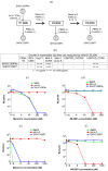
Figure 1
Figure 1
Generation and characterization of SHIV stocks (SHIVC109P4b and SHIVC109P7) which were derived from the parental virus, SHIVC109P4. (a) Schematic diagram showing how SHIVC109P4b was serially passaged in ChRMs to generate a macaque-adapted inoculum stock, SHIVC109P7. The cell tropism of SHIVC109P4 stocks, SHIVC109P4b and SHIVC109P7, for different mammalian cell lines (b) was assessed by measuring the level of viral replication in vitro using SIVp27 ELISA kits. In addition, the co-receptor usage of the SHIVC109P4b stock prior to passage in ChRMs (c,d) and SHIVC109P7 after passage (e,f) was determined using CCR5 (maraviroc) and CXCR4 (JM-2987) inhibitors. The effect of maraviroc (c,e)and JM-2987 (d,f)in a competitive inhibition assay to selectively block the entry of the viruses into TZM-bl cells was compared with control HIV-1 laboratory strains, BalR5 (R5-tropic) and pNL4-3 (X4-tropic). The line graphs show the level of viral replication of each virus in TZM-bl cells in the presence of serial dilutions of the co-receptor inhibitors. (IV: intravenous inoculation; PBMC: peripheral blood mononuclear cells isolated from heparinised whole blood).
Figure 2
Figure 2
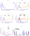
Plasma viral loads and CD4+ T cell counts pre-inoculation and at various time points p.i. (a) Shows the viral loads in the peripheral blood of monkeys 42, 43 and 32 which were inoculated IV with SHIVC109P4b, SHIVC109P5 and SHIVC109P6 respectively. SHIVC109P4b was prepared from the original SHIVC109P4 by in vitro expansion. Plasma viral loads (solid blue lines & symbols) and absolute CD4+ T cell counts (dashed orange lines & symbols) in the peripheral blood of ChRMs inoculated IR with 1:1 dilution (b), 1:10 dilution (c) and 1:100 dilution (d) of SHIVC109P7. Viral loads are also depicted as levels of SIV proviral DNA in the blood of ChRMs inoculated IR with 1:100 dilution of SHIVC109P7 at various time points (e) and both as SIV viral RNA and proviral DNA in the tissues (splenocytes and mononuclear cells extracted from either inguinal or axillary lymph node) at necropsy at the times indicated (f). Each line graph (a to e) represents an individual animal at pre-inoculation and p.i. with SHIV and each bar (f) represents a single tissue, either spleen or inguinal or axillary lymph node.: indicates inoculation times.
Figure 3
Figure 3
IFN-γ ELISpot responses in ChRMs following infection. (a) ChRMs (#32, #42, #43) inoculated IV with the passaged viruses. ChRMs inoculated IR with 1:1 dilution (#35, #36) (b), 1:10 dilution (#37, #38) (c) and 1:100 dilution (#47, #48, #51, #53) (d) of SHIVC109P7. The line graphs represent cumulative ELISpot responses to SIV Gag and HIV-1 Env peptide pools presented as spot-forming units (SFU) per million PBMC at each time point. Individual responses below the cut-off value of 30 SFU per million PBMC were considered negative and assigned a zero value.
Figure 4
Figure 4
Neutralizing antibody titres to Tier 1A pseudovirus MW965.26 elicited in ChRMs following infection. (a) ChRMs inoculated IV with the passaged viruses. (b) ChRMs inoculated IR with 1:1 dilution (35 and 36), 1:10 dilution (37 and 38) and 1:100 dilution (47, 48, 51 and 53) of SHIVC109P7. The line graphs represent the antibody titre that caused a 50% reduction in relative luciferase units. A titre <40 was considered negative for neutralization.
Figure 5
Figure 5
Amino acid sequence comparison of potential N-glycosylation sites (PNGS) in the V1, C2 and V4 regions of SHIVC109F.PB4, SHIVC109P4 and viruses isolated following serial passage of SHIVC109P4 in ChRMs. HIV-1 env single genome amplicons (10 per sample) were prepared from SHIVC109P4b (virus inoculum generated from in vitro expansion of SHIVC109P4 in ChRM PBMC), SHIVC109P5, SHIVC109P6 and SHIVC109P7 (virus from passage 1, 2 and 3 respectively). The numbers of amplicons matching the indicated sequence (*) is shown for the inoculum and each passage (per 10 clones sequenced). PNGS are shown in red font and new PNGS are indicated with red boxes.

Similar articles

Cited by

References

    1. Del Prete G.Q., Lifson J.D. Nonhuman primate models for studies of AIDS virus persistence during suppressive combination antiretroviral therapy. Curr. Top. Microbiol. Immunol. 2018;417:69–109. doi: 10.1007/82_2017_73. - DOI - PubMed
    1. Van Rompay K.K.A. Tackling HIV and AIDS: Contributions by non-human primate models. Lab Anim. 2017;46:259–270. doi: 10.1038/laban.1279. - DOI - PubMed
    1. Nishimura Y., Igarashi T., Donau O.K., Buckler-White A., Buckler C., Lafont B.A., Goeken R.M., Goldstein S., Hirsch V.M., Martin M.A. Highly pathogenic SHIVs and SIVs target different CD4+ T cell subsets in rhesus monkeys, explaining their divergent clinical courses. Proc. Natl. Acad. Sci. USA. 2004;101:12324–12329. doi: 10.1073/pnas.0404620101. - DOI - PMC - PubMed
    1. Cohen J. AIDS research. Vaccine studies stymied by shortage of animals. Science. 2000;287:959–960. doi: 10.1126/science.287.5455.959. - DOI - PubMed
    1. Ling B., Veazey R.S., Luckay A., Penedo C., Xu K., Lifson J.D., Marx P.A. SIV(mac) pathogenesis in rhesus macaques of Chinese and Indian origin compared with primary HIV infections in humans. AIDS. 2002;16:1489–1496. doi: 10.1097/00002030-200207260-00005. - DOI - PubMed

Publication types

LinkOut - more resources